Acta Orthop Traumatol Turc 2013;47(1):68-71 doi:10.3944/AOTT.2013.2831
CASE REPORT
Correspondence: Ali fieker, MD. ‹stanbul Medipol Üniversitesi Ortopedi ve Travmatoloji Anabilim Dal›, Ba¤c›lar, ‹stanbul, Turkey. Tel: +90 532 - 326 22 02 e-mail: [email protected]
Submitted: February 12, 2012 Accepted: August 16, 2012 ©2013 Turkish Association of Orthopaedics and Traumatology
Osteotendinous repair of bilateral spontaneous
quadriceps tendon ruptures with the Krackow
technique in two patients with chronic renal failure
Adnan KARA1, Seçkin SARI2, Ali fiEKER3, ‹rfan ÖZTÜRK4
1
Department of Orthopedics and Traumatology, fiiflli Etfal Training and Research Hospital, ‹stanbul, Turkey;
2
Department of Orthopedics and Traumatology, Baltaliman› Bone Diseases Training and Research Hospital, ‹stanbul, Turkey;
3
Department of Orthopedics and Traumatology, ‹stanbul Medipol University, ‹stanbul, Turkey;
4
Department of Orthopedics and Traumatology, ‹stanbul Medical Faculty, ‹stanbul University, ‹stanbul, Turkey
Available online at www.aott.org.tr doi:10.3944/AOTT.2013.2831 QR (Quick Response) Code:
Although unilateral traumatic quadriceps tendon rupture is a relatively frequent pathology, bilateral non-traumatic spontaneous ruptures are uncommon and are usually associated with chronic renal fail-ure, hyperparathyroidism, gout, and systemic lupus erythematosus. This paper aimed to discuss two patients with chronic renal failure treated with the Krackow suture technique for spontaneous bilater-al quadriceps tendon rupture.
Key words: Bilateral tendon rupture; Krackow technique; quadriceps.
Spontaneous bilateral quadriceps tendon rupture is a rarely seen condition. The majority of patients with spontaneous bilateral quadriceps tendon rupture have systemic disorders such as chronic renal failure, diabetes mellitus, gout, or secondary or tertiary hyperparathy-roidism. Cases of patients with obesity or chronic steroid usage have also been reported.[1,2]
In this paper, we present two patients with sponta-neous bilateral quadriceps tendon rupture. The Krackow technique was used for osteotendinous fixation in both patients.[3]
Case report
Case 1
A 43-year-old male patient with chronic renal failure due to diabetic nephropathy presented to the emergency
department with difficulty in walking after sudden giv-ing way of his knees while descendgiv-ing stairs. He was hemodialysis-dependent for 14 years. In physical exam-ination, the patient was unable to do a straight leg raise and gaps were palpable just superior to both patellas. Bilateral quadriceps tendon ruptures were detected with magnetic resonance imaging (MRI).
Case 2
A 34-year-old woman with chronic renal failure present-ed to the hospital complaining of experiencing a sudden giving way feeling while getting out of the car and the inability to walk afterwards. She was hemodialysis-dependent for 8 years. Gaps were detected superior to the both patella and straight leg raise tests were negative. MRI showed bilateral quadriceps tendon rupture (Fig. 1).
Kara et al. Osteotendinous repair of bilateral spontaneous quadriceps tendon ruptures with the Krackow technique 69
Fig. 1. (a, b) Appearance of the ruptured quadriceps tendon (arrow) in MRI sections of the second patient.
Fig. 2. (a) Intraoperative appearance of the ruptured tendon, (b) the patellar sulcus, (c) sutures passing through tunnels opened in the patella, (d) appearance of the repaired tendon. [Color figure can be viewed in the online issue, which is available at www.aott.org.tr]
Under general anesthesia, pneumatic tourniquets were applied at the level of the thigh. A 15 cm longitu-dinal incision was made from the inferior pole of the patella and extending upward. Following hematoma drainage and tendon debridement, a groove was made on the upper patellar pole. Three longitudinal tunnels
were opened at 1 cm intervals in the patella. Using the Krackow suture technique, two no. 5 Ethibond (non-absorbable polyester; Ethicon, Johnson & Johnson Medical Ltd., Edinburgh, UK) sutures were used to cre-ate double-row locked knots in the medial and lcre-ateral sides of the tendon. The tendon was placed into the
(a) (b)
(a) (b)
70 Acta Orthop Traumatol Turc
groove in the upper patellar pole by migrating the sutures through the tunnels and fastening in the lower pole (Fig. 2). Incisions were closed after application of suction drainages. Cylinder cast was applied with the knees in full extension for 6 weeks.
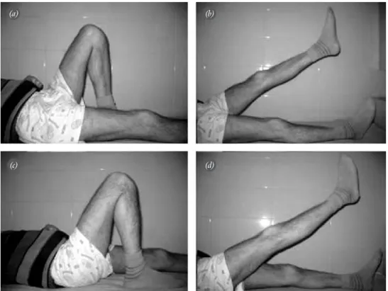
Patients were mobilized with double crutches. After cast removal, weight-bearing was allowed and an active-assisted rehabilitation program was started. Follow-up periods were 51 and 24 months, respectively. Both patients gained full range of motion and there were no occurrence of re-rupture (Fig. 3).
Discussion
Although unilateral traumatic quadriceps tendon rup-ture is a well-known pathology, bilateral non-traumatic spontaneous ruptures are not common. In such cases, delayed diagnosis and misdiagnosis are frequent. Neubaer et al. reported 32 delayed or incorrect diag-noses in 105 bilateral traumatic or spontaneous quadri-ceps tendon ruptures.[1]
Bilateral cases are usually related to chronic systemic diseases. In his series, Shah report-ed 43% renal failure, 16% obesity, 10% diabetes melli-tus, 5% primary hyperparathyroidism and 5% gout dis-ease and 12 patients had no risk factors.[2]
In the healthy patients, ruptures were seen more frequently in males and the elderly more.[1,2]
The mechanism of injury is sudden contraction of the quadriceps tendon of the flexed knee with the foot fixed on the ground. Structural failure of the tendon is the underlying cause.[1]
Uremia has adverse effect on collagen maturation by disturbing the protein-polysaccharide complex and decreased tendon elasticity caused by amyloid accumu-lation. In patients with osteodystrophic renal failure and hyperparathyroidism, osteoporosis causes subperiosteal resorption and decreases tendon endurance. Increased parathyroid hormone secretion causes deposition of cal-cium phosphate in the tendons and decreases elasticity. Fibrinoid necrosis and chronic inflammation in gout disease, long-term steroid treatments and obesity may also cause spontaneous ruptures. Steroids disturb the structure of collagen and obesity increases lipid accumu-lation in the tendons.[1,3,4]
In such cases, surgery is the treatment of choice. In the literature, nonsurgical treatment was chosen for three patients with unilateral ruptures and for only one patient with bilateral ruptures and the healing period was longer for these cases.[5]
There are several surgical treatment options although transosseous suturing through tunnels opened in the patella is more common. Krackow described a transosseous suture technique using a single suture
Fig. 3. (a-d) Images from the 51th month follow-up of the first patient. The patient had a full range of motion.
(a) (b)
Kara et al. Osteotendinous repair of bilateral spontaneous quadriceps tendon ruptures with the Krackow technique 71
material.[6]
This technique is biomechanically superior to the Kessler and Bunnel techniques. With the help of locking nodes, the elongation of tendon grafts was avoided in long periods.[7,8]
Scuderi advises reinforce-ment with wires and triangular flaps of the quadriceps muscle after tendon repair.[9]
Kayali et al. repaired the bilaterally ruptured quadriceps tendon of a patient with chronic renal failure with transpatellar sutures aug-mented with reverse quadriceps tendon and reported satisfactory results.[10]This treatment modality is similar
to our technique although we did not augment after repair. Krackow et al. reported better results with Krackow sutures ended around a screw than with bone-tendon grafts fixed with screws.[11] Parallel double
Krackow sutures were used in our patients and addi-tional reinforcement techniques were not used. There were no complication and complete range of motion was gained at the 3rd postoperative month.
In both cases, complete range of motion was achieved at the end of treatment. No re-rupture occurred during the follow-up. In our opinion, the strength of the repair prevents re-rupture and facili-tates healing despite disturbed collagen maturation. Different repair techniques are suitable for such injuries with good results but studies have not provid-ed long-term results.[1,2,10]
In conclusion, bilateral spontaneous quadriceps ten-don rupture is a rare pathology which may be seen in patients with systemic disorders. The Krackow suture technique, especially with double-rows, can produce good results even without reinforcement of the repair.
Conflicts of Interest: No conflicts declared.
References
1. Neubauer T, Wagner M, Potschka T, Riedl M. Bilateral, simultaneous rupture of the quadriceps tendon: a diagnostic pitfall? Report of three cases and meta-analysis of the litera-ture. Knee Surg Sports Traumatol Arthrosc 2007;15:43-53. 2. Shah MK. Outcomes in bilateral and simultaneous
quadri-ceps tendon rupture. Orthopedics 2003;26:797-8.
3. Shiota E, Tsuchiya K, Yamaoka K, Kawano O. Spontaneous major tendon ruptures in patients receiving long-term hemodialysis. Clin Orthop Relat Res 2002;(394):236-42. 4. Kannus P, Józsa L. Histopathological changes preceding
spontaneous rupture of a tendon. A controlled study of 891 patients. J Bone Joint Surg Am 1991;73:1507-25.
5. Tedd RJ, Norton MR, Thomas WG. Bilateral simultaneous atraumatic quadriceps tendon ruptures associated with ‘pseudogout’. Injury 2000;31:467-9.
6. Krackow KA. The Krackow suture: how, when, and why. Orthopedics 2008;31:931-3.
7. Watson TW, Jurist KA, Yang KH, Shen KL. The strength of Achilles tendon repair: an in vitro study of the biomechan-ical behavior in human cadaver tendons. Foot Ankle Int 1995;16:191-5.
8. Jassem M, Rose AT, Meister K, Indelicato PA, Wheeler D. Biomechanical analysis of the effect of varying suture pitch in tendon graft fixation. Am J Sports Med 2001;29:734-7. 9. Scuderi C. Ruptures of the quadriceps tendon: study on
twenty tendon ruptures. Am J Surg 1958;95:626-34. 10. Kayali C, Agus H, Turgut A, Taskiran C. Simultaneous
bilateral quadriceps tendon rupture in a patient on chronic haemodialysis. (Short-term results of treatment with transpatellar sutures augmented with a quadriceps tendon flap). Ortop Traumatol Rehabil 2008;10:286-91.
11. Krackow KA, Thomas SC, Jones LC. Ligament-tendon fix-ation: analysis of a new stitch and comparison with standard techniques. Orthopedics 1988;11:909-17.