The Relationship between Metabolic Syndrome, Its Components, and Dry Eye: A
Cross-Sectional Study
Sevil Karaman Erdura, Rukiye Aydina, Mustafa Ozsutcua, Oktay Olmuscelikb, Mustafa Eliacika, Goktug Demircia, and Mehmet Selim Kocaboraa
aMedicine School, Department of Ophthalmology, Istanbul Medipol University, Istanbul, Turkey;bMedicine School, Department of Internal Medicine,
Istanbul Medipol University, Istanbul, Turkey
ABSTRACT
Purpose: The aim of this study was to evaluate tear osmolarity and tear film function and ocular surface changes in patients with metabolic syndrome.
Methods: 108 eyes of 64 patients with metabolic syndrome (group 1) and 110 eyes of 55 healthy individuals (group 2) were included in this cross-sectional study. All participants were evaluated using the Ocular Surface Disease Index (OSDI) questionnaire, Schirmer I test, tear film break-up time (TBUT), and tear osmolarity. Main outcome measures were Ocular Surface Disease Index (OSDI) questionnaire, Schirmer I test, tear film break-up time (TBUT), and tear osmolarity values.
Results: Tear osmolarity values and OSDI scores were significantly higher in group 1 (314.4 ± 19.1 mOsm and 38.9 ± 1.1, respectively) compared with group 2 (295 ± 14.3 mOsm and 18.69 ± 17.2, respectively) (p = 0.01 for both). The Schirmer test values and TBUT in group 1 (10 ± 3.7 mm and 14.8 ± 3.6 sec, respectively) were significantly lower compared with group 2 (16.8 ± 2.6 mm and 18.1 ± 0.5 sec, respectively) (p < 0.001 for both). There was significant correlation between tear osmolarity versus waist circumference and fasting blood glucose in the study group (r = 0.364, p = 0.04; and r = 0.542, p≤ 0.001, respectively).
Conclusions: This study showed that metabolic syndrome can influence tear osmolarity and tear film function. Patients with metabolic syndrome showed tear hyperosmolarity and tear film dysfunction.
ARTICLE HISTORY
Received 20 September 2016 Accepted 4 January 2017
KEYWORDS
Dry eye; metabolic syndrome; Schirmer test; tear osmolarity; tear film
Introduction
Metabolic syndrome is becoming a worldwide medical and public health challenge as it has been seen increasing in preva-lence over the years.1This is a syndrome which has components such as dyslipidemia, high blood pressure, and high fasting plasma glucose.2 The etiology of metabolic syndrome mainly consists of two conditions: body fat accumulation which causes adult weight gain and dislocation of fat (central obesity) in intra-abdominal areas including liver, pancreas, and heart. The indi-viduals who have metabolic syndrome are at great risk for diabetes, cardiovascular, and cerebrovascular disease.3,4
Some studies reported associations between components of the metabolic syndrome and eye diseases such as cataract, age-related macular diseases, and glaucoma.5–8 Only one study investigated lacrimal function in patients with metabolic syndrome.9 The authors stated impairment in lacrimal gland functions and tear volume in these patients.
Schirmer I test, tear break-up time (TBUT), and ocular surface fluorescein staining score are the most frequently used tests in diagnosis and follow-up of dry eye disease. Tear osmolarity is valuable method for detecting dry eye. At the present time, by understanding the importance of tear hyperosmolarity in dry eye pathogenesis, tear osmolarity measurement has started to be used in clinical application.10
In this study, we investigated the tear osmolarity and tear film function in patients with metabolic syndrome.
Materials and methods
A total of 128 eyes of 64 patients with metabolic syndrome (group 1) and 110 eyes of 55 healthy individuals (group 2) were included in this single-center, cross-sectional observa-tional study. Right eye data for each patient were assessed.
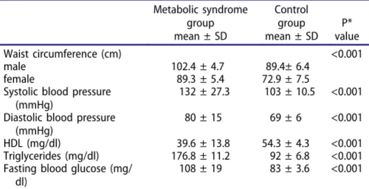
Patients in group 1 were newly diagnosed with metabolic syndrome by an internal specialist (O.O.). The diagnosis of metabolic syndrome was established by the International Diabetes Federation.4 According to the new IDF definition, for a person to be defined as having the metabolic syndrome, they must have: central obesity (waist circumference >80 cm for female and >94 cm for male) and any of the these two followings raised triglycerides;≥150 mg/dl or specific treat-ment for this lipid abnormality, reduced HDL cholesterol; < 40 mg/dL in males, < 50 mg/dL in females or specific treat-ment for this lipid abnormality, raised blood pressure; systolic BP ≥ 130 or diastolic BP ≥ 85 mm Hg or treatment of previously diagnosed hypertension, raised fasting plasma glu-cose;≥ 100 mg/dL, or previously diagnosed type 2 diabetes.
All subjects were excluded if they had a history of smoking, current or recent drug use that could affect the lacrimal
CONTACTSevil Karaman Erdur [email protected] Medicine School, Department of Ophthalmology, Istanbul Medipol University, Goztepe Mah. 309 Sk No. 4 Bagcilar, Istanbul 34214, Turkey.
CURRENT EYE RESEARCH 2017, VOL. 42, NO. 8, 1115–1117
http://dx.doi.org/10.1080/02713683.2017.1280511
functional unit, active ocular infection or allergy, ocular sur-face scarring, previous eye surgery, or current contact lens or any eye drop use. Any systemic disease other than metabolic syndrome was the other exclusion criteria in the study group. Healthy control subjects were first examined by O.O. for routine check-up, after exclusion metabolic syndrome or any other systemic diseases, and were referred to ophthalmology department.
The study was reviewed and approved by the Istanbul Medipol University Ethics Committee, and written informed consent was obtained from each patient before enrollment. The study was conducted in accordance with the tenets of the Declaration of Helsinki.
Initially, patients completed the International Ocular Surface Disease Index (OSDI) survey. All subjects underwent a full ophthalmological examination in the same order, including visual acuity assessment, standardized slit-lamp examination, and fundus examination. Full ophthalmologic examination and evaluation of tear osmolarity were per-formed on the same day but at different sessions.
Tear osmolarity measurements were evaluated using a TearLab osmometer (TearLab Co, San Diego, CA, USA). Tears were collected from the inferior lateral tear meniscus. Three consecutive measurements were obtained, and their mean was used for statistical analysis.11
Tear film break-up time was assessed after instillation of 2% fluorescein staining under a cobalt blue filter. The time interval between the last complete blink and the appearance of the first dry spot was recorded. The mean of three consecutive
measurements was obtained. The Schirmer I test was per-formed with topical anesthesia using a standardized filter strip (Bio-Tech Vision Care, Ahmedabad, Gujarat, India). The amount of wetting was measured after five minutes.
The normality of the distribution of each of the parameters was checked using the Kolmogorov–Smirnov normality test. The tear osmolarity, Schirmer I test, TBUT values, and OSDI scores between groups were compared using independent t-test. The correlations between each component of metabolic syndrome and tear osmolarity were evaluated using the Pearson’s partial correlation controlled for gender. A p value less than 0.05 was considered statistically significant.
Results
The mean subject age was 47 ± 7years (range 32 to 58 years) in group 1 (32 females, 32 males) and 45 ± 7 years (range 31 to 59 years) in group 2 (28 females, 27 males). There were no significant differences between the groups with respect to age or sex (p = 0.741 and p = 0.921, respectively). Comparison of metabolic syndrome components between two groups was given inTable 1.
Summary statistics are shown in the Table 2. The mean tear osmolarity and OSDI scores were significantly higher in group 1 compared with group 2 (p = 0.01). The TBUT and Schirmer measurements for group 1 were significantly lower compared with those for group 2 (p < 0.001).
As regards the correlations between tear osmolarity and metabolic syndrome components, there was significant cor-relation between tear osmolarity versus waist circumference and fasting blood glucose (r = 0.234, p = 0.04; and r = 0.542, p ≤ 0.001, respectively), whereas no significant correlation was found between tear osmolarity versus triglycerides, HDL and hypertension in the study group (r = 0.231, 0.642, 0.356, respectively, p > 0.05). No significant correlations were found among tear osmolarity versus waist circumference, fasting blood glucose, triglycerides, HDL, and hypertension in the control group (r = 0.435, 0.347, 0.566, 0.678, 0.788, respectively, p > 0.05)
Discussion
Metabolic syndrome is becoming a serious worldwide health problem. Its prevalence has been increasing year by year. The IDF estimates that approximately one-quarter of the world population has metabolic syndrome. Although there are dif-ferent definitions of metabolic syndrome, all these definitions basically agree on the major components which are hypergly-cemia, obesity, dyslipidemia, and hypertension.
The pathogenesis of metabolic syndrome is thought to result primarily from obesity. The IDF definition places obe-sity as the required criterion. Therefore, the IDF definition is the most accepted definition worldwide.12 In our study, we also used the IDF definition.
There are numerous studies reported associations between the individual components of the metabolic syndrome and ocular diseases.5–8Several studies showed that metabolic syn-drome was associated with diabetic retinopathy. An
Table 1.Comparison of metabolic syndrome components between two groups. Metabolic syndrome group mean ± SD Control group mean ± SD P* value Waist circumference (cm) <0.001 male 102.4 ± 4.7 89.4± 6.4 female 89.3 ± 5.4 72.9 ± 7.5
Systolic blood pressure (mmHg)
132 ± 27.3 103 ± 10.5 <0.001 Diastolic blood pressure
(mmHg)
80 ± 15 69 ± 6 <0.001
HDL (mg/dl) 39.6 ± 13.8 54.3 ± 4.3 <0.001
Triglycerides (mg/dl) 176.8 ± 11.2 92 ± 6.8 <0.001 Fasting blood glucose (mg/
dl)
108 ± 19 83 ± 3.6 <0.001 SD = Standard Deviation.
*Independentt-test.
Table 2.The comparisons of the mean tear film parameters and OSDI scores between two groups.
Parameter Group 1 Group 2 P value*
Tear osmolarity (mOsm/L) <0.001
Mean ± SD 314.4 ± 19.1 295 ± 14.3 Schirmer test (mm) <0.001 Mean ± SD 10 ± 3.7 16.8 ± 2.6 TBUT (second) <0.001 Mean ± SD Range 14.8 ± 3.6 18.1 ± 0.5 OSDI <0.001 Mean ± SD Range 38.9 ± 1.1 18.69 ± 17.2
SD = standard deviation; TBUT = tear film break-up time; OSDI = ocular surface disease index.
*Independent samples t-test. 1116 S. K. ERDUR ET AL.
association between metabolic syndrome and cataract has also been reported in large series.5
Previously only one study by Kawasaki et al mentioned tear film dysfunction in patients with metabolic syndrome.9 They reported that patients with metabolic syndrome have dry eye risk approximately twofold than controls. In this valuable study, tear volume was measured using the Schirmer 1 method. Patients with metabolic syndrome had significantly lower Schirmer values.
In our study, we also found significantly lower Schirmer values in patients with metabolic syndrome com-pared to controls. Additionally, we assessed tear film func-tions in patients with metabolic syndrome using tear osmolarity, tear film break-up time, and ocular surface disease index. When comparing risk factors for dry eye, among the components of metabolic syndrome, we found that obesity and hyperglycemia were associated with tear osmolarity.
Dry eye has been shown to be associated with inflam-matory changes in the entire ocular surface including the adnexa, conjunctiva, and cornea. It is known that meta-bolic syndrome may cause increased inflammation and oxidative stress.13 This pathologic effect of metabolic syn-drome may provide a potential explanation for tear film abnormalities in patients with metabolic syndrome. The other explanation is decreased tear production in these patients. Both insufficient tear production and excessive tear evaporation cause tear hyperosmolarity. Dry Eye Workshop Report reported tear osmolarity as the single best marker for dry eye disease.14 New instruments such as the TearLab™ Osmolarity System offer quick and reli-able measurements of tear osmolarity at clinical practice.
This study has some limitations such as small number of patients, the quantitative studies of conjunctival goblet cells, and meibography. Further studies with larger num-ber of patients especially will be required to clarify this issue.
Overall, our study shows that metabolic syndrome compo-nents obesity and hyperglycemia are significant risk factors for tear film abnormalities. We conclude that routine eye examinations are necessary in patients with metabolic syn-drome, for early detection of subclinical ocular surface dis-eases. Secondly, tear osmolarity, a noninvasive method for detection of dry eye, can be checked easily to rule out dry eye in patients with metabolic syndrome.
Acknowledgments
None.
Funding
None.
References
1. Poh S, Mohamed Abdul RB, Lamoureux EL, Wong TY, Sabanayagam C. Metabolic syndrome and eye diseases. Diabetes Res Clin Pract 2016;113:86–100.
2. Ritchie SA and Connell JM. The link between abdominal obesity, metabolic syndrome and cardiovascular disease. Nutr Metab Cardiovasc Dis 2007;17:319–326.
3. National Cholesterol Education Program (NCEP) Expert Panel on Detection, Evaluation, and Treatment of High Blood Cholesterol in Adults (Adult Treatment Panel III). Third Report of the National Cholesterol Education Program (NCEP) Expert Panel on detection, evaluation, and treatment of high blood cholesterol in adults (adult treatment panel III) final report. Circulation 2002;106:3143–3421.
4. Alberti KG, Zimmet P and Shaw J. Metabolic syndrome– a new world-wide definition. A Consensus Statement from the International Diabetes Federation. Diabet Med 2006;23:469–480. 5. Park S, Lee EH. Association between metabolic syndrome and
age-related cataract. Int J Ophthalmol 2015;8:804–811.
6. Ghaem Maralani H, Tai BC, Wong TY, Tai ES, Li J, Wang JJ, Mitchell P. Metabolic syndrome and risk of age-related macular degeneration. Retina 2015;35:459–466.
7. Kim M, Jeoung JW, Park KH, Oh WH, Choi HJ, Kim DM. Metabolic syndrome as a risk factor in normal-tension glaucoma. Acta Ophthalmol 2014;92:637–643.
8. Wygnanski-Jaffe T, Bieran I, Tekes-Manova D, Morad Y, Ashkenazi I, Mezer E. Metabolic syndrome: a risk factor for high intraocular pressure in the Israeli population. Int J Ophthalmol 2015;8:403–406.
9. Kawashima M, Uchino M, Yokoi N, Oh WH, Choi HJ, Kim DM. Decreased tear volume in patients with metabolic syndrome: the Osaka study. Br J Ophthalmol 2014;98:418–420.
10. Lemp MA, Bron AJ, Baudouin C, Benítez Del Castillo JM, Geffen D, Tauber J, et al. Tear osmolarity in the diagnosis and manage-ment of dry eye disease. Am J Ophthalmol 2011;151:792–798. 11. Khanal S, Millar T Barriers to clinical uptake of tear osmolarity
measurements. Br J Ophthalmol 2012;96:341–344.
12. Eckel RH, Grundy SM, Zimmet PZ. The metabolic syndrome. Lancet 2005;365:1415–1428.
13. Roberts CK, Sindhu KK. Oxidative stress and metabolic syn-drome. Life Sci 2009;84:705–712.
14. The definition and classification of dry eye disease: report of the definition and classification subcommittee of the International Dry Eye Workshop (2007). Ocul Surf 2007;5:75–92.