R E S E A R C H A R T I C L E
Open Access
Effect of oxytocin administration on nerve
recovery in the rat sciatic nerve damage
model
Bilal Gümüs
1, Ersin Kuyucu
1,5*, Oytun Erbas
2, Cemal Kazimoglu
3, Fatih Oltulu
4and Osman Arslan Bora
1Abstract
Background: Growth factors such as nerve growth factor (NGF) and insulin-like growth factor-1 (IGF-1) have been shown to play a role in the healing process of nerve injury. Recent researches have also shown that oxytocin administration activates these growth factors of importance for the healing of nerve tissue. The objective of the present study was to evaluate the effects of oxytocin on peripheral nerve regeneration in rats.
Methods: Twenty-four male Sprague-Dawley rats were underwent transection damage model on the right sciatic nerve and defective damage model on the left sciatic nerve. The animals were assigned to one of two groups: control group or treatment group (received 80 mg/kg oxytocin intraperitoneally for 12 weeks). The sciatic nerve was examined, both functionally (on the basis of climbing platform test) and histologically (on the basis of axon count), 3, 6, 9, and 12 weeks after the injury. Also, stereomicroscopic and electrophysiological evaluations were carried out.
Results: Significantly greater improvements in electrophysiological recordings and improved functional outcome measures were presented in the treatment group at 12-week follow-up. Stereomicroscopic examinations
disclosed prominent increases in vascularization on proximal cut edges in the oxytocin group in comparison with the control group. Higher axon counts were also found in this group.
Conclusion: Intraperitoneal oxytocin administration resulted in accelerated functional, histological, and electrophysiological recovery after different sciatic injury models in rats.
Keywords: Oxytocin, Sciatic nerve, Rat, Nerve regeneration Background
Treatment of peripheral nerve injuries is still challenging and may lead to considerable disability. Besides the treat-ment of transection injuries, especially those that result in large gaps between nerve ends are particularly trouble-some [1–3]. Pharmacologic agents and immune system modulators have been investigated for the purpose of enhancing nerve regeneration [4, 5–10]. However, there is not any drug in clinical use proved to be effective in nerve regeneration. On the other hand, clinical and experim-ental researches have achieved a remarkable progress in understanding the neurobiology involved in nerve injury
over the last decades. A number of molecules such as nerve growth factor (NGF) and insulin-like growth factor-1 (IGF-1) have been shown to enhance nerve re-generation and promote axonal growth rate [8–10].
Oxytocin has been shown to play an important role in wound healing via modulating stress responses. Besides, well-controlled animal studies suggest that oxytocin has a direct influence on the healing process due to its anti-inflammatory properties and via modulating some import-ant cytokines [10–13]. Recent researches have shown that exogenous administration of oxytocin increases the plasma levels of NGF and IGF-1. These neurotropic factors have shown to improve nerve healing. Therefore, oxytocin may influence the peripherical nerve healing positively by in-creasing the plasma levels of these important cytokines [14–17]. Our hypothesis in the present study was that oxy-tocin would enhance healing following peripheral nerve
* Correspondence:[email protected]
1
Department of Orthopaedics and Traumatology, Izmir Ataturk Training and Research Hospital, Izmir, Turkey
5
Orthopaedics & Traumatology, Istanbul Medipol University, TEM Avrupa Göztepe çıkışı, No: 1 Bağcılar, Istanbul, Turkey
Full list of author information is available at the end of the article
© 2015 Gümüs et al. Open Access This article is distributed under the terms of the Creative Commons Attribution 4.0 International License (http://creativecommons.org/licenses/by/4.0/), which permits unrestricted use, distribution, and reproduction in any medium, provided you give appropriate credit to the original author(s) and the source, provide a link to the Creative Commons license, and indicate if changes were made. The Creative Commons Public Domain Dedication waiver (http://creativecommons.org/publicdomain/zero/1.0/) applies to the data made available in this article, unless otherwise stated.
injury and this may lead to have better outcomes after per-ipheral nerve injuries especially on orthopaedic and trauma surgery.
In this study, we evaluate the effects of oxytocin ad-ministration on peripheral nerve regeneration using an established rat sciatic nerve injury models in male Sprague-Dawley rats.
Material and methods
Twenty-four male Sprague-Dawley rats weighing approxi-mately 200 g were used in the present study. All animal experiments were performed with the approval of the in-stitutional animal care and use committee. All rats were provided with autoclaved pellets and water ad libitum. The rats were permitted 1 week to acclimate to their en-vironment prior to the study. The animals were housed under a 12-h light/dark cycle. All experimental procedures were underwent under aseptic conditions and were com-pleted in accordance with the National Institutes of Health guidelines on the care and use of laboratory animals for research purposes.
The animals were randomly divided into two groups, each group consisting of 12 animals. Group I (treatment group) received 80 mg/kg oxytocin intraperitoneally for 12 weeks (Pituisan 50 ml) [EGE-VET]. Group II (control group) received 1 cm3physiologic serum intraperitone-ally for 12 weeks so as to refrain from effects of injection stress. The animals were handled on a daily basis for 1-week period prior to the study (Fig. 1).
Surgical procedure
All rats were anesthetized with 50 mg/kg Ketamine-HCl (Alfamine®) and 10 mg/kg Ksilazin HCl (Alfazyne) ad-ministered intraperitoneally. The surgical site was shaved and was washed with antiseptic solution prior to posi-tioning for surgery. Thereafter, local anesthetic (0.5 mL of 1 % lidocaine hydrochloride) was injected subcutane-ously at the surgical site. The sciatic nerve on both sides was approached via semitendinosus-biceps femoris (long head) muscle-splitting incision. After nerve dissection were carried out, transection and defect model was per-formed at the right and left limbs, respectively. Excision of 10 mm segment from the sciatic nerve was performed in order to obtain a defective nerve model. The wound was closed with 4-0 sutures for the skin. Follow-ups were done at 0, 1, 3, 9, and 12 weeks and one animal in each group were sacrified at each time interval. The nerve regen-eration and functional recovery were evaluated by stereomi-croscopic observation [SOIF XLB45-B3 + MD30 3.2 MP] and by electrophysiological and histologic analyses.
Histological evaluation: axon count of sciatic nerve tissue
Following nerve stimulation, the rats were sacrificed. Thereafter, sections measuring 0.5 cm in length were
removed from the sciatic nerves, 0.5 cm proximal and 0.5 cm distal to the site of injury. The nerve tissue samples were embedded in paraffin and stained with hematoxylin and eosin blue. Histomorphometric analysis was performed with the use of an SOİF SZM 45 T2 Trinoculer Stero-Microscope with a MD 30 camera for axon counts per unit area. In all animals, the corresponding sections were harvested and processed in a similar manner; the axon counts were performed blinded by a investigator with a light microscope.
Electrophysiologic evaluation
Investigators who were unaware of the treatment pro-cedure performed the electrophysiological investigations. The results of the EMG (electromyography) tests com-prised both amplitude and latency measures. EMG re-cordings were carried out with the use of MP30 data acquisition and analysis system (Biopac Systems Inc., CA, USA). Proximal electrode was placed slightly distal to the sciatic notch. Distal electrode was placed initially
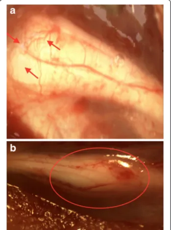
Fig. 1 a Stereomicroscopic photograph of nerve regeneration in oxytocin treatment group. Axonal sprouting (arrows) is pronounced and joins the nerve ends at the third-week follow-up. b Completion of nerve healing and maturation of the axonal sprouting at the 12-week follow-up (circled zone). The distal stump revealed considerable increase in diameter
at the second web space of the foot and later on moved distally at the gastrocnemius (GM) muscle to obtain bi-polar recording.
Functional evaluation
The functional assessments of the sciatic nerve recovery were performed by measuring the maximum angle at which the animals were able to climb upon an inclined plane (climbing degrees) which is sensitive in detecting 5-degree difference. The maximum angle at which an animal could support its weight was measured. Climbing level achieved in each group was recorded periodically.
Statistical analysis
All statistical analyses were conducted using the SPSS 15.0 program. The Studentt and ANOVA tests were used to determine differences in the parametric values when-ever necessary between the groups. The Mann-WhitneyU test was used to evaluate the nonparametric values. The level of significance was set atP < 0.05.
Results
The mean body weight of the rats before the surgery and at follow-ups was similar in both groups revealing no significant difference.
At one, 3 and 9 weeks follow-ups, higher number of axons were counted in the treatment group among the rats that were sacrified in comparison with the control group. After 12 weeks, all the animals were sacrified and underwent histological assessment. The samples obtained at the right sciatic nerve in oxytocin group presented an ongoing degenerative process. The results at 12 weeks revealed significant difference between the treatment and control group. A significantly higher number of axons were counted distal to the injury in the group of rats that received oxytocin treatment in comparison with the group that received normal saline solution. Table 1 presents the comparison of axon counts of two groups at follow-ups and after completion of the study in detail.
Functional assessment of the rats revealed similar re-sults before the operation. Average climbing degree was
70° in each group. Significantly greater climbing degrees were measured at 1 and 12 weeks in the treatment group in comparison of the control group. Table 2 presents the functional assessments in detail at the follow-ups.
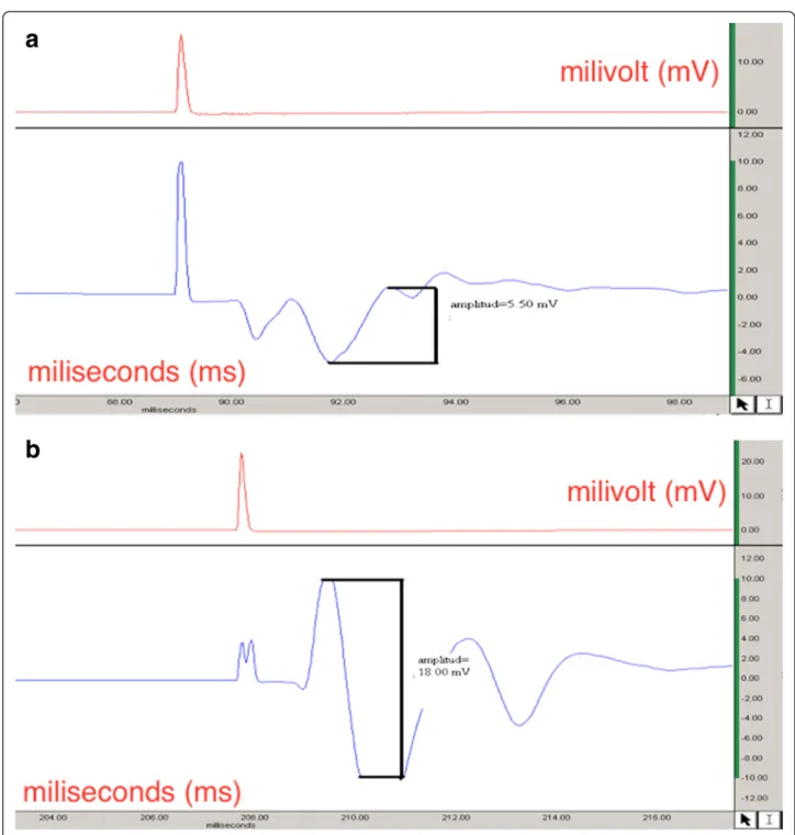
At postoperative, 1, 3, or 6 weeks group patterns were basically very similar, and there was no EMG response when the distal electrode was located at the second web space. The EMG recordings were carried out via positing the distal electrode at the second web space and GM muscle, respectively, after postoperative 9 weeks. There was no EMG response in both groups when the distal electrode was at the second web space. However, EMG responses were noted at the right side recordings (tran-section) in the treatment group after replacement of the electrode at the GM muscle at 9-week follow-up. This difference was obtained with regard to amplitude only whereas latency patterns were similar. After 12 weeks, EMG recordings revealed a significant difference in the treatment group with regard to both amplitude and la-tency in comparison with the control group. The lala-tency recordings were significantly shorter in the treatment group whereas the amplitude measures revealed signifi-cantly higher results (Fig. 2).
On the other hand, similar improvements in EMG recordings were seen at the left side (defect model) in the treatment group revealing shorter latency periods and higher amplitude levels when compared with the control group at 12-week follow-up. The results of the electro-diagnostical evaluation are summarized in Tables 3 and 4.
Stereomicroscopic evaluations of the right sciatic nerve (transection) after the first week revealed that the revascularization at the proximal site of the injury was more prominent. Besides, the diameters of the vessels were larger, and the walls of the vessels were thicker when compared with the control group in the treatment group.
Axonal sprouting was observed in both nerve damage models in both groups after 3 weeks. This sprouting has been observed to reach the distal site of the injury in the transected sciatic nerves in both groups after 9 weeks. Nerve fibers crossed the lesioned region and reached the distal stump joining the nerve stumps in both groups. However, the maturation of the sprouting was more pro-nounced in the treatment group.
Table 1 Body weight measurements (grams)
Follow-up period
Treatment group Control group P value
Mean ± SS Mean ± SS Before surgery 195.83 ± 5.15 197.08 ± 4.64 0.187 1 week 203.33 ± 8.88 206.04 ± 10.53 0.342 3 weeks 219.09 ± 25.08 222.73 ± 21.64 0.203 6 weeks 261 ± 36.95 261.5 ± 35.28 0.969 9 weeks 277.78 ± 47.38 277.22 ± 49.09 0.824 12 weeks 284.29 ± 41.98 272 ± 54.01 0.220
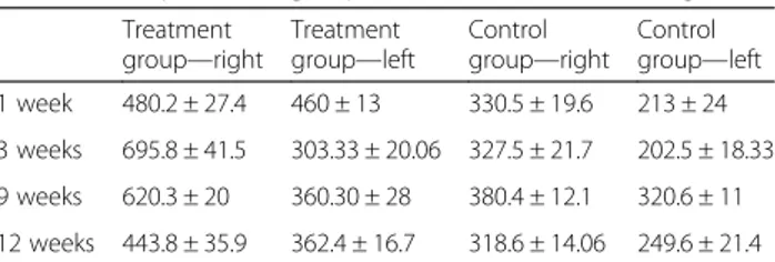
Table 2 Comparison of groups in terms of axon counting
Treatment group—right Treatment group—left Control group—right Control group—left 1 week 480.2 ± 27.4 460 ± 13 330.5 ± 19.6 213 ± 24 3 weeks 695.8 ± 41.5 303.33 ± 20.06 327.5 ± 21.7 202.5 ± 18.33 9 weeks 620.3 ± 20 360.30 ± 28 380.4 ± 12.1 320.6 ± 11 12 weeks 443.8 ± 35.9 362.4 ± 16.7 318.6 ± 14.06 249.6 ± 21.4 P values were P < 0.01 and P < 0.005, respectively, after comparison of right (transection) and left (defect) models among the groups after 12 weeks
Discussion
The functional assessment in the present study revealed that oxytocin group presented significant difference with regard to functional outcome at 12-week follow-up. Al-though we saw a positive effect on nerve regeneration with oxytocin, functional measures failed to demonstrate complete improvement in the recovery from injury. The complete functional recovery might have been observed with prolongation of the experiment time.
Wallerian degeneration is a process of progressive dis-integration and demyelination of the distal axonal segment following the damage to the neuron [1]. A pronounced increase in wallerian degeneration was seen in both groups at the first week whereas epinoral fibrosis was significantly higher in control group. An increase in axon count in com-parison with the control group was noted at 12 weeks in treatment group. Our findings revealed that nerve transec-tion model was superior to gap model at all follow-ups
with regard to histological improvement. The nerve-transection model in oxytocin group presented signifi-cant difference in axon count compared with the control group. This difference was also noted in nerve defect model at the 12-week follow-up. This study herein presented that oxytocin enhances a better histological improvement in two different injury models in rats. This improve-ment was obvious at earlier follow-ups and continued throughout the study.
We assessed that the degree of recovery of sciatic nerve function was assessed via climbing test, which provided a noninvasive and easily quantifiable method in the rat model when both limbs were injured [18]. Goldshmit Y et al. investigated the effect of exercise on axonal regrowth and found positive results with the climbing tests. Our re-sults present the positive effect of oxytocin administration in terms of functional recovery [19]. Our data showed sig-nificant difference at 12 weeks in functional evaluation between group comparisons. Moreover, animals able to climb onto the platform from higher grid levels in the treatment group comparable to the preoperative levels. We can speculate that the animals might have shown more improvements if the study duration was prolonged presenting full functional recovery.
Electrophysiologic measurements reflect the functional behavior of the regenerated nerve [1, 2]. Dose F et al. reported the low doses of oxytocin synergism with electrical afferent stimulation after spinal cord injury and similarly [20], our findings indicate that adminis-tration of oxytocin significantly provides lower latency recordings and higher amplitude measures reflecting a higher number of normally functioning axons in the treatment group.
Taken together, these data indicate that administration of oxytocin promotes functional recovery and enhances nerve regeneration after sciatic nerve damage in the rat. In the present study, we observed that the improvement in functional recovery was accompanied by significant increases in axon counts in the nerve regeneration site as well as close observations via stereomicroscopic eval-uations. At the microscopic level, there was also con-tinuity across the transection site and the defective site as well in the treatment group after 12 weeks. The ob-served improvement in the axon regeneration is in agree-ment with the results.
There is increasing evidence that growth factors may act at multiple levels in the regenerative response of nerve healing [8–11]. One such factor affecting multiple cell processes is IGF-1 and NGF. IGF-1 accelerates glucose uptake in the cells, stimulates mitotic activity and cell pro-liferation, and also inhibits apoptosis. NGF is a member of a family known as neurotrophins that function as signal-ing molecules. These cytokines are important for the growth, maintenance, and survival of neural cells [11, 12].
It has been well documented in the related literature that oxytocin influences plasma levels of some growth factors that play important role in the healing process of damaged tissues [15, 21]. Luppi et al. have reported that an intravenous injection of oxytocin results in a three-fold increase in NGF [15]. Another important study by Gavrilenko et al. revealed that oxytocin resulted in sig-nificant differences of wound process characteristics as compared with those ones in control group in the treat-ment of complex diabetic foot ulcers [19]. The authors presented in their work that oxytocin activates the pro-cesses of neovascularization, proliferation of endothelio-cytes and histioendothelio-cytes, resulting in the effective clearance of the wound, and optimal granulation tissue formation. Peterson et al. revealed in his study that oxytocin en-hances the survival of musculocutaneous flaps via in-creasing plasma IGF-1 levels. The authors revealed that this positive effect blocked by administration of an oxy-tocin antagonist [17]. The present study demonstrated that oxytocin enhances peripheral nerve regeneration in a rat sciatic damage model. This positive impact can be attributed to the benefits of oxytocin in stimulating the nerve growth factors that obviously presented in the
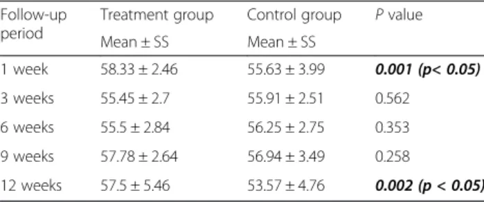
Table 3 Functional evaluation via climbing degrees
Follow-up period
Treatment group Control group P value
Mean ± SS Mean ± SS 1 week 58.33 ± 2.46 55.63 ± 3.99 0.001 (p< 0.05) 3 weeks 55.45 ± 2.7 55.91 ± 2.51 0.562 6 weeks 55.5 ± 2.84 56.25 ± 2.75 0.353 9 weeks 57.78 ± 2.64 56.94 ± 3.49 0.258 12 weeks 57.5 ± 5.46 53.57 ± 4.76 0.002 (p < 0.05)
Table 4 Comparison of EMG recordings between groups
Treatment group Control group P values
Right Left Right Left R/L
(Transection) (Defect) (Transection) (Defect)
9 weeks (amplitude) 12.25 ± 1.57 8.47 ± 1.87 6.99 ± 0.90 6.10 ± 1.53 0.002/0.171
9 weeks (latency) 1.25 ± 0.03 1.31 ± 0.04 1.22 ± 0.02 1.27 ± 0.04 0.27/0.269
12 weeks (amplitude) 12.92 ± 2.86 11.17 ± 1.60 8.34 ± 0.42 7.75 ± 0.68 0.011/0.039
literature such as NGF, as well as substantiating other growth factors such as IGF-1, which has positive effects in nerve tissue healing [8, 10].
The current study is not without limitations. The dur-ation of the study could have been prolonged to draw better conclusions with regard to functional evaluation. Also, we did not examine the effect of the oxytocin on the different cytotoxins that promotes nerve regeneration. Future studies should focus on the exact mechanism that provides nerve healing after oxytocin administration. In spite of these limitations, these data provide evidence that oxytocin administration plays a direct role in nerve regeneration
The strengths of this study are the appropriately pow-ered histological measure (stereomicroscope) and ana-lysis of nerve healing in two different injury models. In addition, assessment of nerve regeneration conducted in three different ways consisting of functional, electrophysi-ologic, and structural evaluations. Understanding these mechanisms may ultimately lead to improvements in per-ipherical nerve damage.
Our findings show that oxytocin promotes functional recovery and enhances nerve regeneration after transac-tional and defective peripheral nerve injury in rats com-pared with controls. The dose response to oxytocin has not been described in the present study Future studies should be performed with different doses to determine clinical application.
In conclusion, these data clearly demonstrated that oxytocin promotes nerve healing in two different rat sci-atic damage models. This healing effect can at least in part be ascribed to the fact that oxytocin activates growth factors of importance for the healing of different tissues.
Competing interests
The authors declare that they have no competing interests.
Authors’ contributions
BG and EK carried out the surgical procedure and writing. OE carried out the electrophysiological evaluation. CK carried out the statistical analysis. FO participated in the histological analysis. AB participated in the coordination and writing. All authors read and approved the final manuscript.
Acknowledgements
All authors declared that they have no funding or any other financial support.
Author details
1
Department of Orthopaedics and Traumatology, Izmir Ataturk Training and Research Hospital, Izmir, Turkey.2Department of Physiology, Ege University,
Izmir, Turkey.3Department of Orthopedics, Katip Celebi University Hospital, Izmir, Turkey.4Department of Histology and Embryology, Ege University,
Izmir, Turkey.5Orthopaedics & Traumatology, Istanbul Medipol University, TEM Avrupa Göztepe çıkışı, No: 1 Bağcılar, Istanbul, Turkey.
Received: 5 August 2015 Accepted: 25 September 2015
References
1. Lee S, Wolfe S. Peripheral nerve injury and repair. J Am Acad Orthop Surg. 2000;8:243–25.
2. Johnson EO, Zoubos AB, Soucacos PN. Regeneration and repair of peripheral nerves. Injury. 2005;36 Suppl 4:S24–9.
3. Noble J, Munro CA, Prasad VS, Midha R. Analysis of upper and lower extremity peripheral nerve injuries in a population of patients with multiple injuries. J Trauma. 1998;45(1):116–22.
4. Xavier AM, Serafim KG, Higashi DT. Simvastatin improves morphological and functional recovery of sciatic nerve injury in Wistar rats. Injury. 2012;43(3):284–9. 5. Javanbakht J, Hobbenaghi R, Hosseini E, Bahrami AM, Khadivar F, Fathi S,
et al. Histopathological investigation of neuroprotective effects of Nigella sativa on motorneurons anterior horn spinal cord after sciatic nerve crush in rats. Pathol Biol (Paris). 2013;61(6):250–3.
6. Hobbenaghi R, Javanbakht J, Sadeghzadeh S, Kheradmand D, Abdi FS, Jaberi MH, et al. Neuroprotective effects of Nigella sativa extract on cell death in hippocampal neurons following experimental global cerebral ischemia-reperfusion injury in rats. J Neurol Sci. 2014;337(1–2):74–9. 7. Hobbenaghi R, Javanbakht J, Hosseini E, Mohammadi S, Rajabian M, Moayeri
P, et al. Neuropathological and neuroprotective features of vitamin B12 on the dorsal spinal ganglion of rats after the experimental crush of sciatic nerve: an experimental study. Diagn Pathol. 2013;8:123.
8. Gao J, Ma S, Ji Y, Wang JE, Li J. Sciatic nerve regeneration in rats stimulated by fibrin glue containing nerve growth factor: an experimental study. Injury. 2008;39(12):1414–20.
9. Mohammadi R, Esmaeil-Sani Z, Amini K. Effect of local administration of insulin-like growth factor I combined with inside-out artery graft on peripheral nerve regeneration. Injury. 2013;44(10):1295–301.
10. Johnson EO, Charchanti A, Soucacos PN. Nerve repair: experimental and clinical evaluation of neurotrophic factors in peripheral nerve regeneration. Injury. 2008;39 Suppl 3:S37–42.
11. Boyd JG, Gordon T. Neurotrophic factors and their receptors in axonal regeneration and functional recovery after peripheral nerve injury. Mol Neurobiol. 2003;27(3):277–324.
12. Boyd JG, Gordon T. The effect of nerve growth factors is known to be dose-dependent. A dose-dependent facilitation and inhibition of peripheral nerve regeneration by brain-derived neurotrophic factor. Eur J Neurosci. 2002;15:613–26.
13. Rabinovski ED. The multifunctional role of IGF-1 in peripheral nerve regeneration. Neurol Res. 2004;26(2):204–10.
14. Vitalo A, Fricchione J, Casali M, Berdichevsky Y, Hoge AE, Rauch SL, et al. Nest making and oxytocin comparably promote wound healing in isolation reared rats. PLoS One. 2009;4(5):e5523.
15. Luppi P, Levi-Montalcini R, Bracci-Laudiero L, Bertolini A, Arletti R, Tavernari D, et al. NGF is released into plasma during human pregnancy: an oxytocin-mediated response? Neuroreport. 1993;48(8):1063–5.
16. Schaeffer HJ, Sirotkin A. The release of insulin-like growth factor-1 by luteinized human granulosa cells in vitro: regulation by growth hormone, oxytocin, steroids and cAMP-dependent intracellular mechanisms. Exp Clin Endocrinol. 1995;103:361–6.
17. Petersson M, Lundeberg T, Sohlström A, Wiberg U, Uvnas-Moberg K. Oxytocin increases the survival of musculocutaneous flap. Naunyn-Schmiedeberg’s Arch Pharmacol. 1998;357:701–4.
18. Rivlin AS, Tator CH. Objective clinical assessment of motor function after experimental spinal cord injury in the rat. J Neurosurg. 1977;47(4):577–81. 19. Goldshmit Y, Lythgo N, Galea MP, Turnley AM. Treadmill training after spinal
cord hemisection in mice promotes axonal sprouting and synapse formation and improves motor recovery. J Neurotrauma. 2008;25(5):449–65. 20. Dose F, Zanon P, Coslovich T, Taccola G. Nanomolar oxytocin synergizes
with weak electrical afferent stimulation to activate the locomotor CpG of the rat spinal cord in vitro. PLoS One. 2014;9(3):e92967.
21. Gavrilenko VG, Esipov VK, Sivozhelezov KG. Morphological characteristic of wound healing process in patients with diabetic purulent-necrotic foot lesion treated with oxytocin. Morfologiia. 2003;124(5):24–7.