CHEMOGENETIC TOOLS FOR CAUSAL CELLULAR
AND NEURONAL BIOLOGY
Deniz Atasoy and Scott M. Sternson
Department of Physiology, School of Medicine and Regenerative-Restorative Medicine Research Center
(REMER), Istanbul Medipol University, Istanbul, Turkey; and Janelia Research Campus, Howard Hughes Medical Institute, Ashburn, Virginia
L
Biology. Physiol Rev 98: 391– 418, 2018. Published December 13, 2017; doi:Atasoy D, Sternson SM. Chemogenetic Tools for Causal Cellular and Neuronal 10.1152/physrev.00009.2017.—Chemogenetic technologies enable selective pharmacological control of specific cell populations. An increasing number of ap-proaches have been developed that modulate different signaling pathways. Selective pharmacological control over G protein-coupled receptor signaling, ion channel conductances, protein association, protein stability, and small molecule targeting allows modulation of cellular processes in distinct cell types. Here, we review these chemogenetic technologies and in-stances of their applications in complex tissues in vivo and ex vivo.I. INTRODUCTION 391
II. GPCR-BASED TOOLS 391
III. LIGAND-GATED ION CHANNEL ... 402 IV. CELL TYPE-SPECIFIC ... 408
V. CONCLUSION 412
I. INTRODUCTION
Electrochemical gradients facilitate basic cellular functions such as nutrient transport, energy production, intracellular signaling, and long-range electrical transmission. Ion chan-nels control cellular electrical activity and are thus prime targets for influencing cellular functions. Ion channels can be perturbed directly with small molecule or peptide ago-nists or antagoago-nists for ligand-gated ion channels (LGICs) or indirectly via G protein-coupled receptor (GPCR) signal-ing (87, 140). Pharmacological control of LGICs and GPCRs has long been used to acutely alter cell function (37, 78, 86, 101). However, the precision of these manipulations has been limited by insufficient selectivity; most LGICs and GPCRs are expressed across multiple cell populations. To address this issue, engineered LGICs and GPCRs have been developed as exogenously applied tools to obtain remote control over cellular electrical activity (62, 180, 199). Che-mogenetic tools (180) are comprised of a transgenic actua-tor for a cellular pathway that is targeted to specific cell populations and can be rapidly switched on or off by deliv-ery of a chemical ligand.
Chemogenetics generalizes chemical control of cellular pathways by engineering a limited set of tunable, modular, and selective receptor/ligand systems that can be applied to virtually any cell population. Optimal chemogenetic tools possess two core properties: 1) the transgenic actuator is normally nonperturbative to cells, i.e., it has low
constitu-tive activity and low responsiveness to endogenous ligands; and 2) the exogenously applied ligand is nonperturbative to cells that lack the actuator transgene. An increasing number of chemogenetic tools have been developed to perturb cel-lular electrical activity in a variety of ways. Here, we will primarily review work focusing on cellular control with LGIC and GPCR tools, along with some additional chemo-genetic strategies that are useful for controlling other as-pects of cell function. Because chemogenetic technologies are used quite extensively, the objective of this review is to highlight the various ways in which these tools are used but not to exhaustively cover all chemogenetic studies.
II. GPCR-BASED TOOLS
GPCRs have extensive functional consequences in cells by influencing signal transduction pathways, gene expression, ion channels, and synapses (181). Intensive investigation over several decades has revealed a number of structural and functional characteristics that can be leveraged to tune the properties of GPCR-based tools (157). Furthermore, many GPCR ligands can have high potency, which is a useful foundation for optimizing receptor selectivity and also for dosing in animal experiments.
A. GPCR Signaling Pathways 1. G␣ signaling
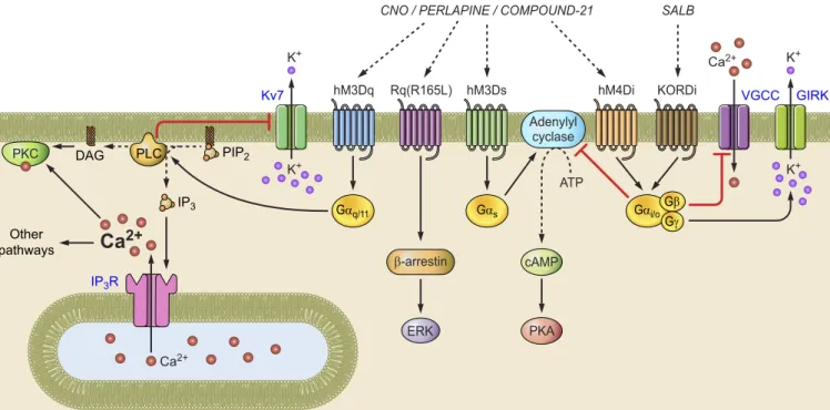
GPCRs interact with a G protein complex comprised of a G␣ subunit bound to guanosine diphosphate (GDP), along with tightly associated G and G␥ subunits(FIGURE 1)(74). Upon agonist binding, GPCRs catalyze exchange of G ␣-GDP for guanosine triphosphate (GTP). G␣-GTP
dissoci-ates from the G␥ complex, and both G␣-GTP and G␥ engage separate downstream signaling processes. There are multiple G protein classes, and GPCRs are often distin-guished by which type of G␣ subunit they interact with. GPCRs that couple to a G␣s(Gs) protein increase the
activ-ity of adenylyl cyclase, which produces cAMP, thereby in-creasing protein kinase A (PKA) activity along with effects on gene expression. GPCRs that couple to Giproteins
in-hibit adenylyl cyclase and reduce PKA activity. GPCRs that couple to Gq proteins activate phospholipase C, which
cleaves phosphatidylinositol 4,5-bisphosphate (PIP2) into
inositol 1,4,5-trisphosphate (IP3) and diacyglycerol (DAG).
IP3binds receptors on the endoplasmic reticulum, leading
to Ca2⫹release from internal stores. DAG and Ca2⫹ acti-vate protein kinase C (PKC), which engages multiple addi-tional intracellular signaling processes.
2. G␥ signaling
The G␥ complex also initiates downstream signaling, no-tably activating G protein-regulated inward rectifier potas-sium (GIRK) channels (122) and inhibiting N-type and P/Q-type voltage-gated Ca2⫹channels (VGCCs) (43, 52). Inter-estingly, these effects seem to be observed selectively with activation of Gi protein-coupled GPCRs despite the fact
that different individual G␥ subunits do not show this selectivity (53). Thus Gi protein-coupled GPCRs activate
GIRKs and inhibit VGCCs, both of which result in reduced cellular excitability. The mechanism by which the G␣ sub-type influences the cellular effects of the G␥ subunits re-mains unclear (102). Nevertheless, different GPCRs have well-characterized, distinct functional consequences that provide a sound basis for chemogenetic tool development.
B. RASSLs
GPCRs have been extensively analyzed by mutagenesis to establish the influence of specific amino acid residues on ligand activation. Structure-function studies identified mu-tated GPCRs with greatly diminished ability to bind an endogenous ligand while preserving the capacity to be acti-vated by synthetic ligands (39, 171, 182). These Receptors Activated Solely by Synthetic Ligands (RASSLs) were used as early tools for cell type-specific perturbations (38, 39). RASSLs are expressed in a desired cell type and are nonper-turbative because they show low sensitivity to endogenous agonists. Once expressed in a desired cell type, RASSLs can be engaged by an exogenous synthetic ligand to selectively perturb cellular function. The major limitation of RASSLs is that the synthetic ligands retain the ability to engage the endogenous receptors.
One striking application of RASSLs is an experiment that examined taste sensory systems to test a hypothesis that sweet taste receptor-expressing neurons were a “labeled line” for the hedonic properties of sweet tastes irrespective of the identity of the receptor. After expressing a RASSL derived from the -opioid receptor (KOR) (152) in sweet receptor-expressing cells in taste buds, mice avidly con-sumed the normally nonpreferred synthetic ligand line at nanomolar concentrations (211). Although spirado-line can activate endogenous KORs, the delivery was lim-ited to the tongue, and control experiments showed no significant effect of the ligand on consumption. Neverthe-less, in many other circumstances, the selectivity profile of this system would be unsuitable, for example, in experi-IP3R Kv7 VGCC GIRK Ca2+
Ca
2+ Ca2+ PIP2 IP3 DAG Other pathways K+ K+ K+ K+ PKC Adenylyl cyclase ATP hM3Dq Rq(R165L) hM3DsCNO / PERLAPINE / COMPOUND-21 SALB
hM4Di KORDi PLC Gαq/11 Gαi/o Gβ Gγ Gαs β-arrestin ERK cAMP PKA
ments involving systemic ligand administration. For that, a synthetic ligand with no endogenous activity is required.
C. DREADDs 1. Design
Development of GPCR tools with low sensitivity to endog-enous ligands and high potency for otherwise inert exoge-nous ligands required more demanding design principles. This was first achieved by engineering a high-potency GPCR interaction with clozapine-N-oxide (CNO), which is an inactive metabolite of the antipsychotic drug clozapine. Clozapine is an agonist for endogenous muscarinic acetyl-choline receptors, and CNO has very low potency at these receptors. With the use of random mutagenesis, a modified muscarinic acetylcholine receptor 3 (hM3) was used to cre-ate a library of mutant receptors that were assayed in a yeast selection system in which growth is dependent on G protein signaling (5). Yeast clones that grew on selective media in the presence of CNO were subjected to additional rounds of random mutagenesis and screening at progressively lower CNO concentrations until a receptor with two mutations was arrived at that exhibited high CNO sensitivity. Two additional criteria were taken into consideration during the clone selection process: minimal sensitivity to acetylcholine and low constitutive activity (5). Application of these mu-tations to hM3 created a Designer Receptor Exclusively Activated by a Designer Drug (DREADD), which was named hM3D (5). Because hM3D couples to Gq-type G proteins, it is often referred to as hM3Dq. CNO treatment of cells expressing hM3Dq increased IP3production with an EC50of 17 nM, which was 480-fold selective over CNO activation of unmutated hM3 (5). Conversely, acetylcho-line potency was reduced by⬎50,000-fold for hM3D. Al-though constitutive activity of hM3D was modestly in-creased by twofold over hM3 (5), heterologous expression of hM3D in cortical neuron cultures did not significantly affect membrane potential (5). The same modification was also applied to human M1, M2, M4, and M5 receptors to generate additional CNO-sensitive DREADDs (5).
2. hM3Dq
hM3Dq was one of the first GPCR-based DREADDs, and it proved well suited to activate neuronal firing. Administra-tion of CNO depolarized neurons and resulted in acAdministra-tion potential firing in a PLC-dependent manner (2). Multiple mechanisms for hM3Dq-mediated neuron depolarization can be predicted from studies on signaling from endogenous Gq-coupled GPCRs. Muscarinic activation reduces the
M-current (22), which is a PIP2-sensitive K⫹conductance
me-diated by Kcnq channels (FIGURE 1) (210). At neuronal resting membrane potentials, K⫹channels reduce neuronal excitability (87). CNO treatment of cells expressing hM3D converts PIP2to IP3, and reduced PIP2decreases M-current
in Kcnq-expressing cells, consequently increasing neuronal excitability (210). However, the ionic mechanisms of hM3Dq signaling are likely more complicated than this. hM3Dq activation also releases Ca2⫹from internal stores (5). Release of internal Ca2⫹ can activate the Na⫹/Ca2⫹ exchanger (18), which is electrogenic and can lead to a depolarizing inward current when internal Ca2⫹is elevated (108, 141), complementing depolarization from M-current suppression. Countervailing receptors can also be engaged, including activation of calcium-sensitive potassium chan-nels, which suppress neuronal activity and thus work against the desired neuron activation (15). Clearly, the ef-fectiveness of hM3Dq for neuron activation reflects the ex-pression of relevant accessory channels (such as Ca2⫹ -sen-sitive K⫹channels) in a cell type of interest. For this reason, verification of cell activation by electrophysiology is impor-tant when using hM3Dq (2, 105).
For such verification purposes, the immediate early gene Fos is often used as an in vivo marker for cellular activation after activating hM3Dq as an alternative to electrophysiol-ogy. One caveat of this validation approach for hM3Dq perturbations in animals is that Fos expression is also sen-sitive to elevated cellular Ca2⫹, which can be derived from internal stores in addition to external sources via VGCCs (69). Consistent with this, Fos expression has been shown, in some cases, to not correlate with independently measured neuronal activity (146). Thus the expression of Fos in re-sponse to hM3Dq activation may be related to pathways that are independent of electrical activity.
3. hM3Ds
To create a Gs-coupled DREADD, the modularity of
hM3Dq signaling was exploited by maintaining the muta-tions that confer CNO sensitivity while altering a region that confers G␣-protein selectivity. By substituting intracel-lular loop sequences from the Gs-coupled-adrenergic
re-ceptor into hM3Dq, hM3Ds was generated, which in-creases cAMP production but not IP3or intracellular Ca2⫹
in the presence of CNO (61, 81). These experiments showed that because GPCRs have separate regions for ligand bind-ing and G protein couplbind-ing, additional chemogenetic tools can be generated with distinct signaling properties while maintaining CNO responsiveness.
4. hM4Di
A Gi-coupled DREADD was generated by applying the
hM3Dq mutations at homologous residues in hM4, which is an endogenous Gi-coupled receptor. hM4Di activation
with CNO reduced forskolin-induced cAMP production (5). Expression of hM4Di in neurons rendered cells sensi-tive to CNO-induced hyperpolarization and reduction of action firing due to activation of GIRK channels (5). Further analysis of hM4Di demonstrated that it is an effective
in-hibitor of synaptic release (131, 173). Stachniak et al. (173) showed that the synaptic silencing function of hM4Di was likely more relevant to inhibiting neuronal output than was the suppression of electrical activity. This was demon-strated by tagging hM4Di with a portion of neurexin recep-tor 1a (hM4Dnrxn) to produce a receptor that was
predom-inantly trafficked to the axonal compartment. hM4Dnrxn
showed minimal effects on somatic conductance with CNO application, but synaptic silencing remained intact as did in vivo behavioral efficacy (173). Thus hM4Di and hM4Dnrxn
are powerful tools for selectively blocking synaptic release from axon projections.
5. DREADD[Rq(R165L)]--arrestin signaling
In addition to G protein activation, GPCRs also influence cell signaling by recruitment of-arrestin. Following ago-nist activation of GPCR signaling, the GPCR is phosphor-ylated, which recruits-arrestin, and this promotes associ-ation with other signaling enzymes, such as extracellular signal-regulated kinase (ERK)(FIGURE 1)(191). Although -arrestin signaling is activated by all DREADDs, a set of hM3Dq mutations that prevent G protein coupling permit-ted creation of a DREADD selective for-arrestin signal-ing, called DREADD[Rq(R165L)] (138). This DREADD stimulated insulin release in response to CNO from a cul-tured pancreatic-cell line (138).
6. Caveats and new ligands
Additional extensions of the DREADD approach include improvements in the chemogenetic agonist. Of note here, CNO administration at commonly used doses and in the absence of any DREADD expression was reported to affect amphetamine-induced dopamine release, locomotion, and acoustic startle reflex (129), possibly due to retro-conver-sion to clozapine (31). Nevertheless, the doses used in this study (1–5 mg/kg) are greater than the recommended in vivo dose of 0.3 mg/kg (158). The potency of behavioral effects in chemogenetic experiments is also affected by the excess receptor expression such that signaling from just a fraction of ligand-bound receptors is sufficient for a behav-ioral response (158). Another concern is a report that, in nonhuman primate brains, CNO is a substrate for the P-gly-coprotein efflux pump, resulting in low partition of plasma CNO into the brain (136, 151). However, different studies in nonhuman primates report contradictory results regard-ing the levels of retro-converted clozapine in the cerebrospi-nal fluid of CNO-treated rhesus macaques (136, 151). More recently, Gomez, et al. (75a) used autoradiography and positron emission tomography with [11C]CNO to show that CNO does not get into the brain in rats. This study provided multiple lines of evidence that CNO is not the active agent for in vivo chemogenetic experiments and that, instead, it is the small amount of retro-converted clo-zapine that is responsible for muscarinic DREADD
activa-tion. Although only 2% of CNO is converted to clozapine, the considerably higher potency of clozapine for hM3Dq and hM4Di relative CNO is suggested to be what permits this chemogenetic system to function in the brain (75a). Due to concern about the metabolic back reaction of CNO to clozapine, a new potent and metabolically stable small mol-ecule agonist, called compound 21, has been identified (34). Compound 21 is a promising tool to replace CNO with DREADDs derived from human muscarinic receptors. The drug perlapine also activates hM3Dq-derived DREADDs but has sedative side effects (34).
7. KORDi
The molecular evolution process for producing DREADDs is applicable to other GPCRs. This was demonstrated for kappa opioid receptor (KOR), which is a Gi-coupled recep-tor that is potently activated by the hallucinogen salvinorin A (SalvA). Directed molecular evolution identified muta-tions in KOR that rendered it sensitive to salvinorin B (SalvB), which is a metabolite of SalvA that is inactive at KOR (193). KORD activation by SalvB inhibited neuron electrical activity and synaptic release in the presence of SalvB (193). With hM3Dq and KORDi, chemogenetic neu-ron activation and silencing could be demonstrated in the same neuron, although neuron inhibition was only ob-served for ~1 h due to the short in vivo half-life of SalvB (193). The development of orthogonal DREADD systems allows for multiple, distinct chemogenetic perturbations in the same organism.
8. Summary
Directed evolution of the hM3 receptor to install sensitivity to the normally inactive drug metabolite CNO led to the identification of two mutations that reduce acetylcholine responsiveness and dramatically enhanced CNO sensitivity. The sites mutated in hM3Dq are conserved across other muscarinic receptors, conferring CNO sensitivity on func-tionally distinct muscarinic receptors. Moreover, existing structure-function analysis of GPCRs can be applied to hM3D receptors to produce receptors with distinct prop-erties such as Gs- and-arrestin-selective signaling. Thus
the discovery of mutations that produce new pharmaco-logical selectivity has been exploited in conjunction with other structure-function information to produce a range of powerful chemogenetic tools for control of different aspects of cellular signaling. Moreover, the platform for discovering DREADD mutations can be applied to mul-tiple GPCRs as evidenced by the KORDi DREADD. This expanding toolbox enables selective pharmacological control over distinct signaling pathways, offering new capabilities for sophisticated analysis of cellular and cir-cuit functions.
D. Neuronal Applications 1. DREADD delivery
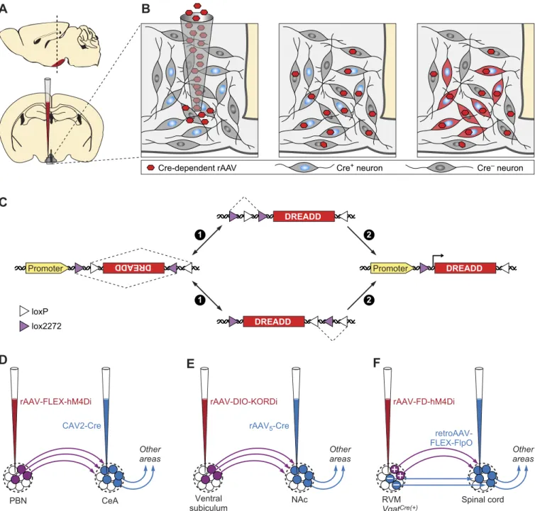
Two of the most commonly employed strategies for genetic targeting of DREADDs involve locally delivered viral injec-tions and expression from genetically engineered animal models. The relatively short DREADD coding sequences allow efficient packaging into viral vectors. Recombinant AAV vectors carrying DREADD transgenes under multiple promoters and fluorescent tags have been developed and are available from plasmid repositories and multiple viral vec-tor core facilities. Stereotactic delivery of DREADD-pressing viral vectors permits anatomically restricted ex-pression. In addition to regionally limited distribution, fur-ther specificity for distinct cell types can be achieved using recombinases, such as Cre and Flp, expressed under cell type-specific promoters in transgenic or knock-in mouse lines (FIGURE 2, A–C). Isolating and manipulating neuron subpopulations with specific projection input/output pat-terns have been achieved using viral vectors that can be transmitted either anterogradely or retrogradely, often with AAV, HSV, or CAV, driving Cre-recombinase expression (FIGURE 2, D–F)(27, 67, 132, 174, 187, 214).
Despite the advantages of speed and flexibility with virally mediated DREADD expression, stereotactic viral delivery strategy leads to variation in transduction efficiency across animal subjects, which requires post hoc evaluation of viral transduction (6). In addition, for loss of function studies, it is frequently important for most of the targeted cells to be silenced, which may be difficult to achieve with viral deliv-ery. Transgenic DREADD-expressing mouse lines have been developed to overcome these issues. An early example was based on a tet-off system, in which removal of doxycy-cline permitted hM3D expression from a CamKII promoter (2). This line had expression reportedly restricted to excit-atory neurons. For targeting DREADD expression to other cell types, two Cre-dependent reporter lines have been de-veloped by knocking DREADD genes into ROSA26 locus under a CAG promoter and Lox-Stop-Lox cassette (84, 212). With the increasing number of Cre-driver choices available, these DREADD lines open a panoply of research applications.
2. Appetite circuits
Energy balance is regulated by a complex interplay of mo-lecularly defined cell types and circuits influencing appetite, satiety, and energy expenditure (179). Thus DREADDs have been used extensively for cell type-specific manipula-tion of appetite-regulating neuroendocrine circuits (178). An early application used acute chemogenetic activation of AGRP neurons with a virally transduced hM3Dq actuator to stimulate food search and consumption behaviors (105). Interestingly, activation of the Gs-coupled DREADD
hM3Ds (61) in AGRP neurons had a slower but much more prolonged hyperphagic effect than activating Gq-coupled
GPCR pathway due to selective upregulation of AGRP re-lease (137). Using hM3Dq activation of AGRP neurons in various knockout mouse backgrounds, researchers have shown that either NPY or GABA (but not AGRP) signaling is sufficient for acutely activating food intake (106). Of note, chemogenetic inhibition of AGRP neurons by hM4Di rapidly reduces food intake in fasted animals (105) as well as overeating induced by ethanol (25), but it is less effective at inhibiting palatable food intake, due to the necessity of other circuits (47).
DREADDs have been used to deconstruct the contributions of distinct cellular nodes in appetite circuits. Based on cir-cuit connectivity, early studies suggested that AGRP neu-rons regulated appetite by antagonizing intermingled POMC neurons (40). However, hM4Di-dependent inhibi-tion of ARCPOMCneurons did not rapidly increase feeding but instead elevated food consumption after 24-h of POMC neuron inhibition (6), suggesting that POMC neuron inhi-bition is not the main mode of action for AGRP neuron-mediated acute activation of appetite. In addition, hM3Dq-dependent activation of ARCPOMCneurons did not acutely
reduce food intake, although chronic activation of these neurons using multiple dose of daily CNO injection signif-icantly reduced food intake (209), a result consistent with an earlier optogenetic activation study (3). Instead, another group of neurons in the arcuate nucleus defined by overlap-ping vGlut2 expression have been shown to be responsible for rapid suppression of feeding (63). Thus acute cell type-specific manipulation of neuron activity was critical for de-termining the relative role of these cells types in appetite and on various timescales.
Several hypothalamic and extrahypothalamic areas receive AGRP axonal input (177). AGRP neuron projections to the PVH were shown to be inhibitory, and hM4Di-dependent or KORDi direct inhibition of PVH neurons (marked by Sim1 expression) resulted in elevated food intake and in-creased motivation to work for food (6, 193). DREADD-dependent inhibition of a subpopulation of PVH neurons expressing MC4R was sufficient to elicit feeding and in-creased motivation for food in sated mice (71). Conversely, chemogenetic activation of PVH neurons marked by NOS1, MC4R, or AVP expression suppressed feeding in food-de-prived mice (6, 71, 144, 183).
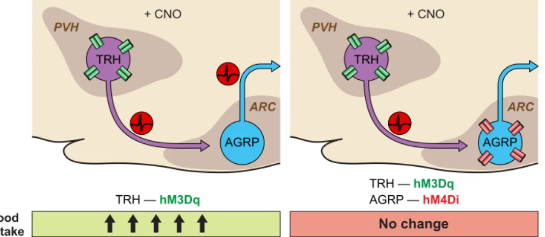
DREADDs have also been used for neural circuit epistasis analysis to examine the functional dependency of two con-nected pairs of neurons. After establishing that a direct excitatory connection exists between TRH neurons in the PVH and AGRP neurons, the behavioral significance of this connection was investigated by activating or inhibiting PVHTRHneurons with hM3Dq or HM4Di, which increased
A
Cre-dependent rAAV Cre+ neuron
C
Cre– neuronB
D
E
F
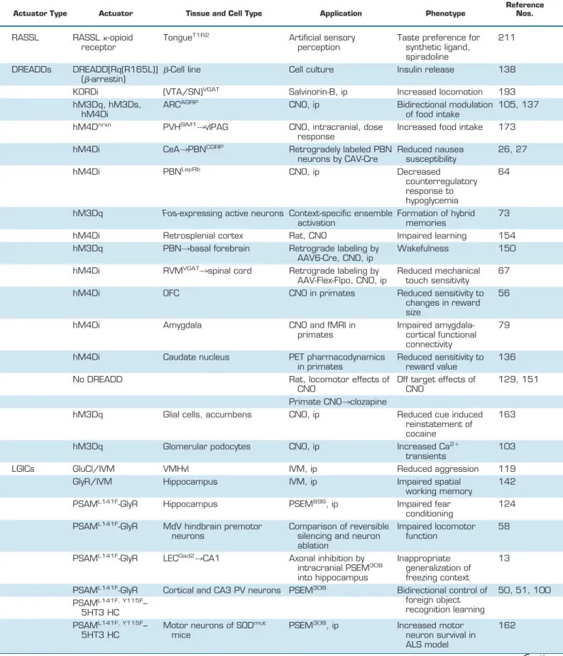
CAV2-Cre CeA PBN Ventral NAc subiculum Other areas rAAV5-Cre retroAAV-FLEX-FlpO Spinal cord RVM VgatCre(+) rAAV-FLEX-hM4Di Other areas rAAV-DIO-KORDi Other areas rAAV-FD-hM4Di lox2272 loxP DREADD DREADD DREADD DREADD Promoter DREADD Promoter DREADD 1 1 2 2FIGURE 2. Cell type-specific or projection-specific transgene expression. A and B: Cre-dependent expression of DREADDs in neurons. A: schematic injection of Cre-dependent virus into brain via stereotactic delivery. B: selective DREADD expression FLEX switch. C: schematic of FLEX-switch used in Cre-dependent viruses and two step Cre-recombination events involving loxP and lox2272 heterotypic recombination sites for permanent inversion of DREADD transgenes. D and E: two-way intersectional viral strategies to express DREADDs in projection-specific neuronal subpopulations. D: axonally targeted CAV2-Cre injected into CeA for retrograde labeling and somatically targeted rAAV-FLEX-hM3Dq injected into PBN for expression only in CeA projecting PBN neurons. E: axonally targeted rAAV5-Cre injected into NAc for retrograde labeling and somatically
targeted rAAV-FLEX-KORDi injected into central subiculum for expression only in NAc projecting subiculum neurons. F: a 3-way intersectional strategy to express hM4Di in a molecularly defined neuron population with a defined axon projection. First, axon-targeted injection of a retroAAV expressing Flp recombinase in a Cre-recombinase-dependent manner (retroAAV-FLEX-FlpO) was made into spinal cord of Vgat-cre mice. Then somatic injection of Flp recombinase-inducible rAAV virus expressing hM4Di (rAAV-FD-hM4Di) targeted to RVM. In this configuration only the spinal cord projecting GABAergic neurons of RVM will be expressing hM4Di.
PVHTRHneuron elevation of food intake on the activity of
AGRP neurons was tested by simultaneous activation of TRH neurons and AGRP inhibition, using dual viral target-ing with hM3Dq and hM4Di, respectively(FIGURE 3). To-gether, these manipulations blunted the food intake-stimu-lating effect of PVHTRHneuron activation, supporting the
conclusion that the appetite stimulating properties of PVHTRHneurons were due to their synaptic connection to
AGRP neurons (107). Such manipulations are also facili-tated by the development of new DREADDs, and KORDi and hM3Dq have been coexpressed in AGRP neurons to selectively activate or inhibit feeding in the same mice (193). Because Gi-coupled DREADDs like hM4Di and KORDi suppress synaptic release, these DREADDs can also be used to functionally map the role of synaptic connections to spec-ified brain regions (173, 193). Appetite regulating projec-tions of PVHSIM1neurons have been traced to a number of
hindbrain locations. Expression of hM4Dnrxn, an
axon-tar-geted Gi-coupled DREADD, in PVH neurons was used to show through a series of intracranial CNO injections that a site in the vicinity of the periaqueductal grey and dorsal raphe led to increased food intake when these axon termi-nals were silenced (173). Dose responses with CNO and injections into nearby sites were used to localize this re-sponse. Importantly, these experiments indicated that hM4Di does not block action potential transmission along axons, making this a useful tool to inhibit synaptic output to a targeted region without blocking transmission through fibers of passage (173).
Visceral signals of satiety and also nausea are integrated through a hindbrain pathway. Hormonal and visceral in-formation enters the nucleus of the solitary tract (NTS) and area postrema (AP). DREADD-mediated activation of two distinct NTS neuronal subpopulations marked by CCK and DBH expression suppress appetite and increase Fos activity in PBN (parabrachial nucleus) neurons (155). Optogenetic mapping confirmed that both NTSCCK and NTSDBH
neu-rons directly project to and excite a subpopulation of PBN neurons marked by calcitonin gene-related peptide (CGRP) expression (PBNCGRP) (155). CNO-mediated activation of
hM3Dq-expressing PBNCGRPneurons suppressed food
in-take and also proved to be aversive (27). Conversely, NTSCCKneurons also send long-range projections to the
PVH, and hM3Dq activation suppressed appetite as well, but unlike those that activate PBNCGRP, stimulating
NTSCCK
¡PVH axons elicited positive valence (45). In addition to DBH and CCK, at least two other NTS neu-ronal subpopulations, expressing POMC and GLP1, are appetite suppressing. Unlike ARCPOMC neurons,
hM3Dq-dependent activation of NTSPOMCneurons
rap-idly and robustly suppresses food intake (209). Chemo-genetic activation of NTSGLP1 fibers with hM3Dq,
tar-geted using Phox2b-Cre mice, with intracranial CNO injections to the VTA selectively suppressed high-fat feeding, which suggests a role for conveying caloric re-ward via this projection (197). Thus DREADDs have been instrumental in teasing apart the distinct role of intermingled hindbrain populations that have been found to have subtly different effects on control of satiety. The hindbrain anorectic circuit defined by PBNCGRP has
been traced to the central nucleus of the amygdala (CeA), a region associated with aversive emotional states. Chemoge-netic inhibition of PBNCGRPneurons that were labeled
ret-rogradely from CeA with a Cre-expressing canine adenovi-rus (CAV) was sufficient to suppress the appetite-lowering effects of satiety and nausea-inducing agents (26, 27) (FIG-URE 2D). Fibers from PBNCGRPneurons directly activated
PKC-␦⫹ neurons in CeA, a connection suggested to be crit-ical for generating an aversive state (83). Activating PKC-␦⫹ neurons suppressed feeding, and conversely, che-mogenetic inhibition of these neurons with hM4Di blunts the anorectic response to nausea- and satiety-inducing agents and increases food intake (24).
Chemogenetic approaches have been used to dissect other hunger-related circuits as well. hM3Dq-dependent activation of lateral hypothalamic VGAT neurons stim-ulated food-seeking and consumption irrespective of ca-loric content (96, 139). Conversely, hM3Dq activation of lateral hypothalamus-projecting septal VGAT neurons suppressed feeding (185). Chemogenetic activation of
TRH — hM3Dq TRH — hM3Dq AGRP — hM4Di Food intake + CNO ARC PVH TRH AGRP + CNO ARC PVH TRH AGRP No change
FIGURE 3. Schematic of neural circuit epistasis for the PVHTRH
¡ARCAGRPfeeding
circuit. TRH activation using hM3Dq drives food intake which is abolished by concomi-tant hM4Di silencing of downstream AGRP neurons.
pyramidal neurons in medial prefrontal cortex (mPFC) enhanced operant performance for food reward without effecting unconditioned hedonic or homeostatic food consumption (198).
3. Glucose homeostasis circuits
Neuronal circuits for appetite regulation are intermingled with neurons that regulate blood glucose levels. Hypogly-cemia induces a counterregulatory response (CRR) to re-store blood glucose to normal levels. A subpopulation of PBN neurons defined by leptin receptor expression was crit-ical for this reflex. hM4Di-mediated inhibition of PBNLepRb blunts the CRR induced by 2-deoxyglucose (2-DG)-medi-ated glucoprivation, whereas PBNLepRbchemogenetic acti-vation with hM3Dq mimicked CRR leading to hyperglyce-mia (64). Many PBNLepRbneurons coexpress CCK, are ac-tivated by glucoprivation, and project axons to the ventromedial hypothalamic nucleus (VMH). With the use of chemogenetic epistasis experiments, PBNCCK¡VMH was shown to be necessary for CRR by simultaneous acti-vation of PBNCCK neurons and silencing of VMHSF1 neu-rons (72).
Manipulations of other neuron populations also impact glucose homeostasis. Activation of hypocretin/orexin neu-rons with hM3Dq acutely increased circulating glucose (90). Glucose-sensitive neurons in the lateral hypothalamus marked by MCH expression are another component of the central glucose regulating system. Chronic MCH overex-pression led to insulin resistance; however, acute hM3Dq-mediated chemogenetic activation of these neurons did not, suggesting that chronic activity would be required (84). In contrast, systemic insulin resistance can be acutely triggered by hM3Dq-mediated activation of AGRP neurons (176). 4. Learning and memory
Innovative chemogenetic methods have facilitated investi-gation of previously intractable problems in the field of learning and memory. Several aspects of memory forma-tion, storage, and retrieval have been studied using chemo-genetic manipulations.
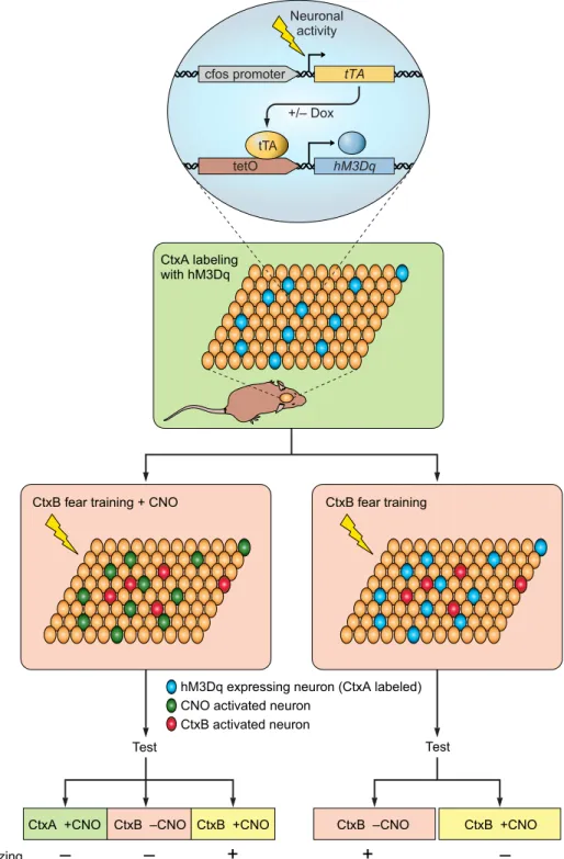
Distributed ensembles of neuronal activity during learning are thought to be necessary for forming association between environmental contexts and emotional states. Within cer-tain brain regions, this activation pattern is suggested to be specific for distinct context-dependent sensory inputs. Che-mogenetics has been used to activate context-specific en-sembles. To achieve this, Garner et al. (73) used transgenic mice expressing hM3Dq under the activity-dependent pro-moter Fos, which allowed selective hM3Dq expression in subset of neurons that were active during a given sensory context(FIGURE 4). This allowed subsequent access to these neurons (which were mainly located in hippocampus,
baso-lateral amygdala, and cortex) and the ability to reactivate them using CNO to investigate how their activation im-pacted memory formation and retrieval. They used a con-textual fear learning task in which a freezing response is stimulated in mice after repeated exposure to an uncondi-tional stimulus (e.g., foot shock) paired with a contextual conditional stimulus. Neurons were marked by hM3Dq ex-pression in context A and then reactivated using CNO dur-ing contextual fear learndur-ing in context B, such that the resulting conditioned fear memory response could not be activated by presenting context B or CNO alone but instead required joint context B and hM3Dq activation of the con-text A-associated ensemble. This result suggests that artifi-cially activated neurons associated with context A formed a hybrid representation with context B that was uniquely as-sociated with representation of the fear memory. If hM3Dq-expressing neurons associated with context A were activated not during context B fear conditioning but during its retrieval, then CNO-induced activation of the competing ensemble interfered with the learned spatial code of context B and thus degraded its recognition. These experiments pro-vided insights into how distinct contextual representations are distributed and retrieved during learning as well as about memorization process of different context.
A similar activity-dependent DREADD expression strategy was also used during Pavlovian fear conditioning (206). Animals form an association between a neutral conditioned stimulus (CS) and a noxious unconditioned stimulus (US), but fear responses to the CS eventually extinguish if pre-sented in the absence of the US. DREADDs were used to investigate whether the original association can be rein-stated by activating the ensemble associated with the fear response (freezing). Mice were trained by Pavlovian fear conditioning while the memory engram was tagged by hM3Dq expression under the control of Fos promoter, which labeled dorsal hippocampal CA1 area, subiculum, the cerebral cortex, and the basolateral amygdala (BLA). After mice underwent extinction training, they no longer responded to the CS; however, when these animals were given CNO, they displayed significant freezing responses. These results suggested that artificial reactivation of origi-nal neuroorigi-nal ensemble was sufficient to recapitulate the fear response and that the original memory engram persisted, even after extinction.
Chemogenetic silencing has also been used in a number of learning and memory studies to replace lesions or common chemical inhibitors of neuron activity such as muscimol or tetrodotoxin. For example, the retrosplenial cortex (RSC) is a structure that receives input from multiple cortical and thalamic sensory areas as well as postrhinal, medial ento-rhinal cortices. The RSC is involved in stimulus-stimulus association such that preconditioning with two cues paired in the absence of reward followed by conditioning with the pairing of one cue with a food reward renders the other
preconditioned cue effective at inducing conditioned re-sponding for food. Examining the role of stimulus-stimulus association in the RSC with reversible inactivation has been challenging due to the large size of this structure, making it difficult to inhibit the entire region with traditional phar-macological inhibitors of neuron activity. Reversible and anatomically encompassing RSC perturbations were achieved after making multiple AAV injections of hM4Di to cover the entire rostrocaudal axis of the RSC in rats (154). Animals with RSC activity inhibited by CNO injection dur-ing preconditiondur-ing were unable to form associations
be-tween neutral sensory stimuli. This study also points to a considerable advantage of DREADDs over existing phar-macological manipulations where perturbations on ana-tomically large or even disconnected regions can be achieved by stereotactic viral delivery. Chemogenetic si-lencing has also been used to investigate fear memory con-solidation in the CA1 region of the hippocampus. Using AAV vectors under control of Ca2⫹/calmodulin kinase II (CaMKII) promoter in dorsal or ventral hippocampal CA1 neurons, hM4Di was expressed selectively in CA1 glutama-tergic neurons (213). Injection of CNO to inhibit CA1
neu-Test Test cfos promoter +/– Dox tTA Neuronal activity tetO hM3Dq tTA
CtxB fear training + CNO
CtxB activated neuron CNO activated neuron
Freezing
–
–
+
CtxB fear training
CtxB –CNO CtxB +CNO
+
–
hM3Dq expressing neuron (CtxA labeled)
CtxA +CNO CtxB –CNO CtxB +CNO CtxA labeling with hM3Dq
FIGURE 4. Summary diagram depicting the experimental strategies to test the im-pact of synthetically activated memory en-grams during fear learning and recall.
rons immediately after contextual fear conditioning dis-rupted fear memory in the ventral but not dorsal hippocam-pus. However, if CNO was given 6 h after training, memory was not affected (213). This suggests that the window for memory consolidation lies somewhere between 0 and 6 h after learning and requires the ventral hippocampus, repre-senting an additional advantage of using AAV vectors to deliver DREADDs to enable anatomical, cell type-specific, and temporal control of neuron activity.
The time period over which consolidated memories require the hippocampus for retrieval has also been examined in fear conditioning. With stereotactic AAV delivery, hM4Di was expressed in the entire length of mouse hippocampus (194). After fear conditioning, the hippocampal depen-dence of memory recall was tested at various time points after training by reversible hippocampal inhibition with CNO. Disrupting hippocampal activity produced a tempo-rally graded amnesia in which recall of recent memories (2 days) was impaired but distant memories (⬎7 wk) appar-ently no longer required intact hippocampal activity. Learning to avoid predators has also been studied in the context of hypothalamic fear encoding. The VMH plays a role in defensive responses to predators. Silva et al. (166) generated a transgenic mouse in which hM4Di is expressed under the control of Nr5a1 promoter to restrict its expres-sion to VMH. They silenced VMH activity during training sessions in which mice were exposed to a predatory rat to test whether this region is involved in acquisition of fear memory. Mice in which VMH activity was suppressed dur-ing predatory fear conditiondur-ing showed significantly de-creased fear response when they were reexposed to the predator-associated context. Furthermore, mice condi-tioned with predatory fear showed impaired fear responses if VMH was silenced during reexposure, suggesting that VMH is involved in both acquisition and recall of predator fear.
5. Sleep
DREADD chemogenetic technology has also been deployed to decipher sleep regulation circuits. Acute activation of parabrachial nucleus neurons in mice with hM3Dq and CNO caused rapid induction of wakefulness and alert state-like electroencephalogram (EEG) activity that can be sus-tained up to 4 days by repeated injections (150). The PBN sends projections to a variety of brain regions, and to deter-mine which downstream target region is responsible, a pro-jection target-specific labeling strategy was used. Each of the potential downstream projection targets was injected with a Cre recombinase expressing AAV serotype 6, which can be taken up by PBN axons and retrogradely trans-ported. By making a second AAV injection into PBN carry-ing a Cre-dependent hM3Dq, they specifically expressed this DREADD in a subset of PBN neurons that send axons to a specific target. The wakefulness response was
recapit-ulated by activating PBN neurons that send projections to basal forebrain or hypothalamus but not thalamus. The role of lateral hypothalamic hypocretin/orexin neurons has also been examined in sleep with chemogenetics. Ex-pression of hM3Dq and hM4Di in Orexin-cre mice was used to measure the impact of chemogenetic activation and inhibition on sleep. hM3Dq-mediated activation of hypo-cretin/orexin neurons significantly increased time spent in wakefulness and reduced the non-rapid-eye-movement (NREM) and rapid-eye-movement (REM) sleep times, while hM4Di-dependent inhibition had an opposite ef-fect on wakefulness and NREM sleep (161). In a related study, DREADD-dependent manipulations were per-formed in basal forebrain (BF) cholinergic neurons in
Chat-ires-cre mice (32). Activating BFCHAT neurons
in-creased wakefulness, whereas inhibition had an opposite effect. This group also took advantage of DREADD ac-tivation of BFCHATneurons and performed a Fos analysis
to mark activated downstream regions. Finally, Chen et al. (33) used DREADD technology and viral lesions to probe the role of various brain stem nuclei in sleep regu-lation and atonia. Acute chemogenetic activation of pre-superior olivary medulla (pSOM) selectively suppressed REM but not NREM sleep, while activation of rostral ventromedial medulla (RVM) increased wakefulness and decreased sleep.
6. Pain
DREADDs have started to find widespread use in itch and pain research. One of the major goals in pain research is to control nociceptive afferent excitability. To silence pain transmitting C-fibers, a Trpv-1-Cre mouse was crossed with Cre-conditional hM4Di mouse line (159). A single dose of CNO rapidly decreased their excitability and significantly increased pain threshold, thereby producing analgesia last-ing up to 3 h (159). Notably, these researchers have also observed CNO-independent effects of hM4Di expression on sensory neurons, including significant alterations in Na⫹ current, increased NaV1.7 expression, and decreased
volt-age-gated calcium channel conductance (159). Further-more, in hM4Di-expressing neurons, endogenous Gi
-cou-pled signaling was also disrupted. Such reports on off-target effects of DREADD expression have been rare, and it is not known whether other neuronal cell types that express DRE-ADDs also have altered functional properties. Nevertheless, these results highlight the need for careful consideration of the consequences of overexpressing any protein, including DREADD-based GPCRs in cells.
Afferent nociceptors were targeted by others as well. In-stead of intersectional transgenic mouse breeding, viral in-jections into sciatic nerve were used to target sensory neu-rons (94). The analgesic effects were compared for chemo-genetic (hM4Di) silencing and optochemo-genetic (step function optogenetic inhibitor SwiChR) inhibition of unmyelinated
DRG neurons that carry pain signal and observed similar increases in mechanical and thermal pain threshold. DREADDs have also been used in pain and itch signaling circuits in the dorsal horn of the spinal cord. Mechanical hypersensitivity was increased when neurons marked by transient expression of vGlut3 in the dorsal horn were ac-tivated by hM3Dq (145). A role of NPY-expressing dorsal horn neurons in light touch mechanical itch was investi-gated by crossing Npy-Cre transgenic mice with a condi-tional hM4Di-DREADD expressing mouse line for acute silencing of NPY neurons (21). After CNO injection, light mechanical stimulation produced robust scratching re-sponses, suggesting that acute NPY neuronal inhibition abolished a cellular circuit node that normally suppressed itch. It should be noted that the intersectional approach to access NPY-expressing neurons described here would drive DREADD expression in other brain regions. Therefore, sys-temic delivery of CNO likely inhibited non-spinal NPY-expressing populations as well.
Pain threshold is modulated, in part, by descending inputs to the spinal cord that affect pain transmission. An impor-tant part of this circuit is thought to be from RVM and local enkephalinergic neurons in the dorsal horn. Francois et al. (67) explored this pathway in detail using DREADDs. They intrathecally delivered AAV expressing Cre-dependent hM4Di to inhibit spinal enkephalinergic neurons in Penk-cre mice, which induced robust mechanical hypersensitivity in the presence of CNO. Viral tracing showed that spinal encephalin neurons receive direct inhibitory input from GABA-releasing RVM neurons. Spinal cord projecting RVMVGAT neurons were targeted with hM4Di using an
intersectional approach. RetroAAV-FLEX-FlpO, an ax-onally transported viral vector expressing FLP-recombinase protein in a Cre-dependent manner, was injected into the dorsal horn of Vgat-Cre mice. In this way, they retrogradely targeted FLP to all VGAT expressing neurons that project to the dorsal horn(FIGURE 2F). By injecting a second AAV into RVM that expresses hM4Di in a FLP-dependent fash-ion, they were able to selectively target hM4Di to spinal cord projecting RVMVGAT neurons. Inhibition of these RVM neurons reduced mechanical sensitivity.
Pain signaling in higher brain centers has also been explored using DREADDs; for example, hM3Dq-mediated activa-tion of periaqueductal gray and dorsal raphe (PAG/DR) tyrosine hydroxylase positive neurons in mice has an anti-nociceptive effect (112). Fos activation mapping for puta-tive downstream mediators of acute pancreatitis-induced visceral pain identified the PVT, PAG, and PFC (97). Selec-tive expression of DREADDs in these regions was used to show that inhibition of PVT neurons or activation of down-stream glutamatergic neurons in mPFC (but not PAG neu-rons) attenuates visceral nociception. Finally, Wakaizumi et al. (196) investigated exercise-induced hypoalgesia and
showed that selective suppression of nucleus accumbens projecting VTA dopaminergic neurons offsets antinocicep-tive effects of low-intensity exercise.
7. Primate applications
The noninvasive and reversible properties of DREADDs have made these tools attractive for primate research as well. In a chemogenetic dissociation study, hM4Di-express-ing viral vectors were delivered to rhinal cortex (Rh) in one hemisphere and orbitofrontal (OFC) cortex in the other hemisphere in rhesus monkeys (56). Silencing these two regions contralaterally disrupts intrahemispheric communi-cation between the two regions, effectively dissociating the two structures. When CNO was injected, these animals had significantly reduced sensitivity to changes in reward size. Related work examined reward value processing by chemo-genetically silencing rostromedial caudate (rmCD), using virally expressed hM4Di in monkeys (136). Inactivating rmCD bilaterally resulted in significant loss of sensitivity to reward value. In most studies verification of DREADD ex-pression is performed through post mortem histological analysis. However, Nagai et al. (136) also developed a method to visualize DREADD expression in vivo. They used a positron emission tomography (PET)-sensitive DRE-ADD ligand,11C-labeled clozapine. Injecting monkeys with this ligand provided information about the size and location of hM4Di expression, and by using a blocking protocol they were able to observe receptor occupancy by CNO. This approach has significant advantages over post mortem analysis, especially in primate research, which requires in-vestment of substantial time and effort for each subject. DREADDs have also been used in conjunction with mag-netic resonance imaging (MRI) in primates. In rhesus mon-keys, chemogenetic silencing of amygdala was combined with functional connectivity MRI, a configuration that al-lowed investigation of the brain-wide functional connectiv-ity in vivo (79). Silencing the amygdala affected not only amygdalo-cortical connectivity, but also corticocortical coupling across multiple functional systems was disrupted, although a high CNO concentration (10 mg/kg) was used. In these studies, DREADD technology has provided signif-icant improvement over classical approaches that have lim-itations imposed by necessity to use implant to manipulate neuronal activity or by compensatory changes that can oc-cur after lesions.
A related technology has been developed by Michaelides et al. (134) termed DREADD-assisted metabolic mapping (DREAMM). This approach is based on making remote-controlled cell type specific perturbations with DREADDs and observing the brain-wide functional consequences in awake-behaving animals under PET scan. In this study au-thors described PET measurement of whole-brain activity by monitoring brain glucose metabolism with injected
[18F]fluorodeoxyglucose (FDG) as they hM4Di-inhibit
dis-tinct nucleus accumbens shell subpopulations. Although this work was performed in rats, a similar approach could be used in other rodents and primates. Furthermore, ent PET probes could be used to monitor changes in differ-ent cellular processes of downstream targets such as neu-rotransmitter dynamics or enzymatic activities. Thus DREAMM has the potential to map global long-range func-tional connectivity networks by combining advantages pro-vided by cell type specificity of DREADD manipulations and noninvasiveness of PET imaging.
Nevetheless, the use of CNO-based DREADD strategies in non-human primates must consider the limited access of CNO to the brain (75a, 136, 151). In addition, retro-version of CNO has been measured to give significant con-centrations of clozapine in cerebrospinal fluid (CSF) in one study (151), but CSF clozapine was not detected in a differ-ent report (136) is the active agdiffer-ent in the brain. Thus alter-native DREADD ligands should be examined for use in non-human primates.
E. Non-neuronal Applications 1. Glial cells
Glial cells are the predominant cell type in the nervous system, and they can be marked by glial fibrillary associated protein (GFAP). Specific manipulation of these cell popula-tions is important for understanding their role in vivo. Transgenic mice expressing hM3D under a GFAP promoter fragment were used to assess physiological impact of glia specific activation. CNO-mediated activation of Gq-cou-pled signaling in glia throughout the body altered auto-nomic nervous system functions by increasing heart rate, blood pressure, saliva formation, and decreasing body tem-perature (1). Glial targeting can be achieved in wild-type animals by using viral vectors expressing DREADDs under a GFAP promoter. Using this approach in rats, hM3Dq activation in glial cells in the nucleus accumbens core in-duced glutamate release, which was sufficient to inhibit cue-induced reinstatement of cocaine (163) and ethanol seeking (23), suggesting a potential in vivo role for glial signaling to influence behavior. CNO-mediated activation of hypotha-lamic astrocytes has been shown to suppress feeding with-out effecting emotional states (184, 203). The influence of in vivo astrocytic Gq signaling on blood flow was investi-gated using hM3Dq and was found to be insufficient to alter cortical hemodynamics (20). Glial cells of the enteric ner-vous system were also activated by hM3Dq, resulting in neurogenic contractions as well as intestinal and colonic motility, showing that gut reflexes can also be regulated by glial cells (133).
A non-neuronal contribution to pain pathways outside the brain have been examined by DREADDs. Grace et al. (77)
investigated the role of spinal cord microglia in morphine-induced neuropathic pain in rats. A common agent for pain management, morphine, induces inflammasome in micro-glial cells. To test causal involvement of microglia, they intrathecally delivered hM4Di under the direction p38 mi-croglial promoter. Activation of mimi-croglial hM4Di inhib-ited proinflammatory signals and reversed morphine-in-duced sensitization, confirming a causal role for these cells. 2. Other cell types
GPCR signaling is important for nearly all cell types. Thus DREADDs can be applied to assess the general function of these cell signaling pathways independently of endogenous GPCRs. For example, the role of Gq-coupled signaling was probed in hepatocytes using hM3Dq, which was shown to stimulate glycogen breakdown and gluconeogenesis (113). Activation of Gsand Gq pathway activation in pancreatic
-cells using transgenic mice showed a protective role for activating Gq pathway against diabetes (81, 95).
hM4Di has been used to interfere with intracellular signal-ing and metastasis in breast cancer cells (202). Researchers have also used hM4Di in T-lymphocytes to engineer che-motaxis toward a CNO source (143). DREADDs have been used in kidney research as well. Gq signaling has been stud-ied in podocytes, specialized cells that wrap glomerular cap-illaries and are thought to influence glomerular filtration. Due to technical limitations it has been difficult to study the role of Ca2⫹signaling in these cells in vivo; therefore, most evidence was based on cultured studies. A mouse line with podocyte specific hM3Dq expression enabled CNO-depen-dent activation of Gq signaling in vivo. This elicited Ca2⫹ transients in podocytes, but surprisingly this manipulation did not have any impact on glomerular perfusion or filtra-tion, suggesting that more prolonged Ca2⫹level alterations might be required to alter these parameters (103).
In summary, a rapidly expanding body of literature, some of which is mentioned here, suggests that DREADDs have already started to transform neuroscience and hold signifi-cant promise for non-neuroscience applications as well.
III. LIGAND-GATED ION CHANNEL CHEMOGENETIC TOOLS
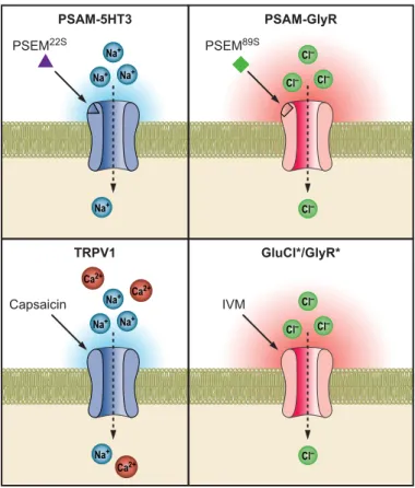
LGICs permit direct pharmacological control over ion con-ductance. The functional properties of ion channels are pri-marily dictated by their ion selectivity. Inward flux of cat-ions or outward flux of ancat-ions depolarizes cells, and corre-spondingly inward flux of anions or outward flux of cations leads to cellular hyperpolarization (98). Some channels also pass Ca2⫹and influence other signal transduction pathways independently of the effect on cellular membrane potential. Additional important characteristics can be tuned with spe-cific mutations, such as conductance magnitude,
localiza-tion, and desensitization properties (87). Several LGIC fam-ilies have been developed as chemogenetic tools, including Cys-loop receptors, ATP-sensitive channels, and transient receptor potential (TRP) channels.
A. Cys-Loop Receptors
The Cys-loop receptors are a family of pentameric ion chan-nels named for conserved vicinal cysteine residues in a loop that is important for channel gating (168, 188). Cys-loop receptors include nicotinic receptors (nAChR), serotonin receptor 3 (5HT3), GABA receptors, glycine receptors (GlyR), and glutamate-gated chloride channels (GluCl), many of which are heteromers that are comprised of multi-ple subunits(FIGURE 5). Several Cys-loop ion channels form as pentameric homomers, which facilitates use of these ion channels as tools for manipulating cellular function because only a single ion channel subunit needs to be expressed. These include␣7 nAChR, ␣9 nAChR, 5HT3, GABA-C, and GlyR. Agonist binding at Cys-loop receptors occurs at the interface between protomer subunits. Binding at the princi-pal subunit face is associated with interaction with the ag-onist pharmacophore, a cationic moiety that is stabilized by interactions with a backbone carbonyl and cation- inter-actions with aromatic tyrosines and tryptophans (111). The complementary face of the ligand binding domain (LBD) is contributed by the adjacent protomer, which largely affects the specificity of ligand-receptor interactions. Ligand
bind-ing leads to a contraction of the LBD, and these structural changes are transmitted to the transmembrane domain to permit ion permeation. Ion selectivity is determined in part by cytoplasmic residues that can be altered to switch cation channels into anion-selective channels and vice versa (147, 169).
1. GABA-C receptor
One of the first chemogenetic ion channel tools involved heterologous expression of the GABA-C (also called GABA rho) receptor in hippocampal neurons. This LGIC is not normally expressed in hippocampal neurons and rendered them selectively sensitive to the GABA-C agonist cis-4-ami-nocrotonic acid (CACA) (36). Therefore, this system re-vealed successful neuronal expression of a heterologous LGIC and used a selective ligand for that channel to control neuron excitability. Nevertheless, this approach was lim-ited by the remaining sensitivity to the endogenous ligand GABA, which was perturbative to neuron activity.
2. GluCl/ivermectin
An improved approach applied GluCl subunits from Cae-norhabditis elegans (41) to potently control neuron excit-ability (170). These channels are sensitive to the natural product ivermectin (IVM), which is also used clinically as an antiparasitic (41). GluCl is a heteromeric channel com-prised of two subunits (GluCl␣ and GluCl); however, more recent results indicate that the GluCl␣ subunit can form homomeric channels (68). A mutation in the gluta-mate binding site reduced glutagluta-mate efficacy by sixfold, although glutamate potency and IVM sensitivity were not substantially changed (114, 170). One limitation of IVM is that it also activates or modulates GABA receptors, glycine receptors,␣7 nAChRs, and P2X4-R albeit with lower po-tency than for GluCl (208), and this is associated with tox-icity in the central nervous system (192). Therefore, low doses of IVM are essential for use as a selective chemoge-netic tool. To aid in more selective usage of IVM, a modified version of GluCl has been developed that is activated with ~10 nM IVM, which is a⬎10-fold improvement in potency (68). Although use of this channel has not yet been reported in vivo, the original modified GluCl system has been used in several studies.
In mice, expression of GluCl in the striatum, an area asso-ciated with motor function, was found to be well tolerated and to not show any obvious changes in locomotor activity. Upon administration of IVM to mice intraperitoneally, the mice displayed elevated rotational behavior in conjunction with amphetamine injection, consistent with loss-of-func-tion in the striatum (110). In another study, this system was used to suppress the function of the ventrolateral portion of ventromedial hypothalamic nucleus (VMHvl) to suppress attack in a resident intruder aggression assay (119). In each
Capsaicin TRPV1 GluCl*/GlyR* Ca2+ Ca2+ Ca2+ Na+ Na+ Na+ Na+ Cl– Cl– Cl– Cl– IVM PSEM22S PSAM-5HT3 PSAM-GlyR Na+ Na+ Na+ Na+ Cl– Cl– Cl– Cl– PSEM89S
FIGURE 5. Chemogenetic LGIC tools for activating and inhibiting neurons.
of these experiments in mice, the pharmacokinetics time-scale of the chemogenetic loss-of-function perturbation was slow to develop and was especially prolonged, with effects reported 1 day following injection and lasting for more than a week (110, 119). This corresponds to the known pharma-cokinetic profile of IVM, which is largely distributed into fatty depots forming a reservoir of the drug that leaches back into circulation over multiday timescales (11). Thus GluCl provides an effective chemogenetic framework, but it has some limitations such as the need for heteromeric re-ceptors, and the toxicity and slow pharmacokinetics of IVM.
3. Glycine receptor/ivermectin
An alternative IVM-sensitive chemogenetic system was de-veloped based on an engineered glycine receptor (GlyR) that has one LBD mutation to eliminate glycine sensitivity and a second IPD mutation to increase IVM potency (127, 128). This channel is activated by 19 nM IVM (126). The engineered receptors are homomeric channels that are se-lective for chloride and can reduce neuron electrical activ-ity. Two Cre-dependent mouse lines based on this channel have been reported, where IVM administration is reported to reduce neuron activity in vivo and ex vivo (89, 142). Another version of this GlyR-based chemogenetic system exploits IVM sensitization of glycine responsiveness. In this channel, IVM sensitivity is enhanced with an IPD mutation, and glycine sensitivity is reduced. However, in the presence of IVM, glycine sensitivity is potentiated, which can sup-press neuron activity (92). The appeal of this system is that it does not lead to a constant chloride shunt of the cell, which in some cases can increase cellular excitability if the chloride gradient is collapsed.
An IVM-sensitive GlyR chemogenetic system has been con-verted to cation selectivity by applying three mutations to the intracellular domain of the channel (91). This system can be applied to depolarize neurons to fire action poten-tials. Thus IVM/GlyR chemogenetic tools can be used as neuron silencing and activation tools, but they require cop-ing with the complex pharmacology of IVM.
4. PSAM/PSEM engineered ion channels and ligands
A) DESIGN.To utilize the functional diversity of the Cys-loop ion channel family, a set of chemogenetic tools was created based on chimeric LGICs derived from␣7 nicotinic acetyl-choline receptor (nAChR) and other Cys-loop family mem-bers. The␣7 nicotinic acetylcholine receptor (nAChR) was chosen because it has been intensively investigated as a drug target for schizophrenia therapies and thus had a number of published small molecule structure activity relationships (49). In addition, mutagenesis studies of ␣7 nAChR had previously identified residues important for binding
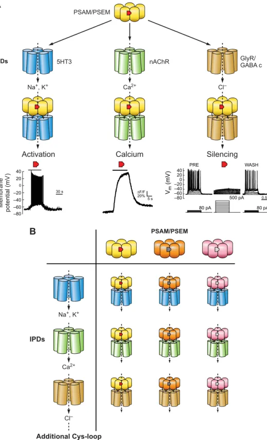
acetyl-choline and there are also crystal structures of the homolo-gous acetylcholine binding protein bound to agonists such as nicotine, carbamoylcholine, and epibatidine (29, 115). ␣7 nAChR forms homopentameric channels, simplifying its use as a tool by requiring expression of only a single sub-unit. Finally, the extracellular ligand binding domain (LBD) of ␣7 nAChR is transferrable to the transmembrane ion pore domains (IPDs) of other members of the Cys-loop LGIC family (55, 80, 130). This property allows the phar-macology of the␣7 nAChR to be maintained while access-ing the ion conductance properties of other LGICs, such as the 5HT3 receptor, GlyR, and the GABA-C receptor. To leverage these characteristics, mutagenesis of the ␣7 nAChR LBD was performed to identify interactions with small molecules that did not activate unmutated ␣7 nAChRs. For this, a chimeric channel consisting of the␣7 nAChR LBD and 5HT3 IPD (␣7–5HT3) was selectively mutated to all other possible amino acids at four sites on the complementary face of the ligand binding pocket formed at the interface of two protomer subunits. These mutant chan-nels were screened in cell-based assays using fluorescent membrane potential dyes against a library of small mole-cules synthesized to have characteristics previously indi-cated to have poor potency against the unmodified ␣7 nAChR (19). This resulted in identification of three muta-tions (W77F, Q79G, L141F) that each conferred selective agonist activity to structurally distinct small molecules with low to no activity against␣7–5HT3 (130). Moreover, ad-ditional molecules could be synthesized that were also se-lective between each mutant channel as well as the unmu-tated channel. Each of these selectivity-conferring muta-tions could be combined with an additional mutation that reduced ACh potency (Y115F, Q139G, L141S). These mu-tated LBDs were shown to be transferrable to four IPDs and were termed pharmacologically selective actuator modules (PSAMs, pronounced as SAMs) (130). The cognate syn-thetic ligands are called pharmacologically selective effector molecules (PSEMs, pronounced as SEMs). Together, PSAM/PSEMs offer flexible tools for selective LGIC phar-macology(FIGURE 6).
PSAMs can be applied to construct a variety of chimeric ion channels with distinct functional properties. PSAM-5HT3 provides prolonged depolarizing currents in the presence of the corresponding PSEM89S, consistent with the functional properties of␣7–5HT3. The addition of three mutations to the IPD of this chimeric channel increased conductance over 100-fold (PSAM-5HT3 HC) (130). PSAMQ79G,L141S -nAChR V13’T allows for primarily Ca2⫹conductance with the V13’T ion pore mutation greatly curtailing the desensi-tization normally associated with ␣7 nAChR channels. Pharmacologically selective inhibitory channels were gen-erated by PSAM-GlyR and PSAM-GABA C channels. Each of these shows large conductances as well as long steady-state window currents to maintain silencing as long as the
agonist is present (130). Cells expressing PSAM-GlyR chan-nels dramatically reduce input resistance by 70%, making it difficult to fire action potentials. Correspondingly, rheo-base (the current required to fire an action potential) in-creases by at least eightfold (130). This makes PSAM-GlyR
channels extremely powerful tools for silencing neuronal activity.
B) EX VIVO APPLICATIONS: NEURAL CIRCUIT ANALYSIS.These engineered chimeric LGICs and their corresponding PSEMs have been
Na+, K+ Ca2+ IPDs PSAM/PSEM Additional Cys-loop channel IPDs
B
Cl– M e mbr ane pot e n tial (mV ) –60 –40 –20 0 20 40 30 s PRE WASH 40 20 0 –40 –60 –80 –20 80 pA 80 pA 500 pA 0.5 s Vm (mV) 5 s ΔF/F 20%Activation Calcium Silencing
A
IPDs PSAM/PSEM 5HT3 Na+, K+ Ca2+ nAChR GlyR/ GABA c –80 Cl–FIGURE 6. Combination of pharmacolog-ical and functional Cys-loop modules to gen-erate engineered chimeric ion channels for controlling ion flux. A: for a given PSAM (yellow)/PSEM (red) pair, combination with different IPDs allows multiple functional out-comes to be achieved ranging from neuro-nal activation, controlling calcium flux, or neuronal silencing. B: multiple orthogonal PSAM/PSEM pairs have been developed, which enables combinatorial generation of diverse chemogenetic ion channel tools.
used in a number of ex vivo and in vivo applications. For ex vivo analysis of circuit functions, PSAM-GlyR has been selected because it strongly suppresses neuron activity. PSAM-GlyR has been used in analysis of interneuron pop-ulations in acute hippocampal microcircuits (12, 125, 156) and retinal (195) ex vivo tissue preparations. These studies reported complete suppression of electrical activity from interneuron populations expressing PSAM-GlyRL141F in
the presence of cognate PSEMs. Effective neuronal silencing facilitates analysis of circuit computations in the presence and absence of signaling through a particular circuit node. C) LEARNING AND MEMORY.The powerful neuronal silencing func-tions of PSAM-GlyR in analysis of cortical and hippocampal circuits have been used to investigate the role of molecularly defined interneuron populations in different learning pro-cesses. Building on ex vivo studies using PSAML141F-GlyR
silencing (125), somatostatin (SOM) interneurons were si-lenced in vivo by intraperitoneal administration of PSEM89S
(124). Consistent with the established fast pharmacokinetics of this molecule (130), in vivo silencing was rapid and recov-ered after 20 –30 min postinjection (124). Silencing SOM in-terneurons but not parvalbumin (PV) inin-terneurons blocked contextual fear conditioning, consistent with the inability to distinguish contextual cues appropriate for fear responding in the absence of firing from this population (124).
In another study (13), the role of long-range inhibitory pro-jections from the lateral entorhinal cortex (LEC) to hip-pocampus in contextual fear conditioning was examined for by axonal silencing with PSAML141F-GlyR. The potent
PSEM308was delivered by intracranial injection to the
dis-tal axon projections of LEC to the hippocampus. Silencing these axons in a contextual fear conditioning assay did not prevent freezing in the conditioned context, but it led to inappropriate generalization such that the mice showed freezing in a distinct context, which is not typically ob-served. These studies demonstrated that the output of local axon projections could be silenced by local PSEM delivery.
Cortical and hippocampal CA3 PV interneuron activity during a foreign object recognition experiment, spatial learning, and contextual fear memory was examined using bidirectional control of PV neuron activity with PSAML141F,Y115F-5HT3 HC and PSAML141F-GlyR (50, 51,
100). Activation of PV neurons increased PV immunoreac-tivity and PV synapse number on CA3 pyramidal cells. Con-versely, inhibition using the same ligand in different mice led to a corresponding reduction in PV immunoreactivity and synapse number. PV neuron inhibition reduced PV ex-pression levels, promoted memory acquisition, and promoted FOR, while PV neuron activation showed opposite outcomes (51). These experiments demonstrate bidirectional control over PV, an dependent gene, along with activity-dependent synaptic plasticity, and associated consequences for the FOR behavioral task. A related set of experiments used PSAML141F-GlyR to investigate SOM and VIP interneuron
influences on cortical integration (135). The use of LGICs to make these perturbations is consistent with the conclusion that the cellular and behavioral consequences of these manipula-tions are activity dependent, which is less certain for perturba-tions with GPCRs that can directly affect gene expression in-dependently of effects on activity.
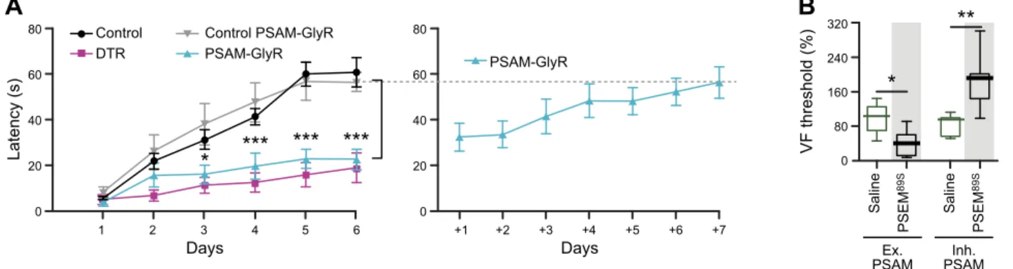
Chemogenetic silencing of a hindbrain premotor nucleus es-tablished its role in learning motor tasks like reaching or ro-torod balancing (58). This study compared the effectiveness of reversible neuronal silencing with PSAML141F-GlyR of acute
neuronal ablation. The behavioral impact on rotorod balance duration or reach success rate was very similar between per-manent neuron ablation and reversible silencing(FIGURE 7A). Moreover, learning was tested over multiple days, and the effectiveness of silencing was maintained with repetitive appli-cation of the ligand, indicating the durability of the PSAML141F-GlyR response. Importantly, however, once the
ligand was no longer administered, mice quickly learned the rotorod task (58). Thus these inhibitory channels show similar neuron suppression efficacy as neuron ablation but allow for reversible, daily control of cell function.
80 60 40 20 0 80 60 40 20 0 1 2 3 4 5 6 +1 +2 +3 +4 +5 +6 +7 Days Days Latency (s) Control DTR Control PSAM-GlyR PSAM-GlyR PSAM-GlyR * *** *** *** 320 240 160 80 0 ** * VF threshold (%) Ex.
PSAM PSAMInh.
Saline PSEM 89S Saline PSEM 89S
A
B
FIGURE 7. Efficacy, durability, and bidirectionality of PSAM/PSEM neuron perturbations. A: daily, reversible silencing of premotor neurons with PSAM-GlyR during rotorod motor learning compared with permanent neuron ablation (left). Subsequent training without PSEM injection led to efficient learning of the task. [From Esposito et al. (58), with permission from Nature Publishing Group.] B: bidirectional activation (Ex. PSAM) or silencing (Inh. PSAM) of D2R neurons in the striatum alters Von Frey (VF) threshold in spared nerve injury (SNI) pain model. [From Ren et al. (153), with permission from Nature Publishing Group.]