109/
Acta Chir Orthop Traumatol Cech. 85, 2018, No. 2p. 109–112
ORIGINAL PAPER
PŮVODNÍ PRÁCE
rare and can be cured mostly by arthroscopic surgery (5, 17).
In the present study, we aimed to investigate the out-comes of arthroscopic surgery in localized giant cell tumors causing knee joint locking.
MATERIAL AND METHODS
This retrospective study includes 7 patients underwent arthroscopic debridement for intraarticular giant cell tumor of tendinous origin (Figs. 2–4). Five patients
INTRODUCTION
Space occupying lesions of knee joint may cause symptoms such as locking or blocking (13, 16, 20, 23, 24, 25). Physical examination reveals sense of blocking, a popping sound during joint movement; and patients typically can’t fully extend their knee. The major causes locking in the knee joint include anterior cruciate ligament (ACL) tear, bucket handle meniscus tear, and intraarticular foreign or loose bodies (3, 4, 15, 18). Rarely, the first symptoms of space occupying lesions of knee may also be blocking or locking. Intraarticular tumors include ganglia, lipoma arborescens, synovial hemangioma, and synovial chondromatosis (7, 9, 11, 22, 24, 26).
Non-osseous giant cell tumors usually originate from periarticular soft tissue (Fig. 1). In 2002, the World Health Organization (WHO) classified giant cell mus-culoskeletal tumors on the basis of their site of origin. Tumors originated from tendon sheath are termed as lo-calized giant cell tendon tumors, and the ones of synovial origin that usually show diffuse localization are referred to as pigmented villonodular synovitis (8).
In diffuse PVNS, radiotherapy is routinely used for disease remission after arthroscopic debulking. It’s cure rate is lower than that of the localized form, which is
Arthroscopic Excision of Tendinous Giant Cell
Tumors Causing Locking in the Knee Joint
Artroskopická exstirpace tendinózních obrovskobuněčných nádorů působících
kolenní blokády
B. GÜLENǹ, E. KUYUCU¹, S. YALÇIN¹, A. ÇAKIR², A. M. BÜLBÜL¹ ¹ Istanbul Medipol University, Department of Orthopaedics and Traumatology, Istanbul, Turkey ² Istanbul Medipol University, Department of Pathology, Istanbul, Turkey
ABSTRACT
PURPOSE OF THE STUDY
Non-osseous giant cell tumors are locally aggressive tumors arising around joints. They are commonly located around synovial joints such as wrist and knee and occasionally cause mechanical symptoms.
MATERIAL AND METHODS
This retrospective case series includes 7 patients operated due to intraarticular lesion. The mean age of the patients was 28.7 (range 22–37) years. Mean follow-up period was 12 months.
RESULTS
All patients underwent arthroscopic debridement. They were followed monthly with clinical examination and magnetic resonance imaging (MRI) was obtained at third month for all patients. Patients were contacted through phone call and evaluated with the WOMAC score retrospectively. No recurrence was detected in any patient.
CONCLUSIONS
Arthroscopic debridement is a safe surgical technique that may replace open surgery in the treatment of intraarticular tendinous giant cell tumors.
Key words: tendinous giant cell tumor, arthroscopy, knee locking.
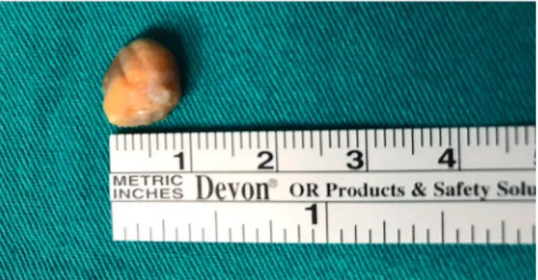
Fig. 1. Morphologic display of a excision material.
were female and two were male (Table 1). Initially, all patients had sense of blocking or locking in the knee joint. Three patients had accompanying pain, while 4 patients didn’t have. The diagnosis of giant cell tumor was confirmed with biopsy before surgery for all patients. (Figs. 5–7). All operations were performed by one senior surgeon (AMB).
110/
Acta Chir Orthop Traumatol Cech. 85, 2018, No. 2ORIGINAL PAPER
PŮVODNÍ PRÁCE
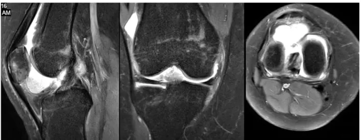
Fig. 2. A mass image seen T2 MRI both coronal,sagittal and axial views.
Fig. 3. 2 cm diameter mass located ahead of anterior cruciate ligament.
Fig. 4. Another giant cell tumor located behind patella seen all MRI views.
Exclusion criteria included: open surgery, patients with missing MRI images before or after the operation, and pathological diagnosis other than a giant cell tendinous tumor. Preoperative data, images, and surgical photos of four patients were retrospectively screened and recorded.
All patients having a pathological diagnosis of tendinous giant cell tumor were ordered a follow-up MRI at third month.
Patients without recurrence on MRI examination returned for a repeat visit at six months and one year post-operatively, and follow-up intervals were lengthened thereafter. No ab-normality was detected at third, six-month and one year follow-ups in any patient. At short term follow-up recurrence was detected in any case.
The mean tumor size at presentation was 2.3*2.4 cm2. All patients were contacted and evaluated with the WOMAC score. The mean WOMAC score was 95,4 (91.1–98,4). None of the patients had tumor recurrence during follow-up. No patient complained of limitation of joint motion or residual pain at the final examination.
All patients had histologic diagnosis of localized giant cell tumor. Histologic features of this tumor include abundant mononuclear histiocytic cells and in three-di-mensional tissue biopsy, hemosiderin within histiocytes. Pathologic evaluation showed osteoclast-like giant cells, foam cells, inflammatory cells and siderophage migra-tion.
DISCUSSION
Giant cell tumors of tendon sheath occur around or sometimes within the joint. Arthroscopic debridement is mostly curative for intraarticular tumors (14). In a retro-spective study involving 30 patients, Loriaut et al reported that the most common symptoms were knee discomfort and swelling. The lesions were located in the gutter region in 45% of patients. All patients were arthroscopically treated and none of the patients experienced recurrence during an average follow-up period of 75 months (14).
In a series (n = 26) reported by Dines et al, the mean age of diagnose was 36 years for pigmented villonodular synovitis with intra-articular localization, and the mass most commonly located at suprapatellar region followed by femoral notch. Unlike our series; 12 patients in that study underwent arthroscopic treatment while the remainders were managed with open surgery (6). Recurrence rates of 16–17% have been reported in the literature for giant cell tumors originated from tendon sheath. The majority of recurrences occur in tumors having diameter greater than 2 cm and patients managed with open surgery (2,21).
In the longest follow-up series reported so far, Zhang et al. followed 12 intra-knee tumors for approximately 15 years. They observed no recurrence, and stated that the outcomes were satisfactory for all patients. They re-ported that a wider tumor resection and the debridement of the whole tunnel through which the tumor passes suffice for surgical management (27).
The most common intraarticular site is the knee joint. Ho et al. reported that intraarticular involvement was seen in 32 of 41 patients, and 18 of these occurred in knee joint. (10)
RESULTS
The mean age of patients was 28.7 (22–37) years. (Table 1) All patients were prescribed post-operative knee range of motion (ROM) and quadriceps muscle strengthening exercises following elastic bandage wrap-ping. Six patients were discharged the following day, and one patient was discharged on second day due to pain. Patients were controlled for knee ROM and strength every second day in first two weeks postoperatively. Wound dressing was removed after a mean of 10 days.
111/
Acta Chir Orthop Traumatol Cech. 85, 2018, No. 2ORIGINAL PAPER
PŮVODNÍ PRÁCE
Fig. 5. HEx40 tumor consisting of monotone cells with diffuse growth pattern. Right upper corner shows focal histiocytic re-sponse.
Fig. 6. HEx100. multinucleated giant cells accompanying mo-nonuclear cells.
Fig. 7. HEx200. Mononuclear, non atypical cells and accom-panying multinucleated giant cells.
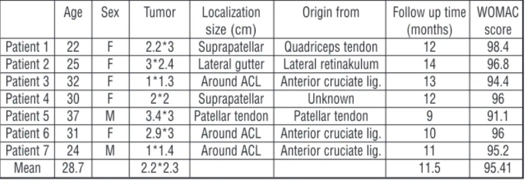
Table 1. General characteristics of the participants
Age Sex Tumor Localization Origin from Follow up time WOMAC
size (cm) (months) score
Patient 1 22 F 2.2*3 Suprapatellar Quadriceps tendon 12 98.4 Patient 2 25 F 3*2.4 Lateral gutter Lateral retinakulum 14 96.8 Patient 3 32 F 1*1.3 Around ACL Anterior cruciate lig. 13 94.4
Patient 4 30 F 2*2 Suprapatellar Unknown 12 96
Patient 5 37 M 3.4*3 Patellar tendon Patellar tendon 9 91.1 Patient 6 31 F 2.9*3 Around ACL Anterior cruciate lig. 10 96 Patient 7 24 M 1*1.4 Around ACL Anterior cruciate lig. 11 95.2
Mean 28.7 2.2*2.3 11.5 95.41
8. Fletcher CDM, Krishnan Unni K, Mertens F (eds). Giant cell tumour of tendon sheath. In: World Health Organization Classification of tumors. Pathology and genetics of tumors of soft tissue and bone. IARC Press, Lyon, 2002, pp 110–111.
9. Hirano K, Deguchi M, Kanamono T. Intra-articular synovial lipoma of the knee joint (located in the lateral recess): a case report and review of the literature. Knee. 2007;14:63–67. 10. Ho CY1, Maleki Z. Giant cell tumor of tendon sheath: cytomor
-phologic and radiologic findings in 41 patients. Diagn Cytopathol. 2012;40(Suppl 2):E94–98.
11. Jose J, O'Donnell K, Lesniak B. Symptomatic intratendinous ganglion cyst of the patellar tendon.. Orthopedics. 2011;34:135. 12. Lee JH, Wang SI. A tenosynovial giant cell tumor arising from fe-moral attachment of the anterior cruciate ligament. Clin Orthop Surg. 2014;6:242–244.
13. Lee KH, Chang PC, Lie DT, Koh JS, Ong KL. An unusual case of knee locking. Singapore Med J. 2010;51:e140–142.
14. Loariaut P, Dijan P, Boyer T, Bonvarlet J, Delin C, Makridis K. Arthroscopic treatment of localized pigmented villonodular sy-novitis of the knee. Knee Surg Sport Traumatol Arthrosc. 2012;20:1550e1553.
15. Logan M, Watts M, Owen J, Myers P. Meniscal repair in the elite athlete: results of 45 repairs with a minimum 5-year follow-up. Am J Sports Med. 2009;37:1131–1134.
16. Napier R, McCormack J. Giant cell tumour of the tendon sheath -an unusual cause for locking of the knee joint. Ulster Med J. 2008;77:130.
17. Rhee PC, Sassoon AA, Sayeed SA, Stuart MS, Dahm DL.Ar-throscopic treatment of localized pigmented villonodular synovitis: long-term functional results. Am J Orthop (Belle Mead NJ). 2010;39:E90–94
18. Say F, Gürler D, Yener K, Bülbül M, Malkoc M. Platelet-rich plasma injection is more effective than hyaluronic acid in the tre-atment of knee osteoarthritis. Acta Chir Orthop Traumatol Cech. 2013;80:278–283.
19. Šimeček K, Látal P, Duda J, Šimeček M. [Comparison of the Ar-throscopic Finding in the Knee Joint and the MRI - Retrospective Study]. Acta Chir Orthop Traumatol Cech. 2017;84:285–291. 20. Snoj Ž, Pižem J, Salapura. Sudden onset of severe anterior knee
pain and knee locking during sleep. V. Skeletal Radiol. 2016;45:407– 408, 431–432.
21. Straka M. [Anterior cruciate ligament injuries in children and adolescents in our patient group]. Acta Chir Orthop Traumatol Cech. 2013;80:155–158.
22. Trompeter A, Servant C. Case report. An unusual cause of a patient presenting with an acutely locked knee: multiple benign fat pad cysts. Arch Orthop Trauma Surg. 2009;129:1123–1125. 23. Tudisco C, Farsetti P, Febo A. Solitary intra-articular lipoma
locking the knee in a young boy. J Pediatr Orthop B. 2008;17:131– 133.
24. Tzurbakis M, Mouzopoulos G, Morakis E, Nikolaras G, Georgilas I. Intra-articular knee haemangioma originating from the anterior cruciate ligament: a case report. J Med Case Rep. 2008;2:254. 25. Yotsumoto T, Iwasa J, Uchio Y. Localized pigmented villonodular
synovitis in the knee associated with locking symptoms. Knee. 2008;15:68–70.
26. Zeman P, Cibulková J, Nepraš P, Koudela K Jr, Matějka J. [Evaluation of the clinical results in patients with symptomatic partial tears of the anterior cruciate ligament diagnosed arthrosco-pically]. Acta Chir Orthop Traumatol Cech. 2013;80:53–59. 27. Zhang WG, Wang LD, Li J, Zhang YF, Liu Y, Wang FS. [Arthroscopic
treatment of the giant cell tumor of tendon sheath in knee joint]. Zhonghua Wai Ke Za Zhi. 2006;44:258–259.
Corresponding author:
Barış Gülenç
Istanbul Medipol University Hospital,
Department of Orthopaedics and Traumatology Tem otoyolu Avrupa Göztepe çıkışı
Bağcılar-Istanbul-Turkey E-mail: [email protected]
112/
Acta Chir Orthop Traumatol Cech. 85, 2018, No. 2ORIGINAL PAPER
PŮVODNÍ PRÁCE
In a case report, Lee and Wang reported that no recur-rence was detected during a 55-month follow-up of a 29-year-old patient after an all-arthroscopic resection using posterimedial and posterolateral portals of a 20*11 mm giant cell tumor originated from ACL (12). Aqarwala et al detected no recurrences six months after arthroscopic resection of a lesion located anterior to the ACL’s insertion site in a 27-year-old man (1).
In our study, tumors were also located in the proximity of the ACL. The most common locations were the suprapatellar pouch and femoral notch. Other locations include patellar tendon sheath, posterior cruciate ligament, and medial gutter. All these sites are accessible by arthroscopy (1, 8, 9).
The strength of this study is that it is one of a few studies performed on the arthroscopic management of intraarticular tumors, and that it also evaluated the final functions of patients.
Its limitations include small sample size, retrospective design, and lack of a control group.
CONCLUSIONS
Arthroscopy rapidly replaces open surgery in the man-agement of tenosynovial intra-knee joint giant cell tendon tumors. Considering patient satisfaction and recurrence rates, the outcomes of arthroscopy are satisfactory. It may minimize the likelihood of serious complications of open surgery, including joint stiffness, quadriceps weakness, and superficial or deep infections. Follow up period should be at least 24–48 months to rule out recurrence. In our study, most of tumors were located around ACL and size of tumors were approximately 2 cm.
Abbreviations
WOMAC: The Western Ontario and McMaster Universities Arthritis Index
PVNS: Pigmented Villonodular Synovitis MRI: Magnetic Resonance Imaging
References
1. Agarwala S, Agrawal P, Moonot P, Sobti A. A rare case of giant cell tumour arising from anterior cruciate ligament: Its diagnosis and management. J Clin Orthop Trauma. 2015;6:140–143.
2. Al-Qattan MM. Giant cell tumours of tendon sheath: classification and recurrence rate. J Hand Surg Br. 2001;26:72–75.
3. Ballas ER, Stillman CA. Anterior cruciate ligament injury and bucket handle tear of the medial meniscus J Orthop Sports Phys Ther. 2009;39:766.
4. Boody BS, Omar IM, Hill JA. Displaced Medial and Lateral Bucket Handle Meniscal Tears With Intact ACL and PCL. Orthopedics. 2015;38:e738–741.
5. De Ponti A, Sansone V, Malcherè M. Result of arthroscopic treatment of pigmented villonodular synovitis of the knee. Arthroscopy. 2003;19:602–607.
6. Dines JS, DeBerardino TM, Wells JL, Dodson CC, Shindle M, DiCarlo EF, Warren RF. Long-term follow-up of surgically treated localized pigmented villonodular synovitis of the knee. Arthroscopy, 2007;23:930–937.
7. Fillingham YA, Coe MP, Hellman M, Haughom B, Adeniran AO, Sparks MB. Report of ganglion cyst in the anterior cruciate ligament of a 6-year-old child. Knee. 2013;20:144–147.