6, Vol. 18, no. 8, 459–466
http://dx.doi.org/10.1080/14764172.2016.1234713
REPORT
CONTACT pelin Üstüner [email protected] department of dermatology, istanbul medipol university, school of medicine, tem avrupa otoyolu Göztepe Çıkışı no: 1 Bağcılar 34214 İstanbul, turkey.
Color versions of one or more of the figures in the article can be found online at http://www.tandfonline.com/ijcl.
© 6 taylor & francis Group, llC
Efficacy and safety of mascara dyeing as an adjunct to Alexandrite and Nd:YAG laser
applications for removing thin and white-colored facial and axillary hair
Pelin Üstüner, Ali Balevi, and Mustafa Özdemir
istanbul medipol university medical faculty, dermatology department, istanbul, turkey
Introduction
Melanin is the main chromophore molecule in hair follicles with a selective absorption spectrum of 600–1100 nm and is therefore used to target hair follicles in laser hair removal (1,2). Alexandrite laser has a wavelength of 755 nm; thus, it can penetrate through the dermis and is less absorbed by the epidermal melanin (2). It is the first-choice laser epilation modality due to the lack of undesirable adverse effects and because it is less painful and more efficient (3). Despite being widely used for more than two decades, the efficacy of the Alexandrite laser largely varies particularly when applied to thin and light-colored facial hair or performed on patients with the Fitzpatrick skin type 3 or higher. The inefficacy of chro-mophore in absorbing laser in this type of hair produces inad-equate results and treatment difficulties (1). Although different lasers such as neodymium-doped yttrium aluminum garnet (Nd:YAG; 1064 nm), diode (810 nm) and intense pulsed light (IPL) have been experimented at different doses and pulses for the removal of white-colored and thin hair, none has so far been accepted as the most efficient and safest option (3–6).
Currently, there is no satisfactory and efficient method for long-term removal of white-colored and thin hair (7). The treatment success of conventional laser removal of female white-colored, thin hair is usually lower than that of thick and
dark black hair. This indicates that hair dye or similar external interventions can be used to improve the efficacy of this treat-ment via darkening or increasing the thickness of hair follicles. Therefore, we consider that exogenous and temporary coloring, by applying a hair dye or mascara directly to the hair root, may not only increase the success rates of Alexandrite and Nd:YAG lasers, but also reduce the required doses (joule/cm2) and
prevent most of the probable adverse effects.
In this clinical trial, we investigated the effects of hair coloring with exogenous pigments immediately prior to the Alexandrite and Nd:YAG laser epilation session as an adjunct on the efficacy and safety of conventional laser removal of facial and axillary light-colored, thin hairs.
Materials and methods
Written informed consent has been obtained.
The sample of this study consisted of 50 patients aged 18–40 years who presented to our dermatology department between December 2015 and June 2016, with a diagnosis of primer (idiopathic) hirsutism and a complaint of abnormal hairy skin. The study was planned as a double-blind, prospective and controlled clinical trial, and approved by the local ethical committee (Institutional Review Board). The patients with the ABSTRACT
Introduction: There is no satisfactory and efficient method for long-term removal of white-colored and
thin hair. Methods: We conducted a randomised clinical trial of hirsute patients with excessive white and/ or thin hair on the face and/or axilla. In Group I (n: 16), the facial hair on one side of the face was painted with a black eyelash mascara immediately before Nd:YAG laser and the other half was only treated by Nd:YAG. In Group II (n: 20), the axillary hair on one side was painted with the mascara before the Alexandrite laser with the other side being only treated by Alexandrite. Results: The terminal hair counts on the painted facial and axillary sides were significantly lower than those on the control sides throughout the study except for the first month. The decrease in the terminal hair count was significant from the beginning of treatment to the second and sixth months on the painted and control facial sides and to the first and sixth months on the axillary sides. Conclusion: Hair coloring with black eyelash mascara is a simple, efficient and safe adjunct to Alexandrite and Nd:YAG laser applications to enhance their clinical efficacy in eliminating white and thin facial or axillary hair.
ARTICLE HISTORY received 8 august 2016 accepted 9 september 2016 KEYWORDS
alexandrite laser; dye; hair removal; lasers; nd:yaG laser
After informed consent was obtained, all the patients were photographed with a digital camera (Nikonâ) under the same
lighting conditions and in the same position before the treat-ment and after each session (Figures 1b,c and 2b,c). The ses-sions were continued at four-week intervals up to six sesses-sions. The patients were asked to score the level of pain they felt dur-ing the procedure from 1 to 10 accorddur-ing to the Visual Analog Scale (VAS) after the first, third, and last sessions. An indepen-dent dermatologist evaluated the patients two weeks after each skin phototype I-III (according to Fitzpatrick) and with thin,
vellus and/or light or white-colored hair on the face and/or axilla were included in the study. The exclusion criteria included being younger than 16 years, not being able to cooperate due to psychological or mental problems, having Koebner-positive skin diseases (e.g., psoriasis and vitiligo) and photoaggravated systemic or cutaneous diseases such as systemic lupus erythe-matosus, and the presence of hormonal diseases; e.g., polycystic ovary syndrome (PCOS) and hyperandrogenism. Patients with an active cutaneous infection, cutaneous wound, abrasion or keloid, those who were on photosynthesisers such as oral con-traceptive drugs, hormonal medication, minocycline, isotretin-ion, chloroquine or its derivatives were excepted. Patients with a history of acute sunburn or tanning, and those with unrealistic or illogical expectations were also excluded from the study.
The study included two treatment arms, to which both the patients and the researchers were blinded. Group I consisted of 16 patients who received the Nd:YAG laser treatment with a wavelength of 1064 nm (Dynamis SP II, Fotona, Ljubljana, Slovenia). One side of the face was designated as the control, and the thin, white-colored hair on the other side was painted with dark black-colored eyelash mascara 10 minutes before the procedure (Figure 1a). The Nd:YAG laser was applied to both sides of the patients’ face with a 6-mm spot size at the following doses: 30–65 Joule/cm2 for 15–25 ms for dark brown–colored
and very thin facial hair; 35–60 Joule/cm2 for 20–35 ms for
dark brown and thin facial hair, 30–60 Joule/cm2 for 15–30 ms
for black-colored and very thin facial hair; and 30–50 Joule/ cm2 for 25–50 ms for black-colored and thin facial hair. In
Group II, 20 patients underwent the 755-nm Alexandrite laser treatment (Deka Synchro, Italyâ), using an appropriate dose of
12–19 Joule/cm2, frequency of 1.5 Hertz, 4–30 ms pulse
dura-tion and 14-mm spot size on bilateral axillae. One of the axillae was designated as the control, and the other side was painted with dark black-colored eyelash mascara 10 minutes before the procedure (Figure 2a). The doses of the Alexandrite laser were as follows: 18–19 joule/cm2 for 20 ms in patients with skin
phototype I-II and black or brown axillar hair, 17 joule/cm2 for
20 ms in patients with skin phototype III and brown axillar hair, and 15 joule/cm2 for 30 ms in patients with skin phototype III
and black hair.
The patients were recommended to shave their body hair superficially five days before the laser treatment and apply a topical anesthetic (Emlaâ cream) at least one hour before the
application. For patients who did not follow these instructions, the body hair was trimmed to 1–5 mm in length in the laser application room. The demographic data such as age, as well as skin phototype (Fitzpatrick), body hair color, type (vellus or terminal), and the number of terminal hairs were noted in the follow-up chart of each patient. Physical examination was per-formed on all patients, and the severity of hirsutism was scored based on the Ferriman–Gallwey scale (8). The facial and axillary areas were drawn with a white-colored guide pencil in order to avoid repeated laser pulses. After the laser procedure, an ice cube was applied to the treated areas for 10 minutes to avoid adverse effects and relieve pain. The perifollicular skin was also protected by applying a topical zinc oxide 20% cream just before coloring it with eyelash mascara. In addition, a cooling device was attached to both Alexandrite and Nd:YAG lasers.
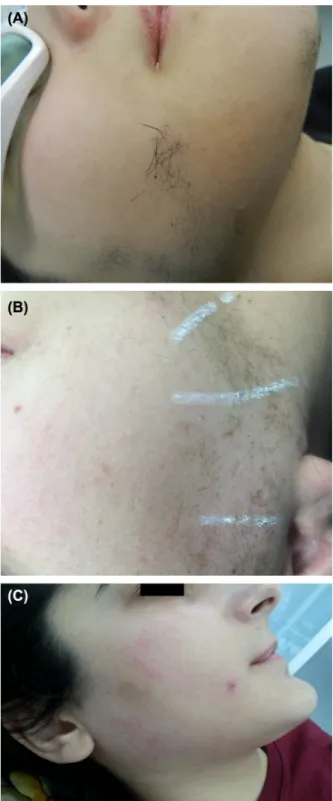
Figure 1. (a) facial hairs just after dyeing with eyelash masquerade. (b) tempo-rary superficial crusting after the dye aided 1 st nd:yaG laser epilation session. (c) significant decrease in the number of facial terminal hairs and a transient erythema after the dye aided 6th nd:yaG laser epilation session.
both control and painted sides by two experienced dermatolo-gists in a blinded manner. In cases of disagreement between the two observers, the average count was recorded and compared with baseline values for both painted and control sides in the two groups. At the end of the study, the clinical responses were evaluated as very good if there was 85% or more decrease in the number of hairs, good if the decrease was 50–85% and unre-sponsive if 50%.
After the first, third, and last sessions, the patients and the clinicians evaluated the improvement on both sides of the laser-treated areas as very bad ( 30%), bad (30–60%), good (60–85%), and very good ( 85%). The assessment of treat-ment efficacy was based on the presence of newly grown hair, and their diameter and density. In this study, patient satisfaction was also determined after the first, third, and last sessions on a scale of 1–4 with 1 being least satisfied and 4 being very sat-isfied. The results obtained from the control and painted sides were statistically compared within each group. Furthermore, every three months, all the adverse effects such as erythema, cutaneous irritation, bullae formation, burned scar, crusting, atrophy, infection, folliculitis, scar, paradoxical hypertrichosis, and hypopigmentation and hyperpigmentation were recorded. Six months after the last session, the recurrence rates were also assessed in both groups and 20% or more increase in the number of terminal hairs was defined as recurrence.
Results
We assessed 50 hirsute women and enrolled 36 into the final randomization. Sixteen patients were excluded from the study due to tanning, use of photosensitizing drug, presence of hormonal disorders such as PCOS, and unrealistic expecta-tions. As a result, a prospective, double-blinded, randomized, and controlled study was conducted, with 16 patients with unwanted white-colored or light facial hair and 20 patients with unwanted axillary hair, who were placed in Groups I and II, respectively. The age of patients in Group I ranged from 19 to 54 years, with the mean age being 31.69 11.46 years. The patients in Group II were aged 20 to 46 years, with the mean age being 26.30 7.67. The Ferriman–Gallwey scores ranged from 1 to 7, with a mean of 4.94 3.82 and 4.65 3.86 in Groups I and II, respectively.
Terminal hair count and treatment efficacy in Group I In Group I, the terminal hair count was similar on the painted and control facial side at the beginning of the study and after the first month (p 0.05) (Table 1). However, the number of terminal hairs on the painted side was significantly lower than that on the control side in the following months (p 0.015 after the first month and p 0.001 after months 2–6; p 0.05) (Table 1, Figure 3). There was a significant decrease in the terminal facial hair count on both sides from the beginning of the treatment to the end of each session.
After the first, third, and sixth sessions, the mean VAS values were noted as 8, 6 and 3, respectively, on the painted facial side and 5, 4.5 and 2, respectively, on the control side. This indi-cated that the mean VAS values of the painted facial side were significantly higher than those of the control side (p 0.005, session. The patients were warned not to use any unprescribed
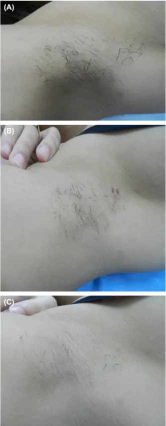
medication or topical cream, not to tan and not to apply any depilation method such as wax or use a depilation device. Before each session, the number of the terminal hairs in the predeter-mined laser application area inside a 3-cm diameter plastic ring was counted monthly using a magnifying illuminated mirror on Figure 2. (a) axillar hairs just after dyeing with eyelash masquerade. (b) temporary superficial crusting in the painted axillar side after the dye aided 1 st alexandrite laser epilation session. (c) significant decrease in the number of axillary hairs after the dye aided 6th alexandrite laser epilation session.
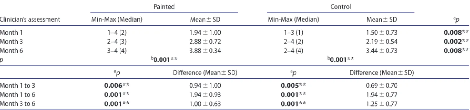
hyperpigmentation, and temporary crusting. On the control side, only slight erythema and folliculitis were observed. For the painted facial side, adverse effects were noted in 8 patients after the first month, 7 patients after the third month, and 1 patient after the sixth month (Figure 4). After the first and third months, the percentage of adverse effects was significantly higher on the painted side than that on the control side (p 0.014 and p 0.014; p 0.05). However, after the last session, no signifi-cant difference was observed between the two sides in terms of the percentage of adverse effects (p 0.05).
Clinical responses of Group I
The clinical responses of Group I were as follows: for the painted side, a very good response in 14 patients (87.5%) and a good response in 2 patients (12.5%); and for the control side, a very good response in 11 patients (68.8%) and a good response in 4 patients (25%). In addition, 1 patient (6.3%) was found to be unresponsive to the control side treatment. The clinical responses in the painted facial side were significantly better than those in the control side (p 0.05). Six months later, clinical recurrences were noted in the painted side; in only 1 patient (6.25%) with a good response and in the control side; in 2 patients (12.5%) with a very good response and in 4 patients (25%) with a good response.
Terminal hair count and clinical efficacy in Group II At the beginning of the study, the terminal hair count in the painted axilla was significantly higher than that on the con-trol side (p 0.032; p 0.05); however, after the first session, the values were similar on both sides (p 0.05; Table 4). In the following months, the number of terminal hairs in the painted axilla was found to be significantly lower than that on the other side (Figure 5). Furthermore, the decrease in the terminal hair count from the beginning of the study to the end of each session was significant both on the painted and control axillary sides (Table 4, Figure 4).
p 0.001 and p 0.001 after the first, third, and sixth sessions, respectively; p 0.01).
The clinician’s score for treatment efficacy was significantly higher for the painted facial side than that for the control side throughout the follow-up period (p 0.008, p 0.002, and p 0.008 after the first, third, and sixth sessions, respectively; p 0.01; Table 2). This indicated a significant improvement on both sides from the first to the third and sixth months, and from the third to the sixth month (p 0.01; p 0.01). Similarly, the patients’ scores for the assessment of treatment efficacy on the painted facial side were significantly higher compared to the those on the control side after the first, third, and sixth ses-sions (p 0.034, p 0.001, and p 0.002, respectively; p 0.01). However, the increase in these scores was not significant on the control side from the first to the third month (p 0.05; Table 3).
The patient satisfaction scores for the painted and control facial sides were similar after the first month (p 0.05), but they were significantly higher for the painted side than for the con-trol side after the third (p 0.034) and sixth (p 0.005) months (p 0.05).
Adverse effects in Group I
The adverse effects seen on the painted facial side of the patients were slight erythema, irritation, folliculitis, transient
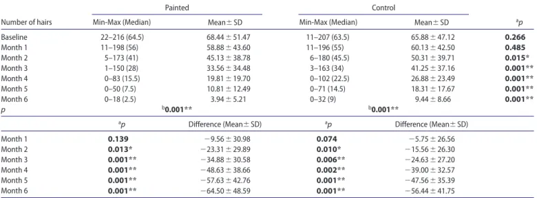
Table 1. number of terminal hairs on the painted and control sides of patients treated with facial laser hair removal.
painted Control
number of hairs min-max (median) mean sd min-max (median) mean sd ap
Baseline 22–216 (64.5) 68.44 51.47 11–207 (63.5) 65.88 47.12 0.266 month 1 11–198 (56) 58.88 43.60 11–196 (55) 60.13 42.50 0.485 month 2 5–173 (41) 45.13 38.78 6–180 (45.5) 50.31 39.71 0.015* month 3 1–150 (28) 33.56 34.48 3–163 (34) 41.25 37.16 0.001** month 4 0–83 (15.5) 19.81 19.70 0–102 (22.5) 26.88 23.49 0.001** month 5 0–50 (7.5) 10.81 12.49 0–71 (14.5) 18.31 17.67 0.001** month 6 0–18 (2.5) 3.94 5.21 0–32 (9) 9.44 8.66 0.001** p b0.001** b0.001**
ap difference (mean sd) ap difference (mean sd)
month 1 0.139 29.56 30.98 0.074 25.75 26.56 month 2 0.013* 223.31 29.89 0.010* 215.56 26.30 month 3 0.001** 234.88 30.58 0.006** 224.63 27.20 month 4 0.001** 248.63 38.66 0.002** 239.00 32.57 month 5 0.001** 257.63 42.76 0.001** 247.56 35.39 month 6 0.001** 264.50 48.59 0.001** 256.44 41.75
aWilcoxon signed-rank test, bfriedman test, *p 0.05, **p 0.01.
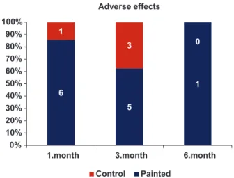
hypopigmentation was also observed. On the painted axillary side, the adverse effects were noted in 6 patients after the first month, 5 patients after the third month, and 1 patient after the sixth month (Figure 6). The percentage of adverse effects on the painted axillary side after the first month was significantly higher than that on the control side (p 0.025; p 0.05); how-ever, it was similar after the third and sixth months (p 0.05). Clinical responses of Group II
For the painted side, all 20 patients (100%) had a very good response. For the control axillary side, 1 patient (5%) had a good response and 19 patients (95%) had a very good response. The clinical responses in painted and control axillary sides were similar (p 0.05). Six months later, patients had no clinical sign of recurrence.
Statistical evaluation
The Number Cruncher Statistical System (NCSS) 2007 (Kaysville, Utah, USA) was used for the statistical analysis. The data were evaluated using statistical methods (mean, standard deviation, median, frequency, ratio, minimum, and maximum). The parameters that did not have a normal distribution on the painted and control sides were compared using the Wilcoxon signed-rank test. For the comparison of follow-up data of three or more parameters that did not have a normal distribution, the Friedman test was performed, while Wilcoxon signed-rank test was used for paired comparisons. The significance was evaluated at p 0.01 and p 0.05.
After the first, third, and sixth months, the mean VAS scores were noted as 7.5, 4, and 1, respectively, on the painted facial side and as 5.5, 3, and 1, respectively, on the control side. These scores were significantly higher for the painted axillary side than those for the control side throughout the duration of the treatment.
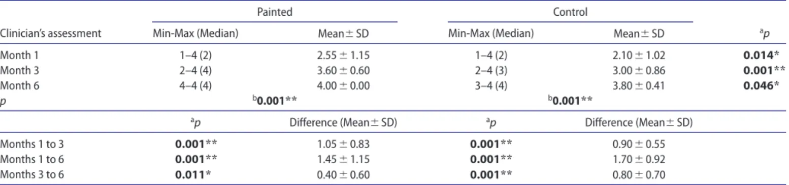
The clinician’s treatment efficacy scores were significantly higher for the painted axillary side than those for the control side after the first, third, and sixth months (p 0.014, p 0.001, p 0.046, respectively; p 0.05) (Table 5). The increase in the clinician’s treatment efficacy scores for both painted and control axillary sides was significant after the third and sixth months compared to that after the first month (p 0.001 and p 0.001), and after the sixth month compared to the third month (p 0.011*; p 0.001) (p 0.05; p 0.01).
The patients’ treatment efficacy scores for the painted axil-lary side were significantly higher than those for the control side after the first, third, and sixth months (p 0.005, p 0.004, and p 0.046, respectively; p 0.05; Table 6). For both axillary sides, the increase in these scores was significant after the third and sixth months compared to the first month, and after the sixth month compared to the third month (p 0.025; p 0.009 and p 0.05; p 0.01). The patient satisfaction score was similar for the painted and control axillary sides throughout the treatment (p 0.05).
Adverse effects in Group II
The adverse effects that were commonly seen on both sides were slight erythema, irritation and folliculitis. On the painted side, Table 2. Clinician’s treatment efficacy for patients treated with facial laser hair removal.
painted Control
Clinician’s assessment min-max (median) mean sd min-max (median) mean sd ap
month 1 1–4 (2) 1.94 1.00 1–3 (1) 1.50 0.73 0.008**
month 3 2–4 (3) 2.88 0.72 2–4 (2) 2.19 0.54 0.002**
month 6 3–4 (4) 3.88 0.34 2–4 (4) 3.44 0.73 0.008**
p b0.001** b0.001**
ap difference (mean sd) ap difference (mean sd)
month 1 to 3 0.006** 0.94 1.00 0.005** 0.69 0.70
month 1 to 6 0.001** 1.94 0.93 0.001** 1.94 0.77
month 3 to 6 0.001** 1.00 0.63 0.001** 1.25 0.77
aWilcoxon signed-rank test, bfriedman test,*p 0.05, **p 0.01.
Table 3. patients’ treatment efficacy scores for facial laser hair removal.
painted Control
patients’ assessment min-max (median) mean sd min-max (median) mean sd ap
month 1 1–4 (2) 2.25 1.13 1–3 (2) 1.88 0.89 0.034*
month 3 2–4 (3) 3.00 0.63 1–4 (2) 2.25 0.77 0.001**
month 6 3–4 (4) 3.94 0.25 2–4 (3) 3.25 0.68 0.002**
p b0.001** b0.001**
ap difference (mean sd) ap difference (mean sd)
months 1 to 3 0.030* 0.75 1.13 0.083 0.38 0.81
months 1 to 6 0.001** 1.69 1.08 0.001** 1.38 0.72
months 3 to 6 0.001** 0.94 0.57 0.001** 1.00 0.63
diode laser treatment by dyeing the hair follicle with liposomal melanin. However, precise information about the study design is not available on the website. The researchers observed 95% hair reduction in 62.5% of the patients. In another study, the efficacy of IPL with a wavelength of 530–1200 nm, 5-ms pulse width, and a fluence of 40 J/cm2 combined with coloring for
the removal of white hair was evaluated (10). The research-ers tested black eyeliner and black hair dye and reported that they had similar efficacy. After six sessions at 4-week intervals, a significant hair reduction (60%) was seen, and 54 patients (88%) were satisfied. The hair elimination rate was more than 80%, and it was similar with the other reported rates of the pre-vious reports of IPL for black hair (50–80%). Furthermore, the researchers evaluated the treatment results based on different skin phototypes and found no significant differences; however, they reported better responses in older women, which may be due to the increased activity of tyrosinase in older patients (10). In the present study, we did not investigate the possible correlation between hair removal success and patients’ skin phototype or age. We also did not include patients with PCOS in the study, which may have improved the efficacy of the treat-ment method. Finally, in another clinical study, the efficacy of photodynamic hair removal using rose bengal–encapsulated liposomal (LRB) gel combined with IPL treatment for facial white hair removal in 15 adult women was compared with that Discussion
Laser removal of thin and white hair is a complicated task with often unsatisfactory results due to the lack of laser-absorbing chromophore (1,6). There are no reports on the use of external coloring of terminal hairs with dye such as eyelash mascara to enhance thin, light-colored, or white hair prior to Alexandrite and Nd:YAG laser procedures for the removal of axillary and facial hair, respectively. Previously, liposomal melanin spray was used, accompanied by three cycles of 800-nm diode laser at 8-week intervals (28–40 J/cm2) (7). In the liposomal
mela-nin group, the mean hair regrowth was found to be 83%, with 14% terminal hair count reduction compared to the 10% reduc-tion in the control group that received physiological saline. The authors reported that liposomal melanin was weak and not cost-effective, and the results on clinical correlation were dis-appointing with no changes regarding hair thickness or color being observed. The researchers had to stop the treatment after three sessions since their patients did not respond as anticipated. However, in that study, the patients were asked to apply the spray on the predefined treatment area by themselves at home before each laser treatment for 12 times a day for 8 weeks. Therefore, the treatment efficacy may have been affected by poor treatment compliance. (7). On the contrary, Leeuw et al. (9) published a non-peer-reviewed report on the Lipoxome manufacturer’s website about successful hair removal with eight sessions of Figure 4. the rate of adverse effects in painted and control facial sides.
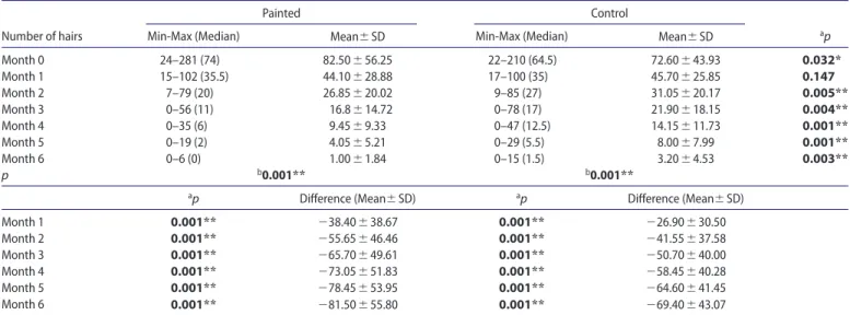
Table 4. number of terminal hairs on the painted and control sides of patients treated with axillary laser hair removal.
painted Control
number of hairs min-max (median) mean sd min-max (median) mean sd ap
month 0 24–281 (74) 82.50 56.25 22–210 (64.5) 72.60 43.93 0.032* month 1 15–102 (35.5) 44.10 28.88 17–100 (35) 45.70 25.85 0.147 month 2 7–79 (20) 26.85 20.02 9–85 (27) 31.05 20.17 0.005** month 3 0–56 (11) 16.8 14.72 0–78 (17) 21.90 18.15 0.004** month 4 0–35 (6) 9.45 9.33 0–47 (12.5) 14.15 11.73 0.001** month 5 0–19 (2) 4.05 5.21 0–29 (5.5) 8.00 7.99 0.001** month 6 0–6 (0) 1.00 1.84 0–15 (1.5) 3.20 4.53 0.003** p b0.001** b0.001**
ap difference (mean sd) ap difference (mean sd)
month 1 0.001** 238.40 38.67 0.001** 226.90 30.50 month 2 0.001** 255.65 46.46 0.001** 241.55 37.58 month 3 0.001** 265.70 49.61 0.001** 250.70 40.00 month 4 0.001** 273.05 51.83 0.001** 258.45 40.28 month 5 0.001** 278.45 53.95 0.001** 264.60 41.45 month 6 0.001** 281.50 55.80 0.001** 269.40 43.07
aWilcoxon signed-rank test, bfriedman test, *p 0.05, **p 0.01.
0 10 20 30 40 50 60 70 80
0.month 1.month 2.month 3.month 4.month 5.month 6.month
Mean
Hair count
Painted Control
further research involving electron microscopic and ultrastruc-tural examinations may provide an explanation for these issues. Dye-aided Alexandrite and Nd:YAG laser hair removal at lower doses and variable pulse widths should also be further evaluated in different groups.
Laser therapy has been reported to produce the best results when dye is applied immediately before the application, which increases the penetration and intensity of the dye (10,12). Coloring terminal hairs with an eyeliner or mascara dye seems to be a better technique than using a hair dye since the pro-tection of the overlying skin is very important for preventing excess chromophore absorption and burning of the skin. We also chose to use eyelash mascara rather than hair dye due to the ease, efficiency, and reliability of this method. We believe that applying topical zinc oxide 20% cream helped avoid the smudging of excess dye on the treatment area as reported in previous studies investigating dye-aided laser hair removal. Accumulated liposomal melanin in nonhair-bearing pores of the laser-treated skin has also been found to have a higher absorption of laser energy, causing a higher rate of erythema after laser treatment compared with the control group (8).
In the literature, studies using hair-dye- or eyeliner-aided IPL have reported leukotrichia, paradoxical hair growth in the periphery of beamed area (previously hair-free), and hyper-trichosis of cured skin as the side effects of the treatment (10). In our study, we did not observe any serious or permanent side effect or complication except for temporary erythema and superficial crusting, which only lasted a week. Compared to the control sides, adverse effects were more common on the painted facial side after the first and third months, and on the painted axillary side only after the first month. In addition, this appli-cation also does not significantly increase the treatment cost. in a control group of 15 patients treated with a placebo gel (10).
At each session, the treatment area was pretreated with a topical LRB gel. Average hair regrowth in the LRB group was 56% after three treatment cycles. After a six-month follow-up, average terminal hair count compared to the baseline score showed a 40% reduction, which indicated a significant difference (11). In our study, we also noted that of the patients treated for facial hair removal, in the painted side, only 6.25% showed clinical recurrence; however, in the control side, 37.5% of the patients had relapsed six months later. We noted that the mascara dyeing as an adjunct to Nd:YAG laser significantly lowered the clini-cal recurrence rates. The contradicting findings obtained from different studies may be attributed to different skin types and hair colors.
We believe that the varying results of studies on dye-aided laser hair removal may be due to the application of different selective coloring methods. Since the depth of dye penetra-tion varies at different stages of the hair cycle, the efficacy of the coloring method may differ (12). As a hair follicle passes from the anagen to the telogen phase, a gap between hair shaft and outer root sheath is formed, allowing for deeper penetra-tion of dye during catagen and telogen phases (12). Therefore, we consider that coloring anagen hair follicle may be inad-equate and may not elicit better results. On the other hand, it is a controversy that hair is most susceptible to some of the newer removal methods during anagen. An important discus-sion point is whether the hair root, bulge, or papillar matrix can be painted by exogenous coloring with an eyelash mascara and whether this would be sufficient to achieve permanent destruc-tion of terminal hair follicles with laser. The study was limited due to the small sample size and lack of histopathological exam-ination before and after the laser applications. We believe that
Table 5. Clinician’s treatment efficacy scores for patients treated with axillary laser hair removal.
painted Control
Clinician’s assessment min-max (median) mean sd min-max (median) mean sd ap
month 1 1–4 (2) 2.55 1.15 1–4 (2) 2.10 1.02 0.014*
month 3 2–4 (4) 3.60 0.60 2–4 (3) 3.00 0.86 0.001**
month 6 4–4 (4) 4.00 0.00 3–4 (4) 3.80 0.41 0.046*
p b0.001** b0.001**
ap difference (mean sd) ap difference (mean sd)
months 1 to 3 0.001** 1.05 0.83 0.001** 0.90 0.55
months 1 to 6 0.001** 1.45 1.15 0.001** 1.70 0.92
months 3 to 6 0.011* 0.40 0.60 0.001** 0.80 0.70
aWilcoxon signed-rank test, bfriedman test,*p 0.05, **p 0.01.
Table 6. patients’ treatment efficacy scores for axillary laser hair removal.
painted Control
patients’ assessment min-max (median) mean sd min-max (median) mean sd ap
month 1 2–4 (3) 2.95 0.89 1–4 (3) 2.55 1.05 0.005**
month 3 3–4 (4) 3.75 0.44 2–4 (3.5) 3.25 0.85 0.004**
month 6 4–4 (4) 4.00 0.00 3–4 (4) 3.80 0.41 0.046*
p b0.001** b0.001**
ap difference (mean sd) ap difference (mean sd)
months 1 to 3 0.001** 0.80 0.70 0.001** 0.70 0.47
months 1 to 6 0.001** 1.05 0.89 0.001** 1.25 0.91
months 3 to 6 0.025* 0.25 0.44 0.009** 0.55 0.76
Declaration of interest
The authors have no conflicts of interest. References
1. Sun TT, Cotsarelis G, Lavker RM. Hair follicular stem cells: the bulge-activation hypothesis. J Invest Dermatol. 1991;96(5):77–78. 2. Patil UA, Dhami LD. Overview of lasers. Indian J Plast Surg.
2008;41(Suppl):101–113.
3. Karaca S, Kaçar SD, Ozuğuz P. Comparison of SHR Mode IPL System with Alexandrite and Nd: YAG Lasers For Leg Hair Reduction. Balkan Med J. 2012;29(4):401–405.
4. Grunewald S, Bodendorf MO, Zygouris A, Simon JC, Paasch U. Long-term efficacy of linear-scanning 808 nm diode laser for hair removal compared to a scanned alexandrite laser. Lasers Surg Med. 2014;46(1):13–19.
5. Mohamed EE, Ahmed AM, Tawfik KM, Ibrahim SM. Trichoscopic changes in hair during treatment of hirsutism with 1064-nm neodymium:yttrium-aluminum-garnetlaser. J Cosmet Dermatol. 2016;15(1):31–35.
6. Al-Dhalimi MA, Kadhum MJ. A split-face comparison of facial hair removal with the long-pulsed alexandrite laser and intense pulsed light system. J Cosmet Laser Ther. 2015;17(5):267–272.
7. Sand M, Bechara FG, Sand D, Altmeyer P, Hoffmann K. A random-ized, controlled, double-blind study evaluating melanin-encapsulated liposomes as a chromophore for laser hair removal of blond, white, and gray hair. Ann Plast Surg. 2007;58(5):551–554.
8. Demir B, Pasa S, Demir S, Tumer C, Atay AE, Gul T, Atamer Y. Hirsutism score and the severity of hyperandrogenism associated with polycystic ovary syndrome in the southeastern region of Turkey. J Int Med Res. 2011;39(4):1529–1535.
9. de Leeuw J, van der Beek N, Neugebauer D. Permanent hair removal of white, grey and light blond hair after laser treatment combined with melanin encapsulated liposomes (Lipoxome), http://www.lipoxome. nl (accessed July 28, 2016).
10. Alijanpoor R, Poorsattar BejehMir A, Mokmeli S. Successful white hair removal with combined coloring and intense pulsed Light (IPL): a randomized clinical trial. Photomed Laser Surg. 2011;29(11): 773–779.
11. Samy N, Fadel M. Topical liposomal Rose Bengal for photodynamic white hair removal: randomized, controlled, double-blind study. J Drugs Dermatol. 2014;13(4):436–442.
12. Genina EA, Bashkatov AN, Sinichkin YP, Kochubey VI, Lakodina NA, Altshuler GB, Tuchin VV. In vitro and in vivo study of dye diffu-sion into the human skin and hair follicles. J Biomed Opt. 2002;7(3): 471–477.
13. Olsen EA. Methods of hair removal. J Am Acad Dermatol. 1999;40(2 Pt 1):143–155; quiz 156–157.
14. Alijanpour R, Poorsattar Bejeh Mir A. The effect of topical glycerol trinitrate on laser-aided facial hair removal: A triple-blinded randomized clinical trial. Photomed Laser Surg. 2015;33(12): 592–597.
15. Loussouarn G, Lozano I, Panhard S, Collaudin C, El Rawadi C, Genain G. Diversity in human hair growth, diameter, colour and shape. An in vivo study on young adults from 24 different ethnic groups observed in the five continents. Eur J Dermatol. 2016;26(2): 144–154.
In this study, the cost of the coloring procedure with eyelash mascara was $5.8 for the entire study duration, which is nearly the same as the cost of hair dye or eyeliner used in the IPL study mentioned previously (10).
Anagen is known as the most susceptible phase during laser hair removal (13). Since nitric oxide (NO) and vascular endothelial growth factor (VEGF)-induced angiogenesis dur-ing the anagen phase, vasorelaxation and perfusion have been shown to improve long-term white hair removal, the efficiency of a new topical gel or dye containing NO or VEFG should be assessed for laser hair removal in the future (14). Such investiga-tion should evaluate the physical properties of hair follicles such as density, color, diameter, length, sensitivity, and growth speed in different body areas (15). Therefore, to improve the removal of particularly thin, light-colored, or white terminal hair, each body area or even different dermatomes of facial skin should be given special consideration.
Conclusion
The selective dyeing of hair follicles with eyelash mascara prior to Alexandrite and Nd:YAG laser applications seems to enhance the success of hair removal without any serious undesirable adverse effects. We particularly recommend combining the Nd:YAG laser with this method to remove facial white hair as a well-tolerated and efficient treatment for reducing terminal hair faster than the conventional Nd:YAG method. It also results in higher patient satisfaction and has temporary adverse effects that only last for approximately three months.
Funding
The authors received no financial support.
6 5 1 1 3 0 0% 10% 20% 30% 40% 50% 60% 70% 80% 90% 100%
1.month 3.month 6.month
Adverse effects
Control Painted