ANALYSIS OF MECP2 GENE MUTATIONS IN RETT SYNDROME PATIENTS
A THESIS SUBMITTED TO
THE DEPARTMENT OF MOLECULAR BIOLOGY AND GENETICS AND
THE INSTITUTE OF ENGINEERING AND SCIENCE OF BILKENT UNIVERSITY
IN PARTIAL FULFILLMENT OF THE REQUIREMENTS FOR THE DEGREE OF MASTER OF SCIENCE
By AYÇA SAYI August, 2001
I certify that I have read this thesis and that in my opinion it is fully adequate, in scope and in quality, as a thesis for the degree of Master of Science.
Prof. Dr. Meral Topçu
I certify that I have read this thesis and that in my opinion it is fully adequate, in scope and in quality, as a thesis for the degree of Master of Science.
Assoc. Prof. Dr. Tayfun Özçelik
I certify that I have read this thesis and that in my opinion it is fully adequate, in scope and in quality, as a thesis for the degree of Master of Science.
Assoc. Prof. Dr. Işık Yuluğ
Approved for the Institute of Engineering and Science
ABSTRACT
ANALYSIS OF MECP2 GENE MUTATIONS IN TURKISH RETT SYNDROME PATIENTS
Ayça Sayı
M.S. in Molecular Biology and Genetics Supervisor: Assoc. Prof. Dr. Tayfun Özçelik
August 2001, 111 pages
Rett Syndrome (RTT) is a progressive X-linked dominant childhood neurodevelopmental disorder, affecting 1/10,000-15,000 girls. The disease-causing gene was identified as MECP2 on chromosome Xq28, and mutations have been found in approximately 80% of patients diagnosed with RTT. We screened for eight recurrent MECP2 mutations (R106W, P152R, T158M, R306C, R168X), one rare mutation (F155S) and one polymorphism (E397K) in 63 RTT patients divided into four groups as classic-RTT (n=43), variant-RTT (n=14), male-RTT (n=4), and familial-RTT (n=2). We identified the recurrent mutations in 18 cases. These are three R106W, two P152R, five T158M, five R306C, and three R270X mutations. R168X and F155S were not detected in our patients. Only one patient had the E397K polymorphism who also had the R306C mutation. All these mutations were confirmed via sequencing analysis. In exon 4 of MECP2, several deletion types of mutations are known. By PCR analysis, two patients were found to have an approximately 44 bp deletion in exon 4. Also, a novel mutation – T197M– was identified in one of the patients. We identified a boy affected by RTT who is mosaic for the R270X mutation, and had a normal male karyotype. This result show that a recurrent MECP2 mutation could lead to a similar phenotype in females and males, if the male is a mosaic for the mutation in his somatic cells. MECP2 mutation frequency for the four groups is as follows: 37.2% for the classic-RTT, 28.57% for the variant-RTT, and 25% for the male-RTT groups. No mutation was found in the familial group. We could not find a consistent correlation between the clinical symptoms and the type of mutations or the X chromosome inactivation patterns of the patients.
ÖZET
TÜRK RETT SENDROMU HASTALARINDA MECP2 GENİNDEKİ MUTASYONLARIN ANALİZİ
Ayça Sayı
Moleküler Biyoloji ve Genetik Yüksek Lisans Tez Yöneticisi: Doç. Dr. Tayfun Özçelik
Ağustos 2001, 111 sayfa
Rett Sendromu (RTT) X'e bağlı dominant kalıtım gösteren, ilerleyici özellikte, bir çocukluk dönemi nöral gelişim hastalığıdır. Büyük bir çoğunlukla kızları etkiler ve 1/10,000-15,000 sıklıkla görülür. Hastalığa neden olan genin Xq28'de bulunan MECP2 olduğu tanımlanmıştır. RTT tanısı konan hastaların yaklaşık %80'inde bu genin mutasyonu bulunmuştur. Biz 63 RTT hastasını klasik-RTT (n=43), RTT-varyant (n=14), erkek-RTT (n=4) ve ailesel-RTT (n=2) olmak üzere dört gruba ayırdık ve bu hastalarda sekiz adet sık gözüken mutasyonu (R106W, P152R, T158M, R306C, R168X), bir adet nadir gözüken mutasyonu (F155S), ve bir adet polimorfizmi (E397K) taradık. Üç hastada R106W, iki hastada P152R, beş hastada T158M, beş hastada R306C ve üç hastada R270X mutasyonu olmak üzere toplam onsekiz sık gözüken mutasyonu saptadık. Bizim hastalarımızda R168X ve F155S mutasyonları bulunamamıştır. R306C mutasyonu olan bir hastada aynı zamanda E397K polimorfizmi saptanmıştır. Tüm bu mutasyonların varlığı sekans analizi ile doğrulandı. MECP2'nin dördüncü eksonunda bilinen birkaç delesyon tipi mutasyon bulunmaktadır. PCR analizi ile iki hastanın ekson 4'ünde yaklaşık 44 baz çifti delesyon bulundu. Hastaların birinde yeni bir mutasyon –T197M-saptanmıştır. Normal karyotipe sahip bir erkek RTT hastasında R270X mutasyonu saptadık. Bu mutasyon normal MECP2 dizisi ile birlikte görüldüğü için hastada somatik mozaisizm olduğu sonucuna varılmıştır. Dört grup RTT hastası için MECP2 mutasyon sıklığı sırasıyla: klasik-RTT için %37.2, RTT-varyant için %28.57, ve erkek-RTT grubu
TO MY PARENTS
BİRSEN , FEVZİ SAYI
AND
TO MY SISTER
NİLAY SAYI
ACKNOWLEDGEMENTS
First of all I would like to express my gratitude to Assoc. Prof. Dr. Tayfun Özçelik for supervising this research and his critical review during the preparation of this thesis. I thank him for his encouragement and guidance.
I would particularly like to thank to Prof. Dr. Meral Topçu from Hacettepe University, for her help in the clinical diagnosis of Rett syndrome and our other collaborators from the same institution, Dr. Mine Cimbiş, Dr. Göknur Haliloğlu, Dr. Dilek Yalnizoglu for their help in obtaining patient samples and clinical data.
I wish to express my thanks to Prof. Dr. Mehmet Öztürk for his support and suggestions.
I would like to thank to all of my instructors for their help and understanding, especially Dr. Uğur Yavuzer for her belief in me during my adaptation period in early days.
I would like to address my very special thanks to my little supervisor and friend Cemaliye Akyerli (Cemo), for sharing her experience with me in science and
I wish to thank to Berna and Emre Sayan (The Sayans), for their closeness and for saving me from difficult situations.
I would like to thank Gökçe Törüner for his help in the dosage analysis experiments. Also many thanks to Tolga Çağatay and Tülay Arayıcı for helping me with the automated sequencing.
I would like to mention the names of Arzu, Deniz, Burcu, Funda, Ahmet, Tuba Dinçer, Tuba Gülbağcı, Tolga, Ebru, Esra, Esin, Hüseyin, Cero (our trainee), Suha and other friends from the lab and thank them for their friendship and help.
I would like to thank to my dear friend Ajda Yılmaz for her optimism, love and supply of courage, Kübra Aysu for sharing my sadness, Berna Aykan and Umut Berberoğlu for sharing good memories with me in Ankara , and my other friends for their support.
Finally, my very special thanks go to my parents and to my sister Nilay for their moral support, love and care. Thank you encouragement and patience during the preparation of this thesis.
TABLE OF CONTENTS Page SIGNATURE PAGE ii ABSTRACT iii ÖZET iv ACKNOWLEDGMENTS v
TABLE OF CONTENTS vii
LIST OF TABLES x
LIST OF FIGURES xi
ABBREVIATIONS xiii
1. INTRODUCTION 1
1.1. Rett Syndrome (RTT) 1
1.1.1. Identification of a new syndrome - RTT 1
1.1.1.1. Natural history of RTT 3
1.1.1.2. RTT variants 5
1.1.2. Hypothesis on the inheritance of the RTT gene 6
1.1.3. Localisation of the RTT locus via exclusion mapping 8
1.1.4. Candidate gene screening 9
1.2. Methyl-CpG binding protein 2 (MECP2) gene 10
1.2.1. Identification of MECP2 gene 10
1.2.2. The structure and function of MeCP2 13
1.2.3. Mutations of MECP2 15
1.2.4. Polymorphisms of MECP2 17
1.2.5. The effects of the mutation on the function of MECP2 18
Page 1.6. Epigenetic regulation of gene expression and RTT 25
1.6.1. X-inactivation 25
1.6.2. Genomic Imprinting 27
1.6.3. Developmental regulation of gene expression 27 1.6.4. Tissue-specific gene expression 28
1.7. Aim and Strategy 29
2. MATERIALS AND METHODS 30
2.1. Materials 30
2.1.1. Patient Samples 30
2.1.2. Oligonucleotides 30
2.1.3. Chemicals and Reagents 32 2.1.4. Restriction enzymes 34 2.1.5. Polymerase Chain Reaction (PCR) materials 35 2.1.6. DNA sequence analysis materials 35 2.1.7. Standard solutions and buffers 36
2.2. Methods 37
2.2.1. DNA isolation from whole blood specimens 38 2.2.2. DNA isolation from hair 40 2.2.3. Polymerase Chain Reaction (PCR) 40 2.2.4. Agarose gel electrophoresis 42 2.2.5. Electrophoresis markers 43 2.2.6. Restriction enzyme digestion 43 2.2.7. Polyacrylamide gel electrophoresis 44
2.2.8. Silver staining 44
2.2.9. DNA sequence analysis 45 2.2.10. Allele-specific X-chromosome inactivation assay 46
3. RESULTS 47
3.1. DNA isolation 47
3.2. Polymerase Chain Reaction 48 3.3. Detection of recurrent MECP2 mutations 48
3.3.1. R106W 49
Page 3.3.3. F155S 55 3.3.4. T158M 58 3.3.5. R168X 61 3.3.6. R306C 64 3.3.7. R270X/V288X 67
3.4. Detection of E397K MECP2 polymorphism 70
3.5. Detection of 3’ deletion by PCR-based approach 74
3.6. Detection of unknown mutation 75
3.7. DNA sequence analysis 76
3.7.1. MECP2 exon 3 R106W mutation 76
3.7.2. MECP2 exon 4.1 P152R mutation 77
3.7.3. MECP2 exon 4.1 T158M mutation 78
3.7.4. MECP2 exon 4.3 R306C mutation 79
3.7.5. MECP2 exon 4.3 R270X mutation 81
3.7.6. MECP2 exon 4.1 T197M mutation 82
3.8. Somatic mosaicism for R270X mutation in a boy with classical RTT 83
3.9. X-inactivation assay 85
3.10. Genotype - Phenotype correlation 87
4. DISCUSSION 90
4.1. Mutation Analysis 90
4.2. X-chromosome inactivation (XCI) 93
4.3. Genotype- Phenotype Correlation 93
5. Future Perspectives 94
6. APPENDICES 97
Appendix 1 98
LIST OF TABLES
Page Table 1 Rett Syndrome Diagnostic Criteria 2
Table 2 MECP2 polymorphisms identified 17
Table 3 Sequence of MECP2 primers 31
Table 4 Sequence of AR primer pair 31
Table 5 Restriction Enzymes Used for Mutation Detection 34 Table 6 Optimum MgCl2 concentrations and Tm for PCR of
MECP2 and AR exons 42
Table 7 MECP2 mutations and polymorphism that are screened 49 Table 8 RTT patients with MECP2 alterations 73 Table 9 X inactivation patterns of RTT patients
with MECP2 mutation 86
Table 10 Genotype-phenotype correlation 88
Table 11 Severity score for RTT 89
LIST OF FIGURES
Page Figure 1 A girl with classical RTT phenotype 3 Figure 2 Age of onset of signs and symptoms in RTT 5 Figure 3 Pedigree for four RTT kindreds, which were used 7
for mapping the locus
Figure 4 The exclusion mapping 9 Figure 5 MECP2 is flanked by IRAK and RCP loci 12 Figure 6 Schematic representation of the interaction
between MeCP2 and histone deacetylase complex 14 Figure 7 Mutations identified in MECP2 16 Figure 8 Schematic representation of normal function of
MeCP2 and the effects of mutations to MeCP2 function 18
Figure 9 MBD protein family 19
Figure 10 The MR genes that are localized on X chromosome 22 Figure 11 Fragment sizes in pUC mix,8 DNA marker and φX174 marker 43 Figure 12 DNA isolation by using phenol/chloroform extraction method 47 Figure 13 Analysis of PCR products 48 Figure 14 Expected NlaIII fragment sizes for wild type, mutant
and heterozygous individuals 51 Figure 15 Detection of R106W mutation by cleavage with NlaIII enzyme 52 Figure 16 Expected NlaIV fragment sizes for wild type, mutant
Figure 20 Expected NlaIII fragment sizes for wild type, mutant
and heterozygous individuals 60
Figure 21 Detection of T158M mutation by cleavage with NlaIII enzyme 61 Figure 22 Expected HphI fragment sizes for wild type, mutant
and heterozygous individuals 63 Figure 23 Detection of R168X mutation by cleavage with HphI enzyme 64 Figure 24 Expected HhaI fragment sizes for wild type, mutant
and heterozygous individuals 66 Figure 25 Detection of R306C mutation by cleavage with HhaI enzyme 67 Figure 26 Expected NlaIV fragment sizes for wild type, mutant
and heterozygous individuals 69 Figure 27 Detection of R270X/V288X mutation by cleavage
with NlaIV enzyme 70
Figure 28 Expected StyI fragment sizes for wild type, mutant
and heterozygous individuals 72 Figure 29 Detection of E397K polymorphism by cleavage
with StyI enzyme 73
Figure 30 Detection of 3' deletion at PCR level 74 Figure 31 Detection of unknown mutation by cleavage
with NlaIII enzyme 75
Figure 32 Electropherogram showing R106W (306 C to T) mutation 77 Figure 33 Electropherogram showing P152R (455 C to G) mutation 78 Figure 34 Electropherogram showing T158M (473 C to T) mutation 79 Figure 35 Electropherogram showing R306C (916 C to T) mutation 80 Figure 36 Electropherogram showing R270X (808 C to T) mutation 82 Figure 37 Electropherogram showing T197M (590 C to T) mutation 83 Figure 38 00-196 sample which is somatic mosaic for R270X 84 Figure 39 X chromosome inactivation pattern of 00-133, 00-188, 00-196 86
ABBREVIATIONS
APS ammonium persulfate
ATP adenine triphosphate
Bisacrylamide N, N, methylene bis-acrylamide
bp base pair
CpG Cytosine guanine pair
cDNA complementary DNA
dATP adenosine deoxyribonucleoside triphosphate dCTP cytosine deoxyribonucleoside triphosphate
del deletion
ddH2O deionized water
dGTP guanosine deoxyribonucleoside triphosphate
DNA deoxyribonucleic acid
DNase deoxyribonuclease
dNTP deoxynucleotide triphosphate dTTP tymine deoxyribonucleoside triphosphate EDTA ethylenediaminetetra-acetic acid
EtBr ethidium bromide
EtOH ethanol g gram kb kilobase M molar min minute ml milliliter mM millimolar µl microliter
PAGE polyacrylamide gel electrophoresis PCR polymerase chain reaction
pmol picomol
rpm revolution per minute
RTT Rett syndrome
SDS sodium dodecyl sulphate
sec second
TBE Tris, Boric acid, EDTA
TEMED N, N, N, N-tetramethyl-1, 2 diaminoethane
U unit
UV ultraviolet
V volt
µg microgram
I. Introduction 1.1. Rett Syndrome
Rett Syndrome (RTT) is an X-linked dominant neurodevelopmental disorder and the second most common cause, after Down syndrome, of severe mental retardation in females (Christodoulou et al., 2001). It affects children of all ethnic groups and has an estimated incidence of 1 in 10,000 to 15,000 females (Kerr et al., 1985, Hagberg et al., 1993, Leonard et al., 1997).
1.1.1 Identification of a new syndrome - RTT
Andreas Rett initially described RTT in 1966 but it was largely ignored until 1983, when Hagberg et al. published, the first description of a series of 35 patients, in English (Rett et al., 1966, Hagberg et al., 1983).
RTT is characterized by the cognitive regression (relating to conscious intellectual activity such as thinking, reasoning), deceleration of head growth, loss of purposeful hand use with the development of stereotypic hand movements, tremors (shaking from physical weakness), gait apraxia (loss of purposeful use of limb), and seizures (a sudden attack, as of disease) occurring after a period of normal development (Schanen et al., 1999) .
There are necessary and supportive criteria to diagnose RTT (Table 1). Normal prenatal and perinatal period with normal developmental progress for the first 6-18 months of life is essential for the diagnosis. After normal head
The distinctive characteristic of RTT is the stereotypical hand movements such as hand wringing, hand washing, clapping, patting or other more bizarre hand automatisms. Gait ataxia (an inability to coordinate voluntary muscular movements that is necessary to walk on foot) is also an important feature. Supportive diagnostic criteria include breathing dysfunction, electroencephalographic abnormalities, spasticity, peripheral vasomotor disturbance, scoliosis (curvature of the spine), and growth retardation. If there is evidence of prenatal onset growth retardation, microcephaly at birth, an identifiable metabolic, degenerative or storage disorder, an acquired neurological disorder, retinopathy or optic atrophy, the clinical diagnosis of RTT is excluded. The nature of the condition is that of an evolving clinical phenotype, making the clinical diagnosis uncomplete until 2 to 5 years of age (Christodoulou et al., 2001). An RTT patient displaying one of the features of the disorder is shown in figure 1.
Table 1 Rett Syndrome Diagnostic Criteria (Christodoulou et al., 2001)
Necessary criteria Supportive criteria Exclusion criteria 1. Apparently normal prenatal
and perinatal period 2. Developmental progress
within normal range for the first 5- 6 months
3. Normal head circumference at birth, with subsequent deceleration
4. Reduction or loss of acquired skills (onset 6 months to 3 years) in particular purposeful hand use, vocalisation or speech (words)
5. Appearance of marked delay in development 6. Acquisition of hand
stereotypes 7. Gait and or truncal
apraxia(loss of purposeful use of hand) (by 4 years)
1. Breathing dysfunction 2. Periodic apnoea during
wakefulness 3. Intermittent
hyperventilation 4. Breath holding
5. Forced expulsion of air or saliva
6. EEG abnormalities 7. Slow wave background
with intermittent rhythmical activity (3-5 Hz)
8. Epileptiform discharges, with or without clinical seizures
9. Spasticity, later with muscle wasting dystonia 10. Peripheral vasomotor
disturbance 11. Scoliosis
12. Growth retardation
13. Hypotrophic, small, cold feet
1. Evidence of prenatal onset of growth retardation or microcephaly
2. Organomegaly or other evidence of storage disorder 3. Retinopathy or optic atrophy 4. Existence of identifiable metabolic or other neurodegenerative disorder 5. Acquired neurological
disorder resulting from severe infection of head trauma
Figure 1. A girl with classical RTT phenotype.
Constant hand wringing, which is a typical symptom of RTT, is observed in this girl. (Patient 00-173's photo was taken with permission from the family)
1.1.1.1. Natural History
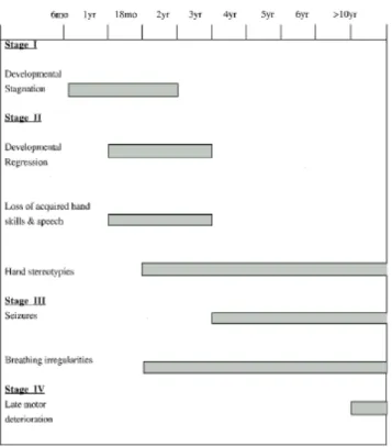
The natural history of a typical patient with classical RTT is characterized by the progression of four stages (see figure 1) (Hagberg et al., 1986). The patients are normal at birth and exhibit normal development between 6-18 months. During stage
I, early onset of stagnation, obtaining of new skills slows and patients frequently
show autistic traits. Head growth slows and hypotonia is seen. After several months,
stage 2, the rapid regression stage slowly develops. Previously acquired skills such
Figure 2. Age of onset of signs and symptoms in RTT (Christodoulou and
Ellaway et al., 2001)
Also several unusual behaviours begin. In Stage 3, the pseudostationary
stage, child's ability about interacting with her environment increases as autistic
features diminish. Patients develop an irregular respiratory pattern while awake. Seizures are most frequent during this stage and hand movements intensify. Somatic growth is poor and many patients develop osteopenia. Stage 4, the late motor
deterioration stage, usually presents by 10 years of age and is characterized by
reduced mobility. However there is no decline in cognition, communication or hand skills. Repetitive hand movements may decrease. Scoliosis is a prominent feature. Rigidity (stiffness) and dystonia (increased muscle tone with abnormal extremity) are characteristic. The majority of RTT patients survive into adulthood.
1.1.1.2. RTT Variants
Females who carry out all of the diagnostic criteria for RTT are classified as having typical or classical RTT. However, the clinical expression of RTT also includes atypical forms that may be more severe or mild in phenotype. Five possible RTT variants were described:
1. Infantile seizure onset type: The characteristic features of this form are predominance of seizures and onset of the disorder before 6 months (Hanefeld et al., 1985).
2. Congenital form: This form appears early without a period of normal development, and involves congenital hypotonia and infantile spasms (Nomura et al., 1985, Rolando et al., 1985).
3. Forme Fruste: This is the milder form that experiences less severe regression and milder mental retardation and does not have seizures (Hagberg et al., 1989).
4. Late childhood regression: In this case, regression has developed later and more gradually than classic RTT (Gillberg et al., 1989).
5. Preserved speech variant (PSV): PSV shares with classic RTT some symptoms like stereotypical hand-washing activities but differs in that
1.1.2. Hypothesis on the inheritance of the RTT gene
The genetic basis of Rett syndrome had been discussed extensively in the literature following the identification of the syndrome. Since 99.5% of all cases are sporadic, it was not easy to define the inheritance pattern (Hagberg et al., 1983, Martinho et al., 1990, Migeon et al., 1995, Comings et al., 1986, Ellison et al., 1992, Zoghbi et al., 1988). There were some clues about the possible genetic origin of the syndrome such as almost exclusive occurrence in females, high concordance rate among monozygotic twins while disconcordance among dizygotic twins, and presence of rare familial cases (Comings et al., 1986, Ellison et al., 1992, Zoghbi et al., 1988, Engerstrom et al., 1992, Schanen et al., 1997, Sirianni et al., 1998, Zoghbi et al., 1990). The first hypothesis by the help of rare familial cases, which indicated inheritance through maternal lines and nonrandom patterns of X chromosome inactivation (XCI) in obligate carrier females, suggested that Rett syndrome is an X-linked dominant disorder caused by mutations in a gene that undergoes X inactivation (Schanen et al., 1997, Sirianni et al., 1998, Zoghbi et al., 1990). There were some argumentative hypotheses against this first one. This inheritance pattern hypotheses included digenic inheritance of X-linked and autosomal loci (Buhler et al., 1990) trinucleotide repeat expansions (Hofferbert et al., 1997), mitochondrial inheritance (Ruch et al., 1989, Dotti et al., 1993, Lappalainen et al., 1994, Haas et al., 1995a, Haas et al., 1995b, Tang et al., 1997), and autosomal dominant inheritance with sex-limited expression (Killian et al., 1986). Careful assessment of the familial cases shown in figure 3, and analysis of the X inactivation patterns in the putative carrier mothers, X-linked dominant inheritance became favourable again (figure 3). In addition, X inactivation studies performed in unaffected and obligate carrier females displayed skewed X inactivation which favours the mutant X, and
thus improves the phenotype (Willard et al., 1996, Puck et al., 1998). II-2 in Rett syndrome kindred 2, I-2 in Rett syndrome kindred 3, and I-2 in Rett syndrome kindred 4 are the examples of the obligate carriers of Rett syndrome who were mildly affected because of the skewed X chromosome inactivation (Migeon et al., 1995, Schanen et al., 1997, Sirianni et al., 1998, Zoghbi et al., 1990). Also in discordant monozygotic twins, the preferential inactivation of paternal X chromosome was reported (Migeon et al., 1995)
Rett Syndrome Kindred 1 Rett Syndrome Kindred 2
Rett Syndrome Kindred 3
I II III 1 2 3 1 2 3 4 1 2 2 1 3 4 5 1 2 1 2 1 2 3 4 5 6 7 8 9
Rett Syndrome Kindred 4
I
II
1 2 3 4
1.1.3. Localisation of the RTT locus via exclusion mapping
In order to map a locus, 3 strategies could be performed. The first one is linkage analysis. This analysis was not proper for mapping RTT locus because 99.5% of all cases were sporadic so there were not enough families to perform linkage analysis. The second is cytogenetic analysis. It was promising, and conducted. But, classical banding techniques, FISH and Southern blotting did not detect any abnormalities such as translocations, microdeletions or duplications, which would be consistent with an X-linked dominant disorder (Fan et al., 1999). The third one, exclusion mapping, was the most suitable method for the localisation of the RTT gene.
The basis of exclusion mapping is that because the related probands inherited the same mutation, the defective gene must lie in a region of the X chromosome that is shared by the probands (Schanen et al., 1999). The first progress in mapping came from the studies of RTT kindreds 1, 2 and 3 (figure 3). These were maternally related half-sisters and aunt-niece pair in these families (Ellison et al., 1992, Anvret et al., 1990, Archidiacono et al., 1991). Polymorphic X-linked markers were typed for RTT kindred 1, 2 and 3, and the region near the marker was excluded if the probands inherited different alleles. Subsequently, a Brazilian family (RTT kindred 4) was identified, which further narrowed the critical region (Figure 3 and 4). In general, using the affected sister pairs for exclusion mapping was risky because the mutation on the putative RTT gene could be transmitted through the paternal lineage if the father was a germline mosaic. But for RTT kindred 4, it was thought that the inheritance was maternal since there were affected and unaffected sisters and skewed X inactivation in the mother. With the help of these four RTT kindreds, the RTT
locus was mapped to Xq28, a particularly gene rich part of the genome, via exclusion mapping (Schanen et al., 1997, Sirianni et al., 1998, Schanen et al., 1998).
Figure 4. The exclusion mapping
A. Pedigree of the Brazilian family and schematic diagram of extended haplotype
of each X chromosome. The only region of X chromosome concordant for RTT is indicated (Sirianni et al., 1998).
B. The regions excluded by the four RTT kindreds is summarized. Filled bars
indicate excluded regions. The loci flanking the discordant and concordant regions are shown on the right. Ideogram shows the approximate cytogenetic location (Schanen et al., 1999).
1.1.4. Candidate gene screening
In many disorders, after genetic mapping, transcripts within critical region were identified and then were screened for mutations in probands. The Human Genome Project was instrumental in the increase of the number of genes that fall into the
“Minimal Critical Region” for RTT
was limited to the central nervous system (CNS) where there was evidence for abnormal development or maintenance of affected neurons. As a note, mosaicism for the mutation was not apparent at the cellular level (Armstrong et al., 1998). Initially, attention was largely directed toward genes expressed predominantly in the CNS, and genes for neurotransmitter and their receptors, neural specific proteins were excluded (Heidary et al., 1998, Percy et al., 1998, Van Den Veyver et al., 1998, Narayanan et al., 1998, Cummings et al., 1998, Wan et al., 1998). However careful examination of female patients and more severely affected male patients indicated that the RTT gene was also important for the function of other tissues (Motil et al., 1994; Haas et al., 1997; Motil et al., 1998). Finally, 14 years of search, Amir et al. and Wan et al. broke the silence in RTT in late summer 1999, and identified MECP2 mutations in RTT probands both in familial and sporadic cases.
1.2. Methyl-CpG binding protein 2 (MECP2) gene 1.2.1. Identification of MECP2 gene
CpG dinucleotides are nonrandomly formed at much of the heterochromatic regions of the chromosomes and the promoter regions of many genes. 60-90% of CpG nucleotides in mammalian genome are modified by methylation at the carbon 5 position. The remaining non-methylated CpGs are found in CpG islands that usually include functional promoters. Methylation of cytosine residues in CpGs is important both in stable silencing of heterochromatin and reversible regulation of gene expression, however, it is not important for the proliferation and in vitro differentiation of embryonic stem (ES) cells (Ng et al., 1999a, Li et al., 1992, Tate et al., 1996).
Two models were suggested for transcriptional repression based on CpG methylation. According to the first model, DNA is bound by proteins, which preferentially interact with methylated CpG sites, and prevent binding of activators or basal transcription factors. Second model suggests that due to preferential interaction between methylated DNA, ubiquitous components of chromatin may alter chromatin structure and lead to transcriptional repression (Mostoslavsky et al., 1997).
Initial efforts to identify protein-mediated CpG methylation-dependent repression led to the identification of MECP1 (Meehan et al., 1989, Boyes et al., 1991). After a year, a second member of MECP family was identified and cloned in rat (Lewis et al., 1992), and in the human (Adler et al., 1995). This member was named as MECP2. Both of the members bind symmetrically methylated CpGs with a sequence-independent manner (Boyes et al., 1991). Although MECP1 required more than 10 CpG pairs to bind DNA (Boyes et al., 1992), MECP2 can bind singly methylated CpG pairs (Lewis et al., 1992). Although there is functional homology between MECP1 and MECP2, different binding specificity and expression patterns suggest that MECP1 does not compensate for the loss of MECP2 function (Dragich et al., 2000).
Mecp2 gene was found to be X-linked in the mouse (Quaderi et al., 1994), and later studies placed MECP2 gene to human X chromosome in Xq28
(figure 5). The 5' non-coding exon was identified recently by Reichwald et al.2000, MECP2 is subjected to X-inactivation (D'esposito et al., 1996).
Figure 5. MECP2 is flanked by IRAK and RCP loci.
The direction of the arrows indicates the orientation of the transcription (Dragich et al., 2000)
Although, expression of MECP2 was at low levels early in development, it was expressed ubiquitously in embryonic and adult tissues (Meehan et al., 1992). MECP2 has three transcripts, which vary in length. Their lengths are respectively 1.8kb, ~7.5kb and ~10 kb. By the different use of polyadenylation signals in 3'UTR, these three transcripts are formed. Although there is tissue-specific variation in expression, short (1.8 kb) and long (10 kb) transcripts are present in most of the tissues. The long transcript is found at higher levels than the shorter one in brain and spinal cord. They are expressed similarly in kidney, thyroid, lung, gastrointestinal tract and adrenal glands. The short one is expressed at higher levels in skeletal and cardiac muscle, lymphoid tissues, liver and placenta (D'esposito et al., 1996, Reichwald et al., 2000). Although, there is a low level of expression of the long transcript in the developing nervous system, the expression is increased in postnatal hippocampus and olfactory bulb (Coy et al., 1999). Because of the identical half-lifes of the short and the long transcripts, the difference between the functions of these transcripts is not fully understood yet (Reichwald et al., 2000).
MECP2
IRAK RCP
1.2.2. The structure and function of MeCP2
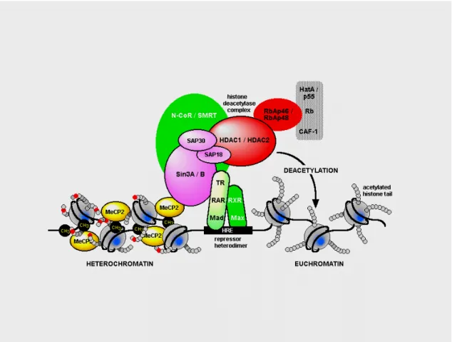
MeCP2 is an abundantly expressed nuclear protein which is associated with 5-methyl-rich heterochromatin (Tate et al., 1996, Nan et al., 1997). Its 486 amino acids consist of four functional domains: (1) a methyl-CpG binding
domain (MBD; 85 amino acids in length) which is necessary to bind 5-methyl
cytosine in the major groove of DNA in the presence or absence of assembled chromatin (Nan et al., 1993, Wakefield et al., 1999); (2) a transcriptional
repression domain (TRD; 104 amino acids in length) which interacts with
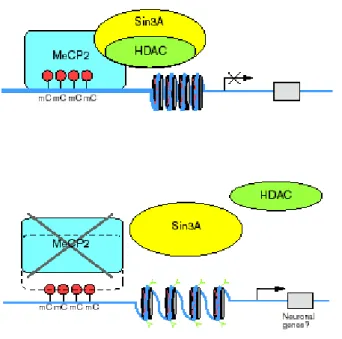
corepressor Sin3A to recruit histone deacetylases 1 and 2 (HDAC1and 2)(figure 6) (Nan et al., 1998b, Jones et al., 1998); (3) a nuclear localization signal (NLS) which may be responsible for the transport of MeCP2 into the nucleus (Nan et al., 1996a, Nan et al., 1996b) and (4) a C-terminal segment which facilitates its binding to the nucleosome core (Chandler et al., 1999).
MeCP2 represses transcription through a mechanism that involves binding to CpGs and recruitment of HDACs to modify chromatin structure. This leads to deacetylation of histones which allows DNA to wind more tightly around the histone, and prevents the access of the transcription machinery to the promoters (Jones et al., 1999, Wolffe et al., 2000). However MeCP2 does not always require deacetylase activity to repress transcription (Nan et al., 1998a, Kaludov et al., 2000, Yu et al., 2000). There is a interaction between MeCP2 and the transcriptional machinery, which comprises TFIIB and E2F (Di Fiore et al., 1999). This interaction
MeCP2’s role in nucleus is complex and mediate transcription through overlapping mechanisms.
Figure 6. Schematic representation of the interaction between MeCP2 and histone deacetylase complex.
1.2.3. Mutations of MECP2
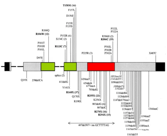
The mutation analyses have been focused on the coding region till now because the untranslated regions of the gene is too long (particularly intron 1, 2 and 3'UTR). MECP2 mutations have been reported in more than 300 RTT patients, and up to %80 of the sporadic and %50 of the familial cases were found to have mutations. As a consequence of the sporadic occurrence of RTT, most mutations are de novo. However, there are eight recurrent mutations namely R106W, R133C, T158M, R168X, R255X, R270X, R294X, and R306C resulting from C to T transitions that account for ~65% of mutations in patients (Amir et al., 1999, Wan et al., 1999, Huppke et al., 2000, Cheadle et al., 2000, Bienvenu et al., 2000, Obata et al., 2000, Hampson et al., 2000, Buyse et al., 2000, Inui et al., 2001, Vacca et al., 2001, Bourdon et al., 2001, Nielsen et al., 2001, Erlandson et al., 2001).
The deletions found in the 3' end of exon 4 which account for 10% of the known mutations are important for the function presumably because they are involved in interaction of MECP2 with nucleosome core (Chandler et al., 1999). More than 64 distinct mutations have been identified; most of them are nonsense (approximately 41) although missense mutations (approximately 23) affecting highly conserved amino acid residues are also seen. Figure 7 shows the majority of the mutations found in MECP2 gene and the mutations already associated with RTT is summarized in appendix 3.
Figure 7. Mutations identified in MECP2.
Exons 2-4 and the mutations of MECP2 is depicted in the diagram. Noncoding regions is in black. MBD is in green and TRD is in red. Missense mutations are listed above and nonsense mutations below the exons. The number of occurrence is written in parenthesis near the recurrent mutations (Amir et al., 2000).
There is a high proportion of C to T transitions at CpG sites because CpG dinucleotides are hypermutable, and germline and somatic mutations are common at these sites (Rideout et al., 1990). The mechanism that causes this transition may involve methylation of 5' cytosine via methyltransferase and spontaneous deamination of 5-methylcytosine to thymine. C to T or G to A (on antisense) transitions constitute ~55% single-nucleotide substitutions (Krawczak et al., 1996). There is a high level of CpG methylation in male germ cells. Also the whole X chromosome, including MECP2, is methylated (Girard et al., 2001). The
coding sequence of MECP2 contains 35 CpGs with a C to T transition possibility. Exon 1 does not contain CpG pairs, which in part explains why no mutation has been found in this exon.
1.2.4. Polymorphisms of MECP2
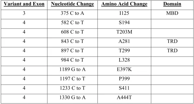
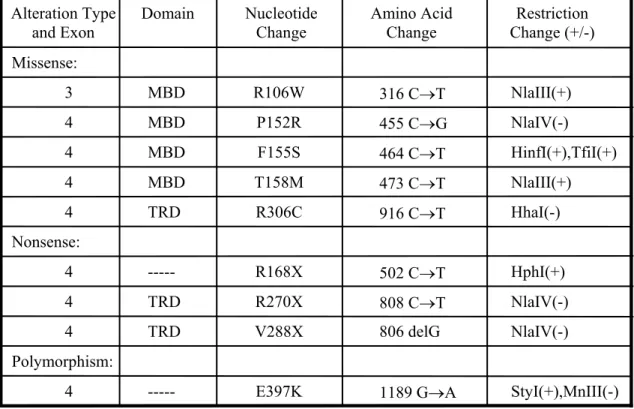
By using familial cases, ten different polymorphisms have been identified (Amir et al., 1999, Wan et al., 1999, Cheadle et al., 2000, Buyse et al., 2000, Amir et al., 2000a, Orrico et al., 2000).
Table 2 MECP2 polymorphisms identified.
Variant and Exon Nucleotide Change Amino Acid Change Domain
3 375 C to A I125 MBD 4 582 C to T S194 4 608 C to T T203M 4 843 C to T A281 TRD 4 897 C to T T299 TRD 4 984 C to T L328 4 1189 G to A E397K 4 1197 C to T P399 4 1233 C to T S411 4 1330 G to A A444T
1.2.5. The effect of the mutations on the function of MECP2
Normal function of MeCP2 includes binding to 5mCs in the promoters, recruiting deacetylase complex, and repressing transcription. When there is a mutation, MeCP2 no longer can bind to 5mC and can not recruit deacetylase complex. So, transcriptional noise from downstream genes appears to be likely (Van Den Veyver et al., 2000) (figure 8).
Figure 8. Schematic representation of normal function of MeCP2 and the effects of mutations to MeCP2 function (Van Den Veyver et al., 2000).
MECP2 is expressed during organogenesis throughout the embryo, and, later most strongly in the hippocampus (Coy et al., 1999). Therefore, mutations of this gene could impair several organs. However, the MECP2 related genes that have transcriptional repression activity and are members of repressor complexes could compensate for MeCP2 dysfunction in some tissues other than the brain. These genes, MBD1, MBD2, MBD3, and MBD4 have been mapped to autosomes; and have similar MBDs, but not TRD domains (Hendrich et al., 1999; Bird et al., 1999; Ng et al., 1999; Wade et al., 1999) (figure 9).
Figure 9. MBD protein family
The MBD protein family shares conserved methyl-CpG binding domains. CxxCxxC domain is found in MBD1. (GR)n represents, glycine-arginine repeats, and (E)
represents glutamic acid repeats. The "repair" domain of MBD 4 is a TG mismatch glycosylase. TRD (transcriptional repression domain) is found only in MeCP2 (Bird et al., 1999).
In general, missense mutations are localized within the MBD domain, except for R306C, which is found within the TRD, and impair selectivity for methylated DNA. Nonsense mutations, which truncate all or some part of the TRD domain, affect the ability to repress transcription, and lead to a decreased level of stability (Yusufzai et al., 2000; Ballestar et al., 2000). Deletions are found within the C-terminus, and affect the stability of the protein.
Majority of the nonsense mutation in the 5' end of the coding sequence of MECP2 are proposed to result in the degradation of the mRNA molecule by a mechanism called nonsense mediated decay (NMD). This process monitors the mRNAs for errors during gene expression and degrades them (Leeds et al., 1992 a,b). In contrast, similar mutations within the last exon may by-pass this pathway and result in the production of a truncated protein (Zhang J., 1998). Since majority of the mutations are nonsense and lie within the last exon, they are expected to escape the NMD pathway. In conjunction with this observation, a decrease in disease severity was noted in cases that had truncating mutations within or downstream of TRD when compared with the mutations within the N-terminal region. The nonsense mutations L138X, R168X, E235X, R255X, R270X, V288X, and R294X were found to lead to the truncation of the TRD, and affect the ability to repress transcription (Yusufzai et al., 2000). A mutation resulting in the most truncated protein was reported from an autistic RTT patient with a relatively mild phenotype. This Q19X mutation led to a gene product of only 19 amino acids. This example indicates that the premature truncation at the beginning of the protein does not have to be the reason for a severe phenotype (Kim et al., 2000; Nielsen et al., 2001).
The three recurrent RTT missense mutations, R106W, R133C, and F155S have a greatly reduced (> 100-fold) affinity to the methylated DNA, which is consistent with the impairment selectivity for binding to methylated DNA (Yusufzai et al., 2000; Ballestar et al., 2000). Another recurrent missense mutation, T158M, which substitutes thr with met on the loop structure outside the DNA-binding domain, shows only a small reduction (2-fold) in affinity to methylated DNA. However, T158 in MECP2 is conserved from Xenopus to human and not present in the other MBD family members, which suggest that this residue has a precise role not related with its methyl-CpG binding activity (Dragich et al., 2000).
C-terminal region of MECP2 is required for protein stability (Yusufzai et al., 2000). Interestingly, this region shows an overall homology of 35% identity, and 50% positivity in a 75 amino acid region with two specific factors, brain-specific factor-1 (BF-1) and fork head 4 (FKH4), which are members of the fork head family. Their role is restricted to developing telencephalon. This subregion overlaps with -COOH terminus of MECP2, which has been shown to facilitate MeCP2 binding to DNA (Chandler et al., 1999).
1.3. MECP2 and X-linked mental retardation
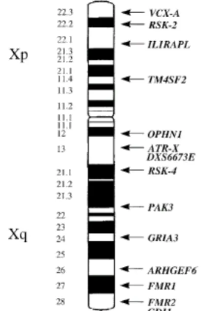
Mental retardation is related with substantial limitations in mental functioning. In OMIM (Online Mendelian Inheritance in Man Database), 937 entries contained the term "MR", corresponding to 220 autosomal dominant, 437 autosomal recessive, 159 X-linked, and 121 non classified conditions. IQ is used for measuring MR; 50<IQ<70 means mild MR, and IQ<50 means severe MR. Approximately 2-3% of the population has an IQ below 70. MR can be subdivided into two: syndromic, which is characterized with consistent and distinctive clinical finding (fragile-X is the most common one); and non specific (MRX) if MR is the only primary symptom among affected individuals. X-linked non specific MR represents 5% of all MRs. Eight genes have been identified which cause X-linked MR when mutated. These genes are OPHN1, GDI1, PAK3, ILRAPL, TM4SF2, VCX-A and ARHGEF6 (figure 10). However, their incidence is very low, being around 0.5-1% of MRX (Toniolo et al., 2000; Castellvi-Bel et al., 2001).
Figure 10. The MR genes that are localized on the X chromosome (MECP2 not included) (Castellvi et al., 2001).
Recently, mutations in MECP2 have been identified in four non specific X-linked mental retardation families with multiple affected individuals. In the first family A140V mutation was identified in all of the four severely affected males and two mildly affected females (Orrico et al., 2000). E406X mutation was identified in a three generation family in which two affected males display severe MR and progressive spasticity (Meloni et al., 2000). E137G mutation was found in another MR family, and finally R167W mutation was found in a three-generation family with four non-specific mentally retarded males (Couvert et al., 2001). So far, these mutations have not been reported in any one of the typical RTT cases.
There is a high frequency of mutations in MECP2 (~2%) when compared with the other non specific X-linked MR genes (%0.5-1%). This finding indicates that MECP2 gene mutations are important for the MR phenotype (Couvert et al., 2001).
In addition to MR, MECP2 mutations have been identified in patients with Angelman syndrome phenotype (Imessaoudene et al., 2001; Watson et al., 2001).
1.4. Male cases
Although RTT exclusively affects females, rare male cases have also been reported. Until now, five reports described mutations in male cases (Wan et al.,
(Orrico et al., 2000). Third described two men affected by severe mental retardation and progressive spasticity (Meloni et al., 2000). Fourth described two brother who died because of severe neonatal encephalopathy (Villard et al., 2000), and the fifth described a sporadic case in which a boy affected by a nonfatal neurodevelopmental disorder who has somatic mosaicism for a MECP2 mutation (Clayton-Smith et al., 2000).
However MECP2 mutations are considered to be lethal for males. In addition, sex-limited expression of RTT can be explained by the occurrence of the de novo X-linked mutations exclusively in the male germ cells which results in affected daughters. With this hypothesis, the absence of affected males can be explained by the fact that sons do not inherit their X-chromosome from their fathers. The frequency of male-germ-line transmission of the mutation was found as 71% (Girard et al., 2001) and 96.3% (Trappe et al., 2001). These findings suggest that male patients are naturally protected from de novo MECP2 mutations.
1.5. Mouse models for RTT
The unresolved issue in the pathogenesis of RTT is whether the disease is the result of a dysfunction of postnatal neurons when the symptoms become apparent or a prenatal developmental abnormality with postnatal phenotypic appearance of the disease (Chen et al., 2001). The most appropriate approach to address this issue appears to be mouse studies.
The first mouse study indicated that Mecp2 is essential for development, and its disruption leads to embryonic lethality (Tate et al., 1996). Recent studies oppose this finding and demonstrated that Mecp2-null mice are
viable, and show some of the symptoms of RTT at approximately six weeks of age (Chen et al., 2001, Guy et al., 2001).
In both of these studies conditional knock-out technology was used to delete exon 3 or exons 3 and 4 of MECP2. Both in Mecp2-null mice and mice in which Mecp2 was deleted in brain showed some of the symptoms of RTT such as tremor, heavy breathing, and cold extremities indicating autonomic abnormalities that are also characteristic for human RTT patients. The most consistent changes with the human RTT in mutant mice were smaller brain size and general reduction of neuronal cell size (Chen et al., 2001, Guy et al., 2001).
1.6. Epigenetic regulation of gene expression and RTT
Methylation of CpG dinucleotides is necessary for transcriptional repression and underlies the processes of X-inactivation, genomic imprinting, tissue-specific and developmental regulation of gene expression (Cross et al., 1995).
1.6.1. X-inactivation
Dosage of X-linked genes is kept equal between females and males by random X inactivation in each cell early in embryogenesis in females (Lyon et al., 1986). The exception for that is the genes in pseudoautosomal region which normally escapes from X-inactivation and have functional homologues on Y chromosome (Schneider-Gadicke et al., 1989, Goodfellow et al., 1984, Fisher et al.,
inactivation leading to functional disomy of an X-linked gene(s), was proposed to explain exclusive occurrence of RTT in females (Riccardi et al., 1986). The existence of male patients argues strongly against this disomy model (Schanen and Francke, 1998).
The second is a more likely model which suggests that X-inactivation patterns influence the phenotypic expression of RTT (Schanen et al., 1999). Random X-inactivation pattern was found in most of the RTT cases (Zoghbi et al., 1990, Anvret et al., 1990, Nielsen et al., 2001, Amir et al., 2000a, Webb et al., 1993, Camus et al., 1996). Skewed X inactivation, which leads to differences in phenotypic expression was also observed in several non penetrant or mildly affected obligate carrier females, and in an unaffected twin. These results indicate that non random pattern of X inactivation protects against the consequences of MECP2 mutations (Migeon et al., 1995, Schanen et al., 1997, Sirianni et al., 1998, Zoghbi et al., 1990, Wan et al., 1999, Amir et al., 2000a, Krepischi et al., 1998, Villard et al., 2001). Recently, a case of RTT with 46,X,r(X) in which complete skewed inactivation of the ring was shown. Interestingly, no mutations were found in the MECP2 gene present in intact X. This finding suggested that, there could be two loci related with RTT, one with the MECP2 locus which mutations predominantly cause sporadic RTT. The second is an unidentified locus in which mutations cause mildly affected or unaffected carriers or familial cases (Rosenberg et al., 2001).
To evaluate the X-inactivation pattern, a rapid PCR methylation assay has been developed for androgen receptor gene locus, where methylation of AR locus correlates with X-inactivation. In AR assay, which is widely used, the paternal
allele can be distinguished from the maternal copy through the polymorphism of the trinucleotide repeats in the locus (Allen et al., 1992)
1.6.2. Genomic Imprinting
Genomic imprinting is an epigenetic form of gene regulation that determines expression or repression of genes according to their parental origin (Reik et al., 1998, Jirtle et al., 1999). This mechanism results in monoallelic expression of the imprinted genes. More than 25 imprinted genes have been identified. These genes have a role in fetal and placental growth, cell proliferation, and adult behaviour (Jirtle et al., 1999, Barlow et al., 1995).
Methylation at CpG sites controls the multi step imprinting process. In this process, the chromosome is methylated during gametogenesis or in the zygote depending on its parental origin. There is continuity in methylated state during cell division and differentiation and the transcriptional machinery recognises the methylated CpGs that result in monoallelic expression (Pfeifer et al., 2000). Since methylation-related transcriptional silencing underlies genomic imprinting, a role for MeCP2 in this process can be thought. However no data has been found about the over or underexpression of imprinted genes as a consequence of MECP2 mutations (Wan et al., 1999).
1.6.3. Developmental regulation of gene expression
good example of this process (Hagman et al., 1994). The second is at the level of chromatin structure. The developmentally regulated changes in histones determines the repression of specific genes (Wolffe et al., 1996).
Since MeCP2 is a global transcriptional repressor, the mutations of MECP2 can disrupt gene regulation in development by affecting trans-acting factors.
1.6.4. Tissue-specific gene expression
Although, the DNA content of all eukaryotic cells is identical, there are different cell types. The thing that makes this difference is the pattern of the genes, which are expressed in the cell. Briefly, the genes that are expressed define the function of the cell. Common housekeeping genes, which are expressed in all cell types, perform the essential cell function. The expression of other genes is restricted to specific cell type with the help of tissue-specific gene expression process (Strachan et al., 1996).
MeCP proteins bind to methylated CpG at a promoter and prevent expression of the gene. This process constitutes the cornerstone of the tissue specificity. It can be thought as the mutations of MECP2 can disrupt the tissue specific gene expression. But this hypothesis is highly unlikely since with impaired tissue- specific gene expression process, the fetus would most probably be not viable. Also there are functional and structural homologues of MeCP2 protein, which can take over the impaired function of MeCP2.
1.7. Aim and Strategy
The Rett syndrome project in our laboratory has the main aim of studying the possible disturbances in DNA-methylation dependent gene silencing which may be a new disease mechanism in human. My specific aims in this project are (1) Conformation of RTT diagnosis by DNA analysis. For this purpose recurrent MECP2 mutations will be analysed by restriction enzyme analysis and confirmed via automated sequencing. (2) Correlation of the phenotype (the symptoms) with the genotype (the mutations). (3) Correlation of the X chromosome inactivation patterns with the clinical severity.
2. Materials and Methods
2.1. Materials
2.1.1. Patient Samples
Collaborating physicians at Hacettepe University Medical Faculty (Ankara, Turkey) referred Rett Syndrome patients to Bilkent University, Faculty of Science, Molecular Biology and Genetics Department (Ankara, Turkey). Blood samples were collected in tubes containing EDTA. Informed consent was obtained from the parents of the patients.
2.1.2. Oligonucleotides
The primers used in the polymerase chain reactions (PCR), and the cycle sequencing reactions were synthesized on the Beckman Oligo 1000 M DNA synthesizer (Beckman Instruments Inc., Fullerton, CA, USA) at Bilkent University, Faculty of Science, Department of Molecular Biology and Genetics (Ankara, Turkey). The primer sequences used for the analysis of MECP2 gene and AR gene are given in Table 3 and 4.
Table 3 Sequence of the MECP2 primers
Exons Name Sequence (5'→3') Expected
Size (bp) [MgCl2] (mM) RTT 3 F GA 526 CCTGGTCTCAGTGTTCATTG RTT 3 R GA 527 CTGAGTGTATGATGGCCTGG 597 1.5 RTT 4.1 F GA 530 TTTGTCAGAGCGTTGTCACC RTT 4.1 R GA 531 CTTCCCAGGACTTTTCTCCA 380 1.5 RTT 4.2 F GA 532 AACCACCTAAGAAGCCAAA RTT 4.2 R GA 533 CTGCACAGATCGGATAGAAGAC 380 1.5 RTT 4.3 F GA 534 GGCAGGAAGCGAAAAGCTGAG RTT 4.3 R GA 535 TGAGTGGTGGTGATGGTGGTGG 366 1 RTT 4.4 F GA 536 TGGTGAAGCCCCTGCTGGT RTT 4.4 R GA 537 CTCCCTCCCCTCGGTGTTTG 414 1.5 RTT 4.5 F GA 538 GGAGAAGATGCCCAGAGGAG RTT 4.5 R GA 539 CGGTAAGAAAAACATCCCCAA 386 1.5
Table 4 Sequence of the AR primer pair
Exons Name Sequence (5'→3') Expected
Size (bp)
[MgCl2] (mM)
2.1.3. Chemicals and Reagents
The chemicals and reagents used in this project were purchased from the following sources:
Reagent Supplier
Acrylamide Sigma, St.Louis, MO, USA
Acetic Acid Carlo Erba, Milano, Italy
Agarose Basica LE, EU
Ammonium persulfate Sigma, St.Louis, MO, USA Bisacrylamide Sigma, St.Louis, MO, USA
Boric acid Sigma, St.Louis, MO, USA
Bromophenol blue Sigma, St.Louis, MO, USA
Chelex BioRad,Hercules, CA, USA
Chloroform Carlo Erba, Milano, Italy
Ethanol Merck, Frankfurt, Germany
Ethidium bromide Sigma, St.Louis, MO, USA Ficoll Type 400 Sigma, St.Louis, MO, USA
Formamide Sigma, St.Louis, MO, USA
Glycerol Carlo Erba, Milano, Italy
Hydrogen peroxide 40% Carlo Erba, Milano, Italy Isoamyl alcohol Carlo Erba, Milano, Italy Metaphor Agarose FMC BioProd, Rockland, USA NuSieve 3:1 Agarose Basica LE, EU
Silver Nitrate Sigma, St.Louis, MO, USA
Phenol Carlo Erba, Milano, Italy
Reagent Supplier
QIAquick PCR purification kit Qiagen, Chatsworth, CA, USA Sodium acetate Carlo Erba, Milano, Italy Sodium chloride Sigma, St.Louis, MO, USA Sodium dodecyl sulfate(SDS) Sigma, St.Louis, MO, USA Sodium hydroxide Sigma, St.Louis, MO, USA
TEMED Carlo Erba, Milano, Italy
TrisHCl Sigma, St.Louis, MO, USA
Trisodium citrate Sigma, St.Louis, MO, USA Xylene cyanol Sigma, St.Louis, MO, USA
2.1.4. Restriction Enzymes
The restriction enzymes used in this project with their recognition and restriction sites and the composition of the recommended buffers are listed in table 5. The enzymes were obtained from the designated suppliers and used according to the manufacturers' instructions.
Table 5 Restriction Enzymes Used for Mutation Detection Restriction
Enzyme
Recognition Site Buffer (1X) Supplier
NlaIII 5’-CATG↓-3’ 3’-↑GTAC-5’ NE Buffer 4 50mM potassium acetate 20mM Tris acetate 10mM magnesium acetate 1mM DTT Biolabs Beverly, MA, USA
NlaIV 5’-GGN↓NCC-3’ 3’-CCN↑NGG-5’ NE Buffer 4 50mM potassium acetate 20mM Tris acetate 10mM magnesium acetate 1mM DTT Biolabs Beverly, MA, USA
HphI 5’-GGTGA (N)8↓-3’ 3’-CCACT (N)7↑-5’ Buffer B+ 10mM Tris-HCl 10mMMgCl2 0.1mg/ml BSA
MBI Fermentas Inc. Amherst,NY,USA HhaI 5’-G↓CGC-3’ 3’-CGC↑G-5’ Buffer Y+ 33mM Tris acetate 10mM magnesium acetate 66mM potassium acetate 0.1 mg/ml BSA
MBI Fermentas Inc. Amherst,NY,USA Hinf I 5’-G↓ANTC-3’ 3’-CTNA↑G-5’ Buffer Y+ 33mM Tris acetate 10mM magnesium acetate 66mM potassium acetate 0.1 mg/ml BSA
MBI Fermentas Inc. Amherst,NY,USA Eco130I (StyI) AA 5’-C↓CTTGG-3’ 3’-GGAAC↑C-5’ TT Buffer Y+ 33mM Tris acetate 10mM magnesium acetate 66mM potassium acetate 0.1 mg/ml BSA
MBI Fermentas Inc. Amherst,NY,USA HpaII 5’-C↓CGG-3’ 3’-GGC↑C-5’ Buffer Y+ 33mM Tris acetate 10mM magnesium acetate 66mM potassium acetate 0.1 mg/ml BSA
MBI Fermentas Inc. Amherst,NY,USA
2.1.5. Polymerase Chain Reaction materials
The kits, which were used in PCR reaction, were obtained from MBI Fermentas Inc. (Amherst, NY, USA). Kits contained Thermus aquaticus DNA polymerase (5U/µl), 10X PCR buffer (100 mM Tris-HCl (pH 8.8 at 25 °C), 500 mM KCl, 0.8% Nonidet P40), 25 mM MgCl2 solution, and 10 mM dNTP mix (one ml of
10 mM dNTP solution contains 10µmol each of dATP, dCTP, dGTP, dTTP). PCR reactions were performed in 0.2 ml ThermowellTM tubes (Corning Costar Corp., Cambridge, MA, England) using the Gene Amp PCR system 9600 (Perkin Elmer, Foster City, CA, USA).
2.1.6. DNA sequence analysis materials
Cycle sequencing reaction was performed using the ABI PRISMTM Ready reaction Dye Terminator Cycle Sequencing Kit (ABI, Perkin Elmer, Foster City, CA, USA). The sequencing kit contained terminator premix with A-dye terminator, C-dye terminator, G-dye terminator, T-dye terminator; dITP, dATP, dCTP and dTTP; Tris-HCl (pH 9.0); MgCl2; thermal stable pyrophosphatase; and AmpliTaq
DNA polymerase, FS (8 U/µl). Each kit also contained a PGEM R 3 Zf(+) control
template (0.2 µg/µl) and -21 M13 forward primer (0.8 pmol/µl). Cycle sequencing reactions were performed in the Gene Amp PCR system 9600. Electrophoresis was performed using the 377 Sequencer (ABI,Perkin Elmer, Foster City, CA, USA).
2.1.7. Standard Solutions and Buffers
Acrylamide:Bisacrylamide stock solution (%40) 39.5 acrylamide
0.53g bisacrylamide
The volume was adjusted to 100 ml by adding ddH2O.
Agarose gel loading buffer (6X) 15 % ficoll 0.05 % bromphenol blue 0.05 % xylene cyanol Developer Solution 1.5 % NaOH 0.1 % formaldehyde Extraction buffer 10 mM Tris HCl, pH 8.0 10 mM EDTA, pH 8.0 Proteinase K 20 mg/ml 0.5 % SDS Fixative Solution 10 % ethanol 0.5 % acetic acid Silver nitrate solution
Sequencing loading buffer
5 parts deionized formamide 1 part EDTA/ blue dextran
25mM EDTA (pH 8.0) 50 mg/ml blue dextran SSC (20X) 3 M NaCl 0.3 M trisodium citrate, pH 7.0 TE Buffer 10 mM Tris HCl pH 8.0 1 mM EDTA
Tris-boric acid-EDTA (TBE) (10 X) (1L) 108 g Tris HCl
55 g boric acid 20 ml 0.5 M EDTA q.s. 1000 ml ddH2O
2.2. Methods
2.2.1. DNA isolation from whole blood specimens
Blood samples have been stored at 40C for one to five days. Before starting DNA isolation, blood was frozen in 700 µl aliquots in 1.5 ml eppendorf tubes at -800C for at least one day.
Biofuge, Osterode, Germany) at 13,000 rpm for 1 minute. The supernatant was removed without disturbing the cell pellet and discarded into disinfectant. Then 1.4 ml 1X SSC was added and the tube was vortexed briefly to resuspend the cell pellet. Again, it was removed, avoiding the pellet. Cell pellet could be washed several times with 1 X SSC if necessary.
Next, 800 µl extraction buffer (10 mM TrisHCl ph 8.0, 10 mM EDTA pH 8.0, 0.5 % SDS) and 10 µl proteinase K (20 g/ml ddH2O) were added. The tube was
vortexed briefly to resuspend the cell pellet. The suspension was incubated at 560C for at least 1 hour. Incubation could be done overnight if necessary to dissolve the cell pellet.
The DNA was then extracted with 400 µl phenol/chloroform/isoamyl alcohol (25:24:1) and vortexed for 60 seconds. This step must be carried out in the fume hood. The tube was spun in a microfuge for 5 minutes at 13,000 rpm. The upper aqueous layer (∼ 700 µl) was removed and placed in a new tube. If DNA supernatant was sticky or if the interface was not clear after this step, the supernatant is not removed. An additional extraction step was performed with 350 µl phenol/chloroform/isoamyl alcohol. The recovered supernatant was separated into two or more tubes (350 µl per tube).
The DNA was then precipitated from the suspension by adding 35 µl NaOAc (3M, pH 5.2) and 700 µl ice-cold absolute ethanol (EtOH) were added to each tube, mixing by inversion and placing at - 200C for 30 minutes. The tubes were spun in a microfuge for 15 minutes at 13,000 rpm. The alcohol was removed and the pellet
was washed with 1.0 ml room temperature 70 % EtOH. The tubes were spun in a microfuge for 5 minutes at 13,000 rpm. All the alcohol was removed with a micropipette and the tubes were left open on the bench (∼30 min) to allow the EtOH to evaporate. The DNA was solubilized in 200 µl TE (pH 8.0) by incubating at 560C
for at least 1 hour. Incubation was done overnight if necessary to solubilize the pellet. The DNA was then stored at - 200C.
The concentration and purity of the double stranded DNA was determined on the Beckman Spectrophotometer Du 640 (Beckman Instruments Inc., Fullerton, CA, USA) using the Beckman Instruments Du Series 600 Spectrophotometer software program. Absorbance readings were taken at wavelengths of 260 nm and 280 nm. The A260 allows calculation of the concentration of nucleic acid in the sample. An
optical density value of one corresponds to approximately 50 µg/ml of double stranded DNA. The A260/A280 ratio provides an estimate of the purity of the nucleic
acid. A pure preparation of DNA will have A260/A280 ratio between 1.8- 2.0. If there
is contamination with protein or phenol, the A260/A280 ratio will be significantly less
than the values given above and accurate quantitation of the amount of nucleic acid will not be possible.
DNA was also checked by horizontal agarose gel electrophoresis to verify that it was high molecular weight. A 1.0 % agarose minigel with 1 X TBE was prepared. Ethidium bromide (1 µl/ml) was incorporated into the gel. DNA samples
2.2.2 DNA isolation from hair
Procedures utilizing Chelex 100 chelating resin have been used for extracting DNA from hair samples for use with the polymerase chain reaction. The procedures are simple, rapid and do not involve organic solvents.
Minimum 5-6 individual pieces of rooted hair was pulled out. Then, it was washed with 1-2 ml ddH2O. Rooted hair was put into an eppendorf tube that
included 200 µl 5% chelex. The mixture was incubated overnight at 56oC. Then
vortexed for 10 seconds.
Next boiled for 8 minutes and vortexed for 10 seconds. The tube was spun at 13.000 rpm for 2-3 minutes. Then for PCR reaction, 10-20 µl was taken from supernatant.
2.2.3 Polymerase Chain Reaction (PCR)
Polymerase chain reaction (PCR) is a technique, which is used to amplify the number of copies of a specific region of DNA, in order to produce enough DNA to be adequately tested. There are three distinct events in PCR, which are repeated for 30 to 40 cycles: template denaturation, primer annealing and DNA synthesis. Template DNA is denatured by heating the reaction to 95-960C. After denaturation,
the primers are allowed to hybridize to their complementary single-stranded target sequences. The temperature of this step depends on the homology of the primers for the target sequence as well as the base composition of the oligonucleotides. The last step is the extension of the oligonucleotide primer by the thermostable polymerase.
720C is the ideal working temperature for the polymerase. Usually, the larger the template, the longer the time required for a proper extension.
Generally, 50- 100 ng DNA was used as a template. A 25 µl PCR reaction contained 2.5 µl 10 X PCR buffer (final concentration 1 X PCR buffer), 1.5- 3.0 mM MgCl2, 200µM dNTP, 20 pmol forward primer, 20 pmol reverse primer and 1
U Taq polymerase. The volume was adjusted to 25 µl by adding ddH2O. Setting up
a series of PCR reactions using a range of MgCl2 concentrations optimised MgCl2 concentration for each primer pair. Table 6 lists the appropriate MgCl2
concentration and melting temperatures for each exon.
Amplification was performed in the GeneAmp PCR with the following parameters: initial denaturation at 950C for 5 min; 30 cycles of 950C for 30 sec (denaturation), 57-620C for 30 sec (annealing), 720C for 30 sec (extension); and a final extension at 720C for 10 min. After the PCR cycles were completed, the tubes were held at 40C for at least 5 minutes or until removal.
Table 6 Optimum MgCl2 concentrations and Tm for PCR of MECP2 and AR
exons
2.2.4 Agarose Gel Electrophoresis
Agarose gel electrophoresis is a commonly used method for DNA analysis. The method is based on the mobility of DNA molecules in the pores of agarose. Agarose is a chain of sugar molecules, which is extracted from seaweed. DNA has a negative charge in solution, so it will migrate to the positive pole in an electric field. The rate of migration will depend on the amount of charge and on the shape or size of the molecule.
Genomic DNA and PCR products were analysed by using agarose gel electrophoresis. Agarose gels included agarose, 1X TBE and ethidium bromide (20 mg/ml). Runs were performed with 1 X TBE at 90 V for 30 minutes.
Gene Exons Primers Tm
(0C) [MgCl2] (mM) Exon 3 GA 526/527 62 1.5 Exon 4.1 GA 530/531 60 1.5 Exon 4.2 GA 532/533 58 1.5 Exon 4.3 GA 534/535 62 1 Exon 4.4 GA 536/537 58 1.5 MECP2 Exon 4.5 GA 538/539 60 1.5 AR Exon 1 GA 542/543 57 1
2.2.5 Electrophoresis Markers
The length of DNA fragments were estimated by comparing to known molecular weight standards that had been run on the same gel. PUC mix,8 was used as DNA marker. The sizes of the fragments were given in figure 11.
Figure 11. Fragment sizes in pUC mix,8 DNA marker and φX174 DNA/HinfI
marker,10.
2.2.6 Restriction Enzyme Digestion
Restriction enzyme digestion of PCR products with HphI, NlaIII, NlaIV, HhaI, HinfI, HpaII and StyI were performed in 20 µl reaction volumes. Reactions were carried out using the reaction buffer and conditions recommended by the manufacturer. One unit of enzyme was used to digest the PCR products. In order to determine the amount of PCR product that would be used in digestion, the PCR samples were run on agarose gel before the digestion. The incubation temperature was 370C for all of the enzymes. After digestion, heat inactivation was performed at
After incubation the cut and uncut PCR fragments were analysed by agarose gel electrophoresis. DNA size markers were used to calculate the sizes of the bands. Electrophoresis was performed using 3 % Nusieve 3:1 agarose or 2 % Metaphor at 5 V/cm in 1X TBE for 2.5 hours. After the electrophoresis, the gel was stained in EtBr (1µg / ml) for 20 minutes and then destained by two 15 minutes washes with distilled water.
2.2.7 Polyacrylamide Gel Electrophoresis (PAGE)
Polyacrylamide gel electrophoresis is a high-resolution technique. This technique is based on the mobility of DNA molecules from negative pole to positive pole upon voltage application through the porous structure of the polyacrylamide gel.
12% nondenaturing gel was used for analysing the restriction enzyme digestion results. In order to prepare the PAGE, acrylamide / bisacrylamide from 40% stock, 10XTBE was mixed and the volume is completed to 40 ml with ddH2O.
10% APS and Temed was added to the mixture and poured to the PAGE apparatus. The sample was loaded to the gel and runs are performed at 60 V for 3 hours.
2.2.8 Silver Staining
Silver staining is a method suitable for detection of double-stranded and single-stranded DNA, and is more sensitive than ethidium bromide.
The steps in silver staining was as follows. The gel was rinsed twice in ddH2O for 1 minute. Then it was incubated in 300 ml fixative solution for 3 minutes