Comparison of amniotic membrane (AM) transplantation and
mesenchymal stem cell (MSC) transplantation in corneal alkaline
burn-induced stem cell deficiency in rabbits
Fatma Eser ÖZGENCİL1, İrem GÜL SANCAK1, Mehmet Eray ALÇIĞIR2, Ercüment OVALI3
University of Ankara, Faculty of Veterinary Medicine, 1Department of Surgical Sciences, 2Department of Pathology, Ankara; 3Karadeniz Technical University, Faculty of Medicine, Department of Hematology, Acıbadem Stem Cell Laboratory, Trabzon,
Turkey.
Summary: The objective of this study was to compare the transformation of genus-specific rabbit bone marrow (BM)-derived MSCs into corneal epithelium with acellular AM and MSC transplantation. Alkaline burn-induced 3rd degree limbal deficiency was created in 24 female rabbit eyes (including the cornea and the limbus). Each eye was administered 1N NaOH for 60 s with the use of 12-mm ring, and was then flushed with isotonic NaCl. The 24 rabbit eyes were divided into 3 groups, as follows: Group 1 (n = 8) had placenta-derived acellular AM, group 2 (n = 8) received no additional treatment, and group 3 (n = 8) had MSC (embedded in amniotic membrane) transplanted into the cornea. Group 3 was divided into 2 subgroups. In subgroup 3a marked MSC with DAPI [4,6,diamidino-2-phenylindole]) were used, and the rabbits were euthanized 12 d after transplantation, and their corneal blocks were examined with immunofluorescence microscopy. Subgroup 3b rabbits were euthanized 2 months after transplantation, and subgroup 3b, group 1, group 2 corneal blocks were evaluated histopathologically and immunohistochemically. The results were evaluated statistically. Conjunctivalization in group 3 was less severe than in groups 1 and 2. MSCs facilitated and accelerated the regeneration process in group 3. DAPI-positive MSCs were observed in the corneal endothelium, as well as in the inner nuclear layer of the retina in subgroup 3a. Cellular and acellular AM used for transplantation acted as barrier that prevented corneal perforation. MSCs had a beneficial effect on the regeneration process, and DAPI-marked MSCs were observed both in retina and cornea.
Key words: Amniotic membrane, cornea, mesenchymal stem cell, rabbit.
Tavşanlarda korneal alkali yanık ile oluşturulan kök hücre yetmezliğinde amniyotik membran (AM) ile mezenkimal kök hücre (MKH) transplantasyonunun karşılaştırılması
Özet: Bu çalışmanın amacı tavşana özgü kemik iliği (KI) kaynaklı MKH’lerin kornea epiteline dönüşümü hücresiz AM ile MKH transplantasyonunun karşılaştırılmasıdır. Yirmi dört adet dişi tavşanın gözünde alkali yanık ile 3. derece limbal yetersizlik oluşturuldu (kornea ve limbusu da içeren). 12 mm çapında halka kullanılarak 1N NaOH her bir göze 60 sn boyunca uygulandı ve izotonik NaCl ile yıkandı. 24 adet tavşanın gözleri 3 gruba ayrıldı. Grup 1’e (n=8) plasenta kaynaklı hücresiz AM uygulaması, grup 2’de (n=8) ek bir tedavi uygulaması yapılmadı, grup 3’te ise (n=8) kornea da MKH (AM’a yerleştirilen) transplantasyonu gerçekleştirildi. Grup 3, 2 alt gruba ayrıldı (altgrup 3a’da bulunan MKH’ler DAPI ile işaretlendi (4,6,diamidino-2-phenylindole)). Altgrup 3a’da bulunan tavşanlara transplantasyondan 12 gün sonra ötenazi uygulandı ve bu tavşanlara ait korneal bloklar immunfloresan mikroskopta incelendi. Altgrup 3b’de bulunan tavşanlara ise transplantasyondan 2 ay sonra ötenazi uygulandı ve altgrup 3b ve 2. grubun korneal blokları 2 ay sonra histopatolojik ve immunhistokimyasal muayene ile değerlendirildi. İstatistiksel değerlendirmede tüm grupların erken ve geç dönem oftalmaskopik muayene bulgularında, ikili orantı (two proportional) testi, histopatolojik bulgularında Kruskal Vallis testi, immunhistokimyasal bulgularında ise varyans analizi kullanıldı. Grup 3’te bulunan konjunktivalizasyon grup 1 ve 2 dekine göre daha az şiddetli olarak izlendi. MKH lerin grup 3 te iyileşmeye olumlu etki ettiği ve süreci hızlandırdığı izlendi. DAPI-pozitif hücrelerin kornea endotelinde ve retinada gözlendiği izlendi. Transplantasyonda kullanılan hücreli ve hücresiz AM‘ların bariyer görevi gördüğü ve korneal perforasyonu engellediği ve MKH’lerin rejenerasyona olumlu etki ettiği izlendi. DAPI ile işaretlenen MKH’ ler retina ve kornea epitelinde gözlendi.
Anahtar sözcükler: Amniyotik membran, kornea, mezenkimal kök hücre, tavşan
Introduction
Some diseases cause a loss of corneal transparency and, as such, may result in visual disturbances. Causes of ocular surface disorders in animals include ulcus cornea, corneal burns, and feline corneal necrosis. Traditionally,
corneal wounds are treated with conjunctival pedicle grafts (CPG) and pharmacologic agents. It is known that limbal stem cell deficiency results in conjunctivalization and corneal opacity. Once stem cell localization in the limbus was proven (19), the treatment of ocular surface
as a proliferative reservoir (3). LSCs have the capacity to reproduce lifelong. The limbus lacks goblet cells, but does contain Langerhans cells and melanocytes, and it inhibits conjunctival epithelial cell migration and growth in the cornea (12, 23, 26, 36). Partial or total destruction of LSCs results in conjunctivalization, chronic inflammation, superficial vascularization, ulceration, and corneal perforation (10, 11, 26, 36). It is reported that goblet cells are indicative of conjunctivalization and can be observed in the tunica fibrosa (9). Hughes’ (16, 17)classification of limbal deficiency in alkaline burns was modified by Ballen (2) and Roper-Hall (34). Stem cells primarily originate from BM cells and over time differentiate into various cell types. Mesenchymal stem cells (MSCs) are multi-potent cells that can differentiate into the 3 germ plates (endoderm, mesoderm, ectoderm) (1, 5, 22). Various tissues are known to contain stem cells, including the cornea and retina (22). Circumferential and centripetal movement of LSCs through the cornea (which occurs following corneal damage) can be observed immunohistochemically. Various markers have been used to observe such LSC differentiation, including keratin 3 (K3) (13, 18, 24, 35, 36), K12 (3, 35, 36), K19 (3, 4), connexin 43 (Cx43) (8), Cx50 (29) and p63 (27, 31, 42, 43); however, these markers have not been proven to definitively show that regeneration and reparation is occurring (3).In bilateral limbal deficiency healthy LSCs are non-existent. As such, Pellegrini(31) reported that a useful alternative to allo-limbal transplantation is autolimbal epithelial cells cultured in AM for maintaining ocular surface integrity. It is also reported that after transplantation of human BM MSCs in rabbit corneas that were experimentally subjected to alkaline burn conjunctivalization was significantly reduced(14, 44).
The study included 24 female New Zealand rabbits aged 4 months under the laws of the ethical committee of Ankara University. Alkaline burn-induced 3rd degree
limbal deficiency was created in 24 eyes (including the cornea and the limbus). Under general anesthesia, each eye was dropped 1N NaOH for 60 s with the use of 12-mm ring and was then flushed with isotonic NaCl (Fig. 1).
The 24 alkaline-burned rabbit eyes were divided in to 3 groups, as follows: Group 1 (n = 8) had placenta-derived acellular AM transplanted into the cornea, group 2 (n = 8) received no additional treatment, and group 3 (n = 8) had MSCs (embedded in AM) transplanted into the cornea. Group 3 was divided into 2 subgroups (MSCs in subgroup 3a were marked with DAPI [(4,6,diamidino-2-phenylindole]), but not in subgroup 3b). Subgroup 3a rabbits were euthanized 12 d after transplantation, and their corneal blocks were examined with immuno-fluorescence microscopy. Subgroup 3b rabbits were euthanized 2 months after transplantation, and their corneal blocks were evaluated using histopathologic and immunohistochemical procedures.
The degree of limbal ischemia due to alkaline burn was evaluated according to the Hughes grading system (1946 a,b). Eyes were evaluated for corneal opacity, perforation, vascularization, and anterior camera clarity using direct ophthalmoscopy.
On day 3 following alkaline burn AM or AM with MSCs were transplanted into each cornea in groups 1 and 3 under operating microscopic guidance with 10/0 monofilament nylon sutures. In all, eight 360°-sutures were used to cover the limbus. All eyes were followed for 12 d in subgroup 3a, and for 2 months in group 1, group 2, group 3, and subgroup 3b. After surgery, 0.3% ofloxacin 5× d–1 was administered for 15 d. Group 2
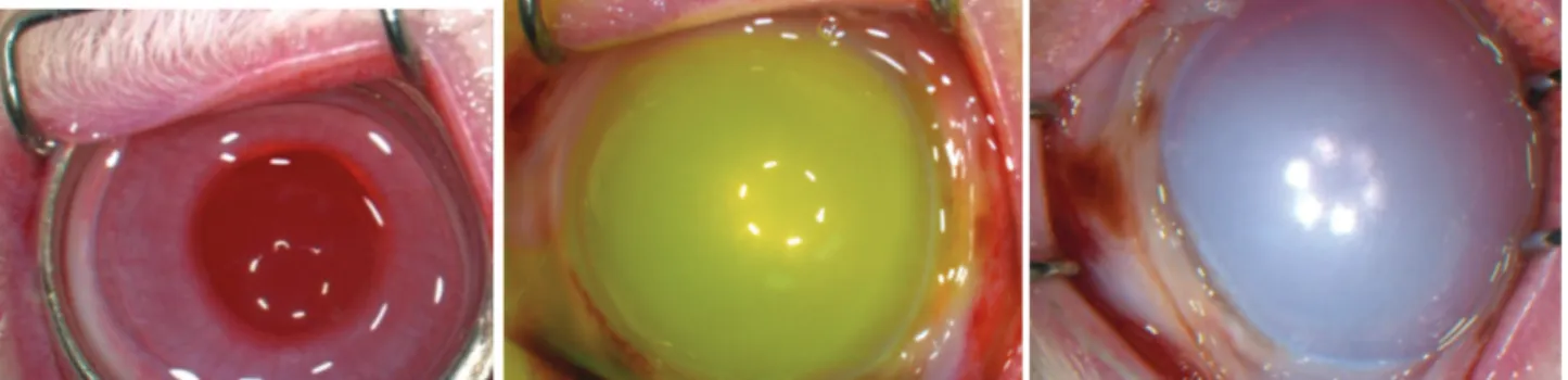
Figure 1. Application of 12-mm ring and 1N NaOH for 60 s in the cornea (including the limbus) (a), fluorescein staining (b) 3rd degree limbal deficiency (c).
Şekil 1. Korneaya 12 mm çemberin ve 1N NaOH’in 60 sn uygulanması (limbusuda içeren şekilde) (a), fluoresein boyama (b), 3. derece limbal yetersizlik (c).
(control group) was divided into 2 subgroups, as follows; subgroup 2a received 0.3% ofloxacin and 0.1% dexamethasone 5× d–1, and subgroup 2b received 0.3%
ofloxacin, 0.1% dexamethasone, and acetylcysteine 5× d–1.
Following macroscopic evaluation, 10% buffered formalin was injected into the anterior camera for improved fixation of the globe. Samples were stored and fixed in the same solution. After fixation, beginning from the corneo-scleral region the eyes was trimmed sagittally through the central corneal tissue. Hematoxylen & eosin (H&E) and periodic acid-Schiff (PAS) stains were used for histopathological examination. The avidin-biotin complex peroxidase (ABC-P) technique was used as an indirect immune peroxidase method for immuno-histochemical analysis. K3/12 (monoclonal mouse anti-rabbit cytokeratin 3,12-C9097-34M, 1:40 dilution [US Biological]), K19 (monoclonal mouse anti-human cytokeratin 19, M0888, 1:50 dilution [DAKO]), p63 (PIN Cocktail-1-P504S/p63, (monoclonal rabbit anti-human P504S and monoclonal rabbit anti-anti-human p63), PIN001-0.5, 1:40 dilution, [BIOLOGO]), and Cx43 (polyclonal rabbit anti-human connexin 43, GJA1, ab65966, [ABCAM]) primer serums were used for immunohistochemical analysis. Results were evaluated via microscopy (Olympus Bx51) and photographed (Olympus DP71). ATI Technologies Stem Cell Laboratories (Ati Teknoloji A.Ş. Trabzon/Türkiye)
provided the male rabbit BM MSCs, and performed decellularization of AM and cultivation of cells in AM.
In statistical evaluation; two proportional test for the early and late phase ophthalmascopic examination findings of all groups, Kruskal-Vallis test for the histopathological examination findings and analysis of variance for the immunohistochemical examination findings were used.
Results
Ophthalmoscopic findings: Corneal opacity covers the iris details; ischemia in the limbus (Hughes grade 3) was seen in all cases 24 hours later after the experiment. In all, 2 of the eyes (20%) in group 1 had pythisis bulbi due to perforation at the end of the 2nd postoperative
month, 30% (n = 3) had 360° corneal vascularization, a descematocele, and a visible anterior camera, and 60% (n = 6) had 360° corneal vascularization and a visible anterior camera. In group 2 at the end of the 2nd
postoperative month 50% of the patients (n = 5) had phthisis bulbi due to perforation, 20% had 360° corneal vascularization, descematocele, and iris prolapse, and 20% (n = 2) had a visible anterior camera. In, subgroup 3a on the 12th day following extirpation there was no
corneal perforation, and 100% of the eyes had 360° corneal vascularization and a visible anterior camera (Fig. 2). In subgroup 3b 100% of the eyes had 360° corneal
Figure 2. Prevention of globe perforation, visibility of the anterior camera and 360° superficial corneal vascularization all eyes in Group 3.
Şekil 2. Grup 3’te tüm gözlerde göz küresi perforasyonun engellenmesi, ön kamara görünürlüğü ve 360 derece yüzeyel korneal vaskülarizasyon.
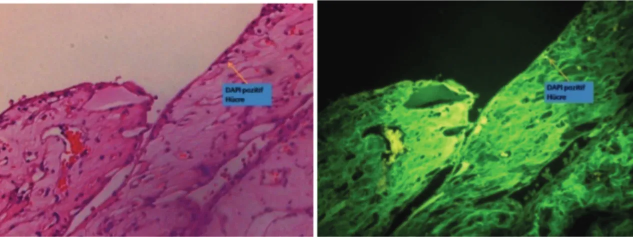
Figure 3. DAPI-positive cells in the posterior cornea Şekil 3. Posterior korneada DAPI pozitif hücreler
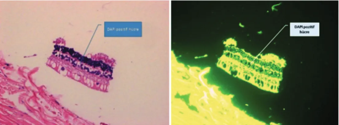
Figure 4. DAPI-positive cells in the inner retinal nuclear layer Şekil 4. Retina iç tabakasında DAPI pozitif hücreler
Figure 5. Group 1 migration of basal cells and TAC’s from peripheral to central cornea, x400, HE (1), Group 1 cell proliferation in central cornea and stromal inflammation, x400, HE (2), Group 2 limbal basal cell proliferation in peripheral cornea and stromal inflammation, x100, HE (3), Group 1 limbal basal cell proliferation in peripheral cornea and inflammatory cells between epithelial cells x100, HE (4), Group 2 proliferation in limbal basal cells and hidropic degeneration x400, HE (5), Group 3 necrotic changes and dense fibrosit and fibroblasts in peripheral cornea, x100, HE (6), Group 3 conjunctivalization throughout the central cornea, x100, HE (7), Group 3 peripheral corneal goblet cells, x100, PAS (8)
Şekil 5. Grup 1’de periferden kornea merkezine doğru bazal hücreler ve TAC’lar, x400, HE (1), Grup 1’de kornea merkezinde hücre proliferas-yonu ve stromada yangı, x400, HE (2), Grup 2’de kornea periferinde limbal bazal hücre proliferas-yonu ve stromada yangı, x100, HE (3), Grup 1’de kornea periferinde limbal bazal hücre proliferasyonu ve epitel hücreler arasında yangı hücreleri, x100, HE (4), Grup 2’de limbal bazal hücrelerde proliferasyon ve hidropik dejenerasyon, x400, HE (5), Grup 3’de nekrotik değişiklikler ve kornea periferinde yoğun fibrosit ve fibroblastlar, x100, HE (6), Grup 3’de kornea merkezi boyunca konjunktivalizasyon, x100, HE (7), Grup 3’de kornea periferinde Goblet hücreleri, x100, PAS (8).
vascularization and a visible anterior camera, 10% (n = 1) had a descematocele, and none of the corneas were perforated. In subgroup 3a DAPI-positive cells were observed in the posterior cornea (Fig. 3) and in the inner retinal nuclear layer (Fig. 4).
Histopathology and immunohistochemistry: In group 3 H&E and PAS staining was highly positive for regeneration and inflammation, and slightly positive for conjunctivalization, whereas in groups 1 and 2 the staining was highly positive for conjunctivalization (Fig. 5). With PAS staining goblet cells in the cornea indicate conjunctivalization. Conjunctivalization in group 3 was less severe than in groups 1 and 2.
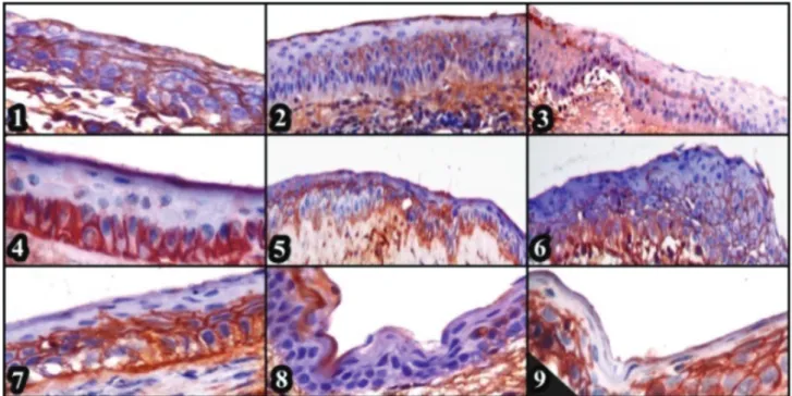
Anti-connexin 43: Anti-connexin 43 reaction was noted predominantly in cell membranes. In group 1 positive anti-connexin 43 reactions were observed in the limbal basal membrane and in post mitotic cells (PMC’s) in 5 of the eyes; in addition, all layers of the central cornea gave a positive anti-connexin 43 reaction. In group 3 positive reactions were detected in peripheral
limbal basal cells in 4 eyes. In group 3 only the central corneal basal and suprabasal cells gave a positive reaction in 4 eyes. Stem cells were positive in only 1 eye in group 3. In group 2 all layers of the central and peripheral cornea were positive (Fig. 6).
Anti-cytokeratin 3/12: Positive reactions for anti-cytokeratin 3/12 were observed predominantly in cell cytoplasm and membranes. In group 1 all limbal basal cells in the peripheral and central cornea, TACs and PMCs gave positive reactions in 3 eyes. In group 3 limbal basal cells in the peripheral cornea and all cells in the central cornea gave positive reactions in 4 eyes. In group 2 positive reactions were noted in the limbal basal and suprabasal cells of the peripheral cornea in all eyes. Additionally, in group 2 all layers of the central cornea, including necrotic cells, gave a positive reaction in three eyes (Fig. 6).
Anti-cytokeratin-19: Anti-cytokeratin-19-positive reactions were observed predominantly in cell cytoplasm. In group 1 limbal basal cells in the peripheral cornea
Figure 6. CK3-12 primary sera, Group 1, positive reaction in central cornea from basal cells throughout the suprabasal cells, x400, ABC-P (1), K3-12 primer serum, Group 2, positivity of limbal basal cells from peripheral to central cornea, x400, ABC-P (2), K19 primer serum, Group 2, positivity of limbal basal cells TAC’s and PMC’s in peripheral cornea, x100, ABC-P (3), CK19 primary sera, Group 2, positivity of basal cells in central cornea, x400, ABC-P (4), Cx-43 primary sera, Group1, positivity of basal and suprabasal cells in central cornea, x400, ABC-P (5), Cx-43 primary sera, Group 1, positivity of basal cells throughout suprabasal cells in central cornea, x400, ABC-P (6), Cx-43 primary sera, Group 2, positivity of epithelial suprabasal cells and cell islets in the regeneration zone in central cornea, x400, ABC-P (7), p63 primary sera, Group 1, positivity of peripheral limbal basal cells, x400, ABC-P (8), p63 primary sera, Group 2, positivity of all layers in central cornea, x400, ABC-P (9).
Şekil 6. K3,12 primer serumu, Grup 1’de kornea merkezindeki bazal ve suprabazal hücreler boyunca pozitif reaksiyon, x400, ABC-P (1), K3-12 primer serumu, Grup 2’de kornea periferinden merkezine kadar limbal bazal hücrelerde pozitiflik, x400, ABC-P (2), K 19 primer serumu, Grup 2’de kornea periferindeki limbal bazal hücreler, TAC’lar ve PMC’in pozitifliği, x100, ABC-P (3), K 19 primer serumu, Grup 2’de kornea merkezindeki bazal hücrelerin pozitifliği, x400, ABC-P (4), Cx-43 primer serumu, Grup 1’de kornea merkezindeki bazal ve suprabazal hücrelerin pozitifliği, x400, ABC-P (5), Cx-43 primer serumu, Grup 1’de kornea merkezindeki bazal ve suprabazal hücrelerde pozitiflik, x400, ABC-P (6), Cx-43 primer serumu, Grup 2’de kornea merkezindeki rejenerasyon hattında suprabazal hücreler ve hücre adacıklarının pozitifliği, x400, ABC-P (7), p63 primer serumu, Grup 1’de periferal limbal bazal hücrelerin pozitifliği, x400, ABC-P (8), p63 primer serumu, Grup 2’de kornea merkezinde tüm katmanların pozitifliği, x400, ABC-P (9).
similar to the group 3 in all eyes (Fig. 6).
Anti-p63: Anti-p63-positive reactions were observed predominantly in cell nuclei. In group 1 limbal basal cells in the peripheral cornea and in the suprabasal and superficial cells in the central cornea in 4 eyes stained positive. In group 3 (all layers of the central cornea) and in group 2 (all layers of the central cornea, including the necrotic cells) the results were similar to those in group 1 (Fig. 6).
Discussion
In the present study cellular and acellular AM used for transplantation acted as barriers that prevented corneal perforation, and MSCs had a positive effect on regeneration. In group 3 MSCs differentiated and prevented corneal perforation, as well as minimized local inflammatory reaction during the early phase. In group 3-severe vascularization was observed in all the corneas, which may have been related to late phase MSC reactions. A recent study on Crohn’s disease (40) reported that MSCs retard the inflammatory process during the early phases, but when they begin to differentiate they express self-tissue antibodies, cause foreign cell reaction, and result in relapse. As such, research in the future must investigate and compare autolog MSC, immunosuppression, and allogenic MSC. Comparison of the acellular AM and AM with MSCs showed us that healing rate and quality is improved in the AM group containing MSCs.
Corneal inflammation and neovascularization occur due to severe LSC deficiency in the cornea. It is reported that molecular mechanisms, including major pro-angiogenic and anti-pro-angiogenic factors (14) and such factors as vascular endothelial growth factor (VEGF) play a role in the development of neovascularization (30). In the treatment of ocular surface injury the AM is used as an external support. In LSC-deficient eyes the membrane acts as a basal membrane, preventing cell migration and adhesion to the injury site (28, 38). It is also known that stem cell transplantation induces angiogenesis (15). In the present study the degree of vascularization was significantly higher in group 3, which might have been due to induction of angiogenesis by MSCs.
Partial or total destruction of LSCs results in conjunctivalization, chronic inflammation, superficial vascularization, ulceration, and corneal perforation (10, 11, 26, 36). When the corneal surface is covered with
neovascularization must be prevented (30). For future research we recommend that such vascularity must be prevented by using anti-VEGFs such as pegaptanib, ranibizumab, and bevacizumab together with stem cells.
In autoimmune diseases and transplantation MSCs have an immunosuppressive effect, but the mechanism remains unknown (39). In the present study severe vascularity in subgroup 3b was observed, which we think was related to the decrease in the anti-inflammatory effect of stem cells during late phase, which is in agreement with the results of a study on GVH diseases (40). Some studies have suggested that immune suppression decreases tissue response to stem cells and accelerates regeneration (7, 33). If immune suppression is inadequate vascular dilatation and edema can be seen, which results in tissue rejection (25). In the present study vascularization in all groups was observed; therefore, we think that immune suppression must be maintained in order to lower the risk of late phase rejection.
Prevention of globe perforation and visibility of the anterior camera are clinically important parameters for surgeons treating corneal diseases (44); the best results were obtained in group 3, and followed by groups 1 and 2.
The standardized limbal deficiency model, as proposed by Hughes (16, 17) suggests that the degree of corneal clarity and level of limbal ischemia must be detected during the diagnosis of limbal deficiency due to corneal alkaline burn. This model was modified by Ballen (2) and Roper-Hall (34), who reported that the degree of limbal ischemia is related to the degree of stem cell damage. As we know, spontaneous healing can be observed in 1st-degree limbal deficiency and ocular
necrosis can be seen in 4th-degree limbal deficiency. In
the present study administration of 1N NaOH for 60 s in the cornea including the limbus was used to create 3rd
-degree limbal deficiency.
Cx43 is a gap junction protein that binds epithelial cells to each other. It is important for terminal amplified cell (TAC) differentiation (8). One study reported that Cx43 does not react positively to immunohistochemical staining (37). The marker K3 has low positivity or is absent in limbal basal cells, but had higher positivity in differentiated central corneal cells (13, 24, 35). K12 is a keratin marker used for corneal and limbal differentiation (3, 35). Studies on rabbits show that K3 and K12 react positively to immunohistochemical staining in corneal basal epithelium (4, 35, 41). These findings indicate that cells originating in the limbus migrate through the central
cornea (4, 13, 24, 35, 36, 41). K19 is an intermediary protein similar to vimentin, which forms the corneal epithelial structure and has a positive effect on cell proliferation in a uniform manner (6, 32). It is reported that the K19 marker stains positive in limbal basal cells and in cells that originate from limbal basal cells (3, 4). Label-retaining cells (LRC) are produced in stem cells during the replication period and K19 is localized in LRCs (3, 4). p63 is a marker of stem cells that become keratinocytes and is primarily used to identify TACs and LSCs (27, 31, 42).
One study reported that p63 positivity was highest in the limbus, followed by the peripheral cornea and central corneal (3).Generally, in the present study K3/12 and Cx-43 was positive in all cases, and stem cells in group 3 accelerated regeneration, as compared to group 1; these findings confirm those of previous reports (3, 35). K19 was highly positive in the limbal peripheral cornea, central basal cornea, and TAC’s, and was slightly positive in superficial cells in group 1, in contrast to previous reports (3, 4). Additionally, in group 1 p63 was positive in the limbal and peripheral regions of the cornea, and the basal-suprabasal cells in the central cornea, and highly positive in suprabasal cells, as previously reported.
MSCs can be marked and followed with DAPI, and green fluorescence can be observed for 3 weeks in tissues (44). DAPI positive MSCs give a positive reaction in posterior corneal endothelial cells and in the anterior corneal surface. Corneal endothelial cells do not renew; therefore in addition to anterior corneal positivity, MSC positivity in the posterior corneal endothelium and in the inner nuclear retinal layer was also noted in the present study. MSCs can migrate to the inner nuclear retinal layer interstitially. As is known, when these cells are multipotent they can differentiate into retinal cells. MSCs that are transplanted into the cornea are beyond multipotent they are pluripotent. As such, it is thought that these cells are expected to differentiate into retinal tissue. Transplanted cells can reach retinal tissue as a result of corneal injury and micro perforations that occur during corneal surgery can cause stem cells to migrate to the retina. This phenomenon might be due to an increase in the expression of CxC4 in damaged tissue, which results in stem cells migrating to the damaged tissue.
References
1. Alberts B, Bray D, Lewis J, Raff M, Roberts K, Watson JD. (1994): Differentiated cells and the maintenance of
tissues 966-982. In: B Alberts (Ed), Molecular Biology of the Cell. Garland Publishing, New York.
2. Ballen PH (1963): Mucous membrane grafts in chemical (lye) burns. Am J Ophthalmol, 55, 302-312.
3. Chee KY, Kicic A, Wiffen SJ (2006): Limbal stem cells: The search for a marker. Clin Experiment Ophthalmol, 34, 64-73.
4. Cotsarelis G, Cheng SZ, Dong G, Sun TT, Lavker RM (1989): Existence of slow-cycling limbal epithelial basal cells that can be preferentially stimulated to proliferate: implications on epithelial stem cells, Cell, 57, 201-209. 5. Darnell JE, Lodish HF, Baltimore D (1989):
Development of cell specificity 1026-1028. In: H Lodish (Ed), Molecular Cell Biology. W.H. Freeman and Company, New York.
6. Daya SM, Ilari L (2001): Living related conjunctival limbal allograft for the treatment of stem cell deficiency, Ophthalmology, 108, 126-134.
7. Deda H (2008): Stem Cell Therapies for Neurological Diseases. J Nervous Sys Surgery, 1: 142-152.
8. Dong Y, Roos M, Gruijters T, Donaldson P, Bullivant S, Beyer E, Kistler J (1994): Differential expression of
two gap junction proteins in corneal epithelium, Eur J Cell Biol, 64, 95-100.
9. Dua HS (1998): The conjunctiva in corneal epithelial wound healing. Br J Ophthalmol, 82 (12), 1407-1411. 10. Dua HS, Azuara-Blanco A (1999): Allo-limbal
transplantation in patients with limbal stem cell deficiency. Br J Ophthalmol, 83, 414-419.
11. Dua HS, Azuara-Blanco A (2000): Limbal stem cells of the corneal epithelium. Surv Ophthalmol, 44, 415–425. 12. Dua HS, Gomes JA , Singh A (1994): Corneal epithelial
wound healing, Br J Ophthalmol, 78, 401-408.
13. Espana EM, Grueterich M, Ti SE, Tseng SC (2003): Phenotypic study of a case receiving a keratolimbal allograft and amniotic membrane for total limbal stem cell deficiency. Ophthalmology, 110, 481-486.
14. Guo T, Wang W, Zhong J, Chen X, Li BZ, Li LS (2006): Experimental study on repairing damage of corneal surface by mesenchymal stem cells transplantation. Zhonghua Yan Ke Za Zhi, 42, 246-250.
15. Hosseini H, Nejabat M (2007): A potential therapeutic strategy for inhibition of corneal neovascularization with new anti-VEGF agents. Med Hypotheses, 68, 799-801. 16. Hughes WF (1946): Alkali burns of the eye; clinical and
pathologic course. Arch Ophthal, 36, 189-214.
17. Hughes WF (1946): Alkali burns of the eye; review of the literature and summary of present knowledge, Arch Ophthal, 35, 423-449.
18. Kasper M, Moll R, Stosiek P, Karsten U (1988): Patterns of cytokeratin and vimentin expression in the human eye, Histochemistry, 89, 369–377.
19. Kenyon KR, Rapoza PA (1995): Limbal allograft transplantation for ocular surface disorders, Ophthalmology,
102 (suppl), 101-102.
20. Kenyon KR, Tseng SC (1989): Limbal autograft transplantation for ocular surface disorders, Ophthalmology,
96, 709-722.
21. Kim JC, Tseng SC (1995): Transplantation of preserved human amniotic membrane for surface reconstruction in severely damaged rabbit corneas, Cornea, 14, 473-484. 22. Kirschstein R (2001): Stem Cells: scientific progress and
future research directions report. National institutes of health, June ES-2/ES-6.
23. Lauweryns B, Van den Oord J.J, Missotten L (1993): The transitional zone between limbus and peripheral cornea. An immunohistochemical study, Invest Ophthalmol Vis Sci, 34, 1991–1999.
the treatment of steroid-resistant, severe, acute graft-versus-host disease: a phase II study. Lancet, 371, 1579-1586. 26. Li W, Hayashida Y, Chen YT, Tseng SC (2007): Niche
regulation of corneal epithelial stem cells at the limbus. Cell Res, 17, 26-36.
27. Lohrum MAE, Vousden KH (2000): Regulation and function of the p53-related proteins: same family, different rules. Trends Cell Biol, 10, 197-202.
28. Ma DHK, Chen JK, Zhang F, Lin KY, Yao JY, Yu JS (2006): Regulation of corneal angiogenesis in limbal stem cell deficiency, Prog Ret Eye Res, 25, 563-590.
29. Matic M, Petrov IN, Chen S, Wang C, Dimitrijevich SD, Wolosin JM (1997): Stem cells of the corneal
epithelium lack connexins and metabolite transfer capacity. Differentiation, 61, 251-260.
30. Ocarino NM, Bozzi A, Pereira RD, Breyner NM, Silva VL, Castanheira P, Goes AM, Serakides R (2008):
Behavior of mesenchymal stem cells stained with 4', 6-diamidino-2-phenylindole dihydrochloride (DAPI) in osteogenic and non osteogenic cultures. Biocell, 32, 175-183.
31. Pellegrini G, Dellambra E, Golisano O, Martinelli E, Bondanza S, Ponzin D, McKeon F, De Luca M (2001):
p63 identifies keratinocyte stem cells. Proc Natl Acad Sci U S A, 98, 3156-3161.
32. Rao SK, Rajagopal R, Sitalakshmi G, Padmanabhan P (1999): Limbal allografting from related live donors for corneal surface reconstruction. Ophthalmology, 106, 822-828.
33. Ren G, Zhang L, Zhao X, Xu G, Zhang Y, Roberts AI, Zhao RC, Shi Y (2008): Mesenchymal stem cell-mediated
immunosuppression occurs via concerted action of chemokines and nitric oxide. Cell Stem Cell, 2, 141-150. 34. Roper-Hall MJ (1965): Thermal and chemical burns.
Trans Ophthalmol Soc U K, 85, 631-653.
35. Schmer A, Galvin S, Sun TT (1986): Differentiation-related expression of a major 64K corneal keratin in vivo and in culture suggests limbal location of corneal epithelial stem cells. J Cell Biol, 103, 49-62.
38. Şaroğlu M (1999): Tavşanlarda deneysel olarak oluşturulan kornea alkali yanıkların sağaltımında bazı antikollajenik ilaçların karşılaştırılması üzerine çalışmalar (Ph.D), İÜ Sağlık Bilimleri Enstitüsü, Cerrahi Anabilim Dalı. 39. Taylor RJ, Wang MX (1998): Rate of re-epithelialisation
following amniotic membrane transplantation. Invest Ophth Vis Sci, 39, 1038.
40. Tseng SC, Tsubata K (1997): Important concepts for treating ocular surface and tear disorders. Am J Ophthalmol,
124, 825- 835.
41. Wang DY, Hsueb YJ, Yang VC, Chen JK (2003): Propagation and phenotypic preservation of rabbit limbal epithelial cells on amniotic membrane. Invest Ophthalmol Vis Sci, 44, 4698-4704.
42. Yang A, Kaghad M, Wang Y, Gillett E, Fleming MD, Dotsch V, Andrews NC, Caput D, Mckeon F (1998):
P63 a p53 homolog at 3q27-29, encodes multiple products with transactiving, death-inducing and dominant negative activities. Mol Cell, 2, 305-316.
43. Yang A, Schweitzer R, Sun D, Kagdat M, Walker N, Bronson RT, Tabin C, Sharpe A, Caput D, Crum C, McKeon F (1999): p63 is essential for regenerative
proliferation in limb, craniofacial and epithelial development. Nature, 398, 714-718.
44. Ye J, Yao K, Kim JC (2006): Mesenchymal stem cell transplantation in a rabbit corneal alkali burn model: engraftment and involvement in wound healing. Eye, 20, 482-490.
Geliş tarihi: 01.07.1013 / Kabul tarihi: 03.01.2014
Address for correspondence:
İrem Gül Sancak University of Ankara,
Faculty of Veterinary Medicine, Department of Surgical Sciences, Ankara.