Diffusion-Weighted MRI in Cranial Bone Marrow Metastasis
Kraniyal Kemik İliği Metastazlarında Difüzyon Ağırlıklı MR Görüntüleme
Özlem Alkan1, Burçak Çakır Peköz1, Naime Altınkaya1 1
Baskent University Medical School, Department of Radiology, ADANA
Cukurova Medical Journal 2015;40(3):474-482.
ABSTRACT
Purpose: Diffusion-weighted MR imaging (DW-MRI) is generally applied to diagnose intra-axial brain pathologies such as parenchyma metastasis, abscesses, and infarcts in patients with systemic cancer. This study aimed to evaluate the DW-MRI findings in cranial bone marrow metastases to facilitate diagnosis.
Material and Methods: Conventional MR imaging and DW-MRI findings of 57 patients with cranial bone marrow metastases were evaluated retrospectively. These images were evaluated by two neuroradiologists for lesion detection and lesion consciousness. Reader agreement was assessed by κ statistics.
Results: Fifty-seven patients exhibited 113 cranial bone marrow metastases. The sensitivities of DW-MRI and the apparent diffusion coefficient to detect cranial bone metastasis for all types of primary malignancy were 86% and 90%, respectively. The sensitivities of T1-weighted imaging (T1WI), fluid attenuation inversion recovery, contrast-enhanced fat-suppressed (CE-FS) T1WI, and T2WI sequences to detect cranial bone metastasis were 93%, 90%, 89%, and 84%, respectively. Regarding lesion conspicuousness, DW-MRI was equivalent to CE-FS T1WI and equivalent or superior to non-contrast conventional sequences in most of the cranial bone marrow metastases. Interobserver agreement was excellent.
Conclusions: DW-MRI is generally indicated not only to assess the diagnosis of parenchyma involvement, but also to discover the cranial bone marrow metastasis in patients with systemic cancer.
Key words: Cranial bone marrow, metastases, brain MRI, diffusion-weighted MRI
ÖZET
Amaç: Difüzyon ağırlıklı MR (DAG-MR) görüntüleme genellikle sistemik kanserli hastalarda parankim metastazı, abse ve infart gibi intraaksiyel beyin patolojilerinin tanısında kullanılmaktadır. Bu çalışmada DAG –MR’nin kranial kemik iliği metastazlarının tanısındaki değeri araştırılmıştır.
Materyal ve Metod: Kranial kemik iliği metastazı saptanan 57 hastanın konvansiyonel MR ve DAG-MR görüntüleri geriye dönük olarak değerlendirildi. Görüntüler 2 nöroradyolog tarafından lezyon saptanması ve göze çarpıcılığı açısından incelendi. Radyologların kappa uyumuna bakıldı.
Bulgular: Elli yedi hastada toplam 113 kranial kemik iliği metastazı saptandı. DAG-MR ve ADC’ nin lezyon saptama sensitivitesi sırasıyla %86 and %90 saptandı.T1, FLAIR, kontrastlı yağ baskılamalı T1 ve T2 ağırlıklı sekansların lezyon saptanmasında sensitivitesi sırasıyla %93, %90, % 89, ve %84 saptandı. DAG-MR’nin lezyon göze çarpıcılığı kontrastlı T1 ağırlıklı görüntülere eşit, diğer konvansiyonel sekanslara eşit ya da üstündü. Radyologların kappa uyumu mükemmeldi.
Sonuç: DAG-MR sadece parankim tutulumunu değerlendirme değil, kemik iliği metastazlarını da değerlendirmede kullanılabilir
Anahtar kelimeler: Kraniyal kemik iliği, metastaz, Beyin MR, Difüzyon MR
475
INTRODUCTION
Neurologic complications occur frequently in patients with systemic cancer. Brain metastases are the most common neurologic complication of systemic cancer in adult patients. Indirect effects of systemic cancer include vascular disorders, infections, metabolic abnormalities and paraneoplastic syndromes1. MR imaging plays a major role in the evaluation and management of patients with metastatic brain tumors. Although conventional MR imaging remains an excellent screening tool, diffusion-weighted MR imaging (DW-MRI) is more sensitive for detecting neurologic complications2. DW-MRI is applied for the diagnosis of parenchyma metastasis, abscesses, and infarcts in patients with systemic cancer, in addition to routine conventional sequences in most radiology centers.
Cranial bone marrow metastasis occurs most frequently in carcinomas of the breast, lung, prostate, and kidney in adults; however, in pediatric patients, neuroblastoma and sarcoma are observed more frequently. The best imaging modality to evaluate bone marrow is MR imaging due to its high resolution3. Normal fatty bone marrow in the adult population is hyperintense on T1-weighted images. Cranial bone marrow metastases may be easily detected on T1-weighted images because they appear as hypointense lesions. On contrast-enhanced, non-fat-saturated sequences, they may be obscured by bone marrow signals. Contrast-enhanced fat-saturated (CE-FS) MR imagings are superior for detecting bone marrow lesions and involvement of the adjacent dura or brain2. However, for the inexperienced reader, cranial bone marrow metastasis may be overlooked on routine, conventional MRI examination.
Although DW-MRI is a well-known diagnostic tool for evaluation of central nervous system diseases, it has had limited use in cranial bone marrow pathologies. On DW-MRI, the background
normal skull is markedly hypointense: the intradiploic fat is hypointense because echo-planar sequences are typically obtained with fat suppression. Increased tumor cell packing may lead to a smaller and more restricted extracellular space. This decreased amount of free water, as well as its restricted mobility due to hypercellularity, may result in increased signal intensity on DW-MRI4. We investigated the role of DW-MRI in the assessment of cranial bone marrow metastasis and compared DW-MRI with conventional MRI.
MATERIALS and METHODS
Patients
Institutional review board approval was obtained for the present retrospective study. A database search was performed to identify all patients with cranial bone marrow metastases among those who underwent imaging at our institution between January 2010 and April 2014. Fifty-seven patients (27 females and 30 males) with an age range of 2–89 years (mean age, 55.7 years) were identified. These patients were referred for MR imaging of known or clinically suspected intracranial metastases with primary neoplasm. Diagnoses of bone marrow metastasis were made based on biopsy findings, bone scintigraphy, positron emission tomography (PET) or follow-up MRI. Twenty patients had undergone bone scintigraphy, and 15 of them had abnormal uptake on bone scintigraphy. Fourteen patients had undergone PET, and six of them showed abnormal uptake. Histological verification was obtained in four patients. For remaining patients, the diagnosis was confirmed at the follow up by MR imaging. Primary neoplasms included breast cancer (n=15), lung cancer (n=13), prostate cancer (n=10), renal cell carcinoma (n=3), nasopharynx cancer (n=2), neuroblastoma (n=2), neuroendocrine tumor (n=2), adrenocortical carcinoma (n=1), cholangiocellular carcinoma (n=2), urinary bladder cancer (n=2), cervix cancer
476 (n=1), ovarian cancer (n=1), thyroid cancer(n=1), primitive neuroectodermal tumor (n=1), and squamous cell carcinoma of the neck (n=1).
Imaging Parameters
Brain MRI was performed in all patients using a 1.5-Tesla MR scanner (Magnetom Avanto or Symphony; Siemens Medical Solutions, Erlangen, Germany). All of the sequences were performed using identical parameters for the number of slices, matrix size (256 × 256), field of view (230 mm), slice thickness (5 mm) and intersection gap (1.5 mm). Sagittal and axial T1-weighted spin echo (SE) images (repetition Time (TR)/echo time (TE)/number of excitations (NEX): 500 ms/11 ms/2, respectively), coronal and axial T2-weighted turbo SE images (TR/TE/NEX: 4500 ms/110 ms/2, respectively), axial fluid attenuation inversion recovery (FLAIR) images (TR/TE/NEX: 9000 ms/110 ms/2, respectively), axial contrast-enhanced fat-suppressed T1-weighted imaging with gadolinium (CE-FS T1WI) with SE, and coronal and sagittal T1-weighted SE images with gadolinium (TR/TE/NEX: 429 ms/12 ms/2, respectively) were acquired as part of the routine imaging protocol. All of the patients underwent DW-MRI in the transverse plane, using a single-shot, SE echo-planar readout. Diffusion-sensitizing gradients were applied in three orthogonal directions with diffusion sensitivities of b = 0 and 1000 s/mm² (TR/TE/NEX: 4200 ms/115 ms/2, respectively). Apparent diffusion coefficient (ADC) maps were generated using commercially available software.
Image Assessment
Two independent experienced neuroradiologists (O.A. and N.A. with 12 and 6 years of experience in interpreting head and neck MR images, respectively) blinded to the clinical histories of the patients and diagnoses reviewed the MR images, including conventional MR images and DW-MR images (trace and ADC images). The number, location, size and involvement type
(focal/diffuse) of calvarial lesions, and the presence of dural, scalp and parenchymal metastases were assessed. The signal intensity of the lesions was categorized as hypointense, isointense, or hyperintense with respect to the presumed normal marrow. Enhancement of the lesions was evaluated on CE-FS T1WI. Conventional MRI and DW-MRI were read as positive for hypointense and hyperintense images and negative for isointense images. In the first reading session, the two readers assessed independently the images in terms of lesion conspicuousness. The images were compared side by side to determine which sequence was better in terms of the conspicuousness of lesions from adjacent structures. Comparison between DW-MRI and routine sequences was performed only for the axial images. We used a combination of trace and ADC images for lesion conspicuousness. Lesion conspicuousness was classified as superior to DW-MRI, equivalent to DW-MRI or inferior to DW-MRI. At the consensus reading, the lesion conspicuousness and detection were determined to form a standard of reference.
Statistical Analysis
Statistical analysis was performed using the statistical software package SPSS (version 17.0; SPSS Inc., Chicago, IL, USA). The categorical variables were analyzed using chi-squared test or Fisher’s exact test. Interobserver reliability was applied using Cronbach’s alpha reliability value. Correlations between observers were tested using the intra-rater correlation test. A p value of 0.05 was deemed to indicate statistical significance. The diagnoses made by the reviewers were characterized as true positive, false negative, or false positive. For each imaging modality, the sensitivity was calculated using the following equation: sensitivity (%) = [number of true positives/ (true positives + false negatives)] ×100. Specificity could not be calculated because the true negatives could not be accurately defined.
477
RESULTS
One hundred and thirteen lesions were detected in 57 patients: six lesions (5.3%) showed diffuse involvement and 107 (94.7%) showed focal involvement. Nineteen of one hundred seven focal lesions (17.8%) were located in the skull base, and 88 of them (82.2%) were found in the calvarium. Axial diameters of the lesions ranged from 3 to 50 mm (mean, 14.09 mm). We observed concomitant parenchymal metastases in 12 lesions (10.6%). Thirty-four lesions (30.1%) had dural metastatic components, and 20 lesions (17.7%) were together with scalp metastases.
Table 1 summarizes the signal intensities of cranial bone marrow metastases. After intravenous contrast material administration, 101 lesions (89.4%) were enhanced, whereas 12 lesions were not enhanced on CE-FS T1WI. No MR image showed false-positive findings in the absence of pathologic findings. The sensitivities of DW-MRI and ADC for detecting cranial bone metastasis for all types of primary malignancies were 86% and 90%, respectively. The sensitivity of ADC for detecting cranial bone metastasis was superior to DW-MRI. The sensitivities of T1WI, FLAIR, CE-FS T1WI, and T2WI sequences for detecting cranial bone metastasis were 93%, 90%, 89%, and 84%, respectively.
The sensitivity of DW-MRI for detecting focal lesions was 90% and that for detecting diffuse lesions was 16.6%. The sensitivity of DW-MRI for detecting focal lesions was superior to that of DW-
MRI for detecting diffuse lesions. The sensitivities of DW-MRI for detecting calvarial and skull base lesions were 89.7% and 94.7%, respectively. The sensitivity of DW-MRI for detecting cranial bone metastasis was 95% with scalp metastasis and 84.9% without scalp metastasis.
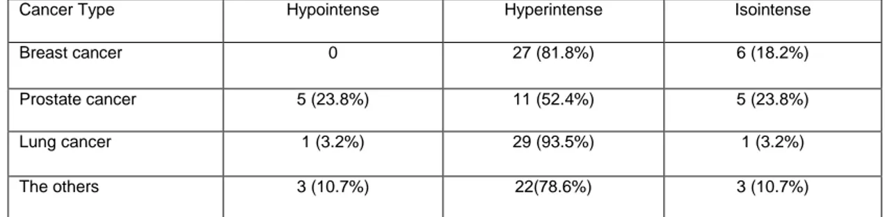
The signal intensities of cranial bone marrow metastases on DW-MRI according to cancer type are shown in Table 2. The sensitivities of DW-MRI for detecting cranial bone metastasis of the lung, breast, and prostate cancers were 96.7%, 81.8%, and 72.7%, respectively.
Table 3 summarizes the comparison of DW-MRI with conventional DW-MRI in terms of the lesion conspicuousness of cranial bone marrow metastasis. Regarding lesion conspicuousness, DW-MRI was equivalent or superior to T1WI and FLAIR images in most of the lesions. DW-MRI was superior to T2WI in lesion conspicuousness. DW-MRI was equivalent to CE-FS T1WI in most of the lesions (Figs. 1, 2 and 3).
Lesion conspicuousness was not significantly different among all primary neoplasm types. When all of the sequences were compared, no significant difference in lesion conspicuousness was found according to lesion size, location, and diffuse or focal involvement. Interobserver correlation was used to evaluate agreement between the observers, and an excellent interobserver agreement was detected (80.0%). The intraobserver correlation was also excellent (κ=0.85).
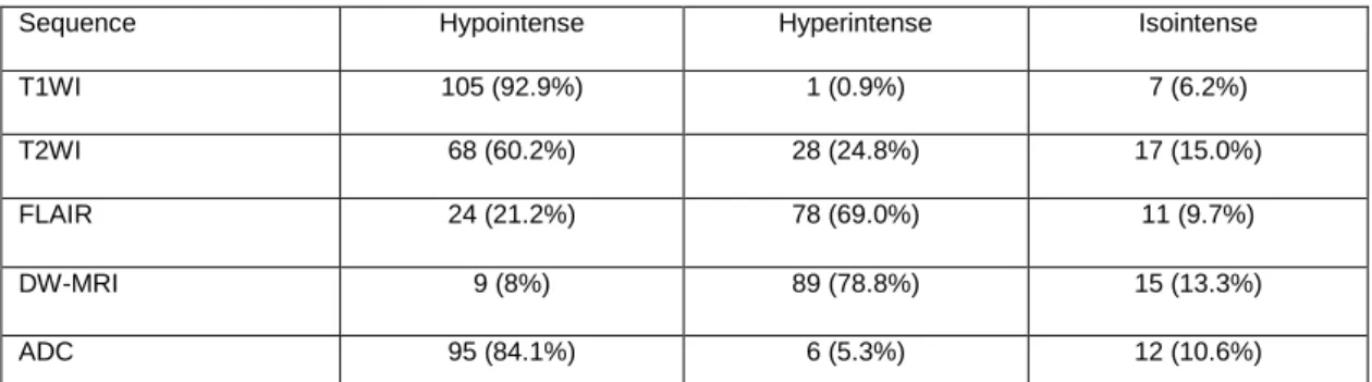
Table 1. Signal intensities of cranial bone marrow metastases
Sequence Hypointense Hyperintense Isointense
T1WI 105 (92.9%) 1 (0.9%) 7 (6.2%)
T2WI 68 (60.2%) 28 (24.8%) 17 (15.0%)
FLAIR 24 (21.2%) 78 (69.0%) 11 (9.7%)
DW-MRI 9 (8%) 89 (78.8%) 15 (13.3%)
ADC 95 (84.1%) 6 (5.3%) 12 (10.6%)
Note. Diffusion-weighted MR imaging =DW-MRI, T1WI= T1-weighted images, T2WI= T2-weighted images, FLAIR= Fluid Attenuated Inversion Recovery, ADC= Apparent diffusion coefficient.
478
Table 2. Signal intensities of cranial bone marrow metastases on DW-MRI according to Cancer Type
Cancer Type Hypointense Hyperintense Isointense
Breast cancer 0 27 (81.8%) 6 (18.2%)
Prostate cancer 5 (23.8%) 11 (52.4%) 5 (23.8%)
Lung cancer 1 (3.2%) 29 (93.5%) 1 (3.2%)
The others 3 (10.7%) 22(78.6%) 3 (10.7%)
Note. Diffusion-weighted MR imaging=DW-MRI; the others= renal cell carcinoma, n=5; nasopharynx cancer, n=4; neuroblastoma, n=4; neuroendocrine tumor, n=3; adrenocortical carcinoma, n=3; cholangiocellular carcinoma, n=2; urinary bladder cancer, n=2; cervix cancer, n=1; ovarian cancer, n=1; thyroid cancer, n=1; primitive neuroectodermal tumor, n=1; squamous cell carcinoma of neck, n=1.
Table 3. Comparison of DW-MRI and conventional MR examination in terms of the lesion conspicuousness of cranial bone marrow metastasis.
Superior to DW-MRI Equivalent to DW-MRI Inferior to DW-MRI
T1WI 10 (8.8%) 49 (43.4%) 54 (47.8%) T2WI 4 (3.5%) 28 (24.8%) 81 (71.7%) FLAIR 10 (8.8%) 51 (45.1%) 52 (46.0%) CE T1WI 22 (19.5%) 61 (54.0%) 30 (26.5%)
Note. Diffusion-weighted MR imaging =DW-MRI, T1WI= T1 weighted images, T2WI= T2 weighted images, FLAIR= Fluid Attenuated Inversion Recovery, CE T1WI= contrast enhanced T1 weighted images.
479
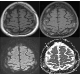
Figure 1.a-d. A 56-year-old woman with breast cancer. Axial T2- weighted (a), T1- weighted (b), diffusion-weighted MR images(c) and ADC map (d) show multiple cranial bone marrow metastases. The consciousness of the lesion is better on ADC map (arrows) than on other images.
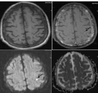
Figure 2. a-d. A 43-year-old woman with lung cancer. Axial T1- weighted (a), fat-saturated contrast enhanced T1- weighted (b), diffusion-weighted MR images(c) and ADC map (d) show skull base metastases (arrow). Diffusion-weighted MR images(c) and ADC map images superior to conventional MR images for the consciousness of the lesion.
480
Figure 3. a-d. A 61-year-old woman with lung cancer. Axial T1- weighted (a), fat-saturated contrast enhanced T1- weighted (b), diffusion-weighted MR images(c) and ADC map (d) show diffuse involvement of cranial bone marrow and parenchyma metastases(arrow).
DISCUSSION
Our study showed that DW-MRI was equivalent to CE-FS T1WI and equivalent or superior to non-contrast conventional sequences for lesion conspicuousness in most of the cranial bone marrow metastases. Moon et al.5 showed that isotropic DW-MRI provided better lesion conspicuousness than T1WI or b0 images. T1WI had the highest sensitivity for detecting cranial bone marrow metastasis in our study. However, DW-MRI and other conventional MR sequences had similar sensitivity for detecting cranial bone marrow metastasis. The sensitivity of ADC for detecting cranial bone metastasis was superior to that of DW-MRI in our study. Therefore, ADC and DW-MRI can be used together to improve lesion detection and conspicuousness. Bone scintigraphy is a valuable screening test to detect bone marrow metastasis. However, it has low spatial resolution and specificity. Therefore, a combination of bone scintigraphy and other imaging methods is required for accurate diagnosis. MR sequences such as T1WI and CE-FS T1WI are valuable for the assessment of cranial bone marrow because
they have high spatial resolution and tissue contrast. DW-MRI has been applied to vertebral bodies to differentiate between benign and malignant vertebral compression fractures6. Because DW-MRI with fat suppression is used; cranial bone marrow shows a hypointense signal on DW-MRI. Malignant infiltrations with enlarged nuclei, hypercellularity, and a reduced extracellular matrix generally produce reduced ADC values and result in increased signal intensity on DW-MRI7.Normal cranial bone marrow is hypointense, and the lesions show hyperintense signals on DW-MRI. This contrast can increase lesion detection and conspicuousness. Our results also reflect these changes.
We observed that DW-MRI had the highest sensitivity for detecting cranial bone marrow metastasis of lung cancer, whereas the sensitivity of DW-MRI for detecting cranial bone marrow metastasis of prostate cancer demonstrated the lowest value. Nemeth et al.8 showed that DW-MRI is a useful sequence for identifying focal skull metastases of breast and lung malignancies compared with conventional MRI. However,
DW-481 MRI is insensitive for the detection of sclerotic skull metastases of prostate carcinoma.
We revealed that DW-MRI had higher sensitivity for detecting focal cranial bone marrow metastasis than for detecting diffuse metastasis. The sensitivity of DW-MRI for detecting focal lesions was 90% and that for detecting diffuse lesions was 16.6%. Our results are also consistent with the report by Nemeth et al.8 that DW-MRI provides improved detection of focal skull metastasis.
The sensitivities of DW-MRI for detecting calvarial and skull base lesions were 89.7% and 94.7%, respectively. The skull base is a challenging region for imaging with DW-MRI due to its contents, such as air, bone, and fat. Thus, severe susceptibility artifacts can be evident, particularly near air-bone interfaces. Using a parallel imaging technique such as sensitivity encoding (SENSE) or generalized autocalibrating partially parallel acquisitions (GRAPPA) can markedly reduce magnetic susceptibility artifacts in the skull base, as was seen in our study.
The limitations of the present study must be acknowledged. First, the gold standard modality— bone scintigraphy—was not used in the current study. Although bone scintigraphy is the standard method for the detection of bone metastases, no consensus exists that it is the best modality to diagnose bone metastases. Second, a relatively small number of cranial bone marrow metastases from each primary neoplasm were assessed in the present study. Additional studies with more homogeneous tumor groups may be required. We could not measure ADC values in the current study. Susceptibility artifacts are more pronounced within the bone, a find that can compromise correct ADC measurements. Thus, the authors decided not to obtain ADC measurements of the bone marrow. ADC measurements may be obtained from scalp metastases or the dural metastatic component of cranial bone marrow metastases in further studies. Malignant neoplasms have lower ADC values, whereas benign neoplasms have
higher ADC values. The latter findings could be used to investigate the role of DW-MRI in differentiating between malignant and benign cranial bone marrow lesions.
In conclusion, DW-MRI is applied for the diagnosis of parenchymal metastasis, abscesses, and infarcts, in addition to routine conventional sequences in most radiology centers. When evaluating cranial bone marrow using DW-MRI and ADC maps, focusing not only on the diagnosis of parenchymal metastasis but also on cranial bone marrow metastasis can be helpful. Additionally, the assessment of DW-MRI and ADC maps together provides improved conspicuousness and detection of cranial bone marrow metastasis.
REFERENCES
1. Newton H, B.Neurologic complications of systemic cancer. Am Fam Physician. 1999;59:878-86. 2. Lee EK, Lee EJ, Kim MS, Park HJ, Park NH, Park
SII, Lee YS. Intracranial metastases: spectrum of MR imaging findings. Acta Radiol. 2012;53: 1173-85. 3. Silva JR Jr, Hayashi D, Yonenaga T ,Fukuda K,
Genant HK, Lin C,et al. MRI of bone marrow abnormalities in hematological malignancies.Diagn Interv Radiol. 2013;19:393-9.
4. Castillo M, Arbelaez A, Smith JK, Fisher LL. Diffusion-weighted MR Imaging Offers No Advantage over Routine Noncontrast MR Imaging in the Detection of Vertebral Metastases AJNR Am J Neuroradiol. 2000;21:948–53.
5. Moon WJ, Lee MH, Chung EC. Diffusion-Weighted Imaging with Sensitivity Encoding (SENSE) for Detecting Cranial Bone Marrow Metastases:Comparison with T1-Weighted Images. Korean J Radiol. 2007;8:185-91.
6. Baur A, Stabler A, Bruning R,Bartl R, Krödel A, Reiser M, Deimling M. Diffusion-weighted MR imaging of bone marrow: differentiation of benign versus pathologic compression fractures. Radiology. 1998;207:349-55
7. Le Bihan D, Breton E, Lallemand D, Aubin ML, Vignaud J, Laval-Jeantet M. Separation of diffusion
482
and perfusion in intravoxel incoherent motion MR imaging.Radiology. 1988;168:497-505.
8. Nemeth AJ, Henson JW,Mullins ME, Gonzalez RG, Schaefer PW. Improved Detection of Skull Metastasis
with Diffusion-Weighted MR Imaging AJNR Am J Neuroradiol. 2007;28:1088 –92
Yazışma Adresi / Address for Correspondence: Dr. Özlem Alkan
Başkent University Medical Scholl Department of Radiology E-mail: [email protected]
Geliş tarihi/Received on : 22.01.2015 Kabul tarihi/Accepted on: 26.02.2015