ORIGINAL ARTICLE
Methylation of cation
–chloride cotransporters NKCC1 and KCC2
in patients with juvenile myoclonic epilepsy
Fatma Genç1 &Murat Kara2&Yasemin Ünal3&Elif Uygur Küçükseymen4&Yasemin Biçer Gömceli1&Taner Kaynar5&
Kürşad Tosun6
&Gülnihal Kutlu7
Received: 18 October 2018 / Accepted: 30 January 2019 / Published online: 13 February 2019 # Fondazione Società Italiana di Neurologia 2019
Abstract
The etiology of juvenile myoclonic epilepsy (JME) is still unknown and the process of elaboration of multiple genetic mechanisms is ongoing. The aim of this study was to investigate the potential role of NKCC1 (SCL12A2) and KCC2 (SCL12A5) in JME by comparing their DNA methylation status in patients with JME versus healthy controls. Forty-nine patients with JME and 39 healthy individuals were compared for DNA methylation at the 5CpG islands. A total of 71 (81%) samples were found to have methylation in the NKCC1 gene, 36 (73%) from patients and 35 (90%) from healthy individuals. Out of the KCC2 samples, 50 (57%) were found to have methylation, 33 (67%) from patients and 17 (44%) from healthy individuals. In patients with JME, methylation of NKCC1 (73%) was lower than its methylation in the controls (90%) (p = 0.047). On the other hand, methylation of KCC2 in patients with JME (67%) was greater than the methylation in the controls (44%) (p = 0.022). Twenty-eight patients were treated with VPA and ongoing medications were not found to be associated with methylation (p > 0.05). In the present study, we determined significantly lower NKCC1 DNA methylation and significantly higher KCC2 DNA methylation levels in patients with JME compared with the healthy controls. This implies that NKCC1 expression can be higher and KCC2 expression can be reduced in affected people. Further studies that investigate the potential effect of DNA methylation mechanisms regulating gene expression on seizure activity and how they change JME network activity will be helpful.
Keywords Juvenile myoclonic epilepsy . Epigenetic . DNA methylation . Cation–chloride cotransporters NKCC1 (SCL12A2), KCC2 (SCL12A5) https://doi.org/10.1007/s10072-019-03743-4 * Fatma Genç [email protected] Yasemin Ünal [email protected] Elif Uygur Küçükseymen [email protected] Yasemin Biçer Gömceli [email protected] Taner Kaynar [email protected] Kürşad Tosun [email protected] Gülnihal Kutlu [email protected]
1 Antalya Training and Research Hospital, Department of Neurology,
Antalya, Turkey 2
Istanbul, Turkey
3 Faculty of Medicine Department of Neurology, Muğla Sıtkı Koçman
University, Muğla, Turkey 4
Neuromodulation Center, Spaulding Rehabilitation Hospital, Harvard Medical School, Boston, MA, USA
5
Sitogen Biomedical and Laboratory Systems Industrial Trade Limited Company, Zümrütevler mah. Hanımeli cad. Aktunç İşmerkezi No:13/1 Maltepe, İstanbul, Turkey
6 Siena College, Loudonville, NY, USA
7
Faculty of Medicine Department of Neurology and Clinical
Neurophysiology, Muğla Sıtkı Koçman University, Muğla, Turkey
Introduction
Juvenile myoclonic epilepsy (JME) is characterized by myo-clonic jerks, generalized tonic–myo-clonic seizures (GTCS), ab-sence seizures, and widely recognized idiopathic generalized epilepsy syndrome. The prevalence of JME is estimated to be 5 to 10% of all epilepsies and around 18% of idiopathic gen-eralized epilepsies [1,2]. Although JME used to be considered as idiopathic generalized epilepsy (IGE), recent understanding of JME has instead focused on a system disorder of the brain and the ictogenesis of bilateral networks located primarily at the thalamus and the selective areas of the neocortex [3]. The clinical spectrum of JME is substantially broad, with a still unknown etiology; the process of the elaboration of multiple genetic mechanisms is ongoing [4]. Although single gene mu-tations have been identified at high penetrance rates among crowded families that have members with epilepsy, certain members who indeed bear the causative mutation do not de-velop the disease expression in addition to the remarkable phenotypic heterogeneity of the specific epilepsy syndrome among the individual family members [5]. Recently, a mega-analysis identified 16 new loci in common epilepsies, specif-ically the STX1B gene at locus 16p11.2 that was associated with JME [6]. Possible reasons for this might be modifying genes, environmental causes, and induced epigenetic modifi-cations [5].
Epigenetic modifications play an essential role in various nervous system disorders, including epilepsy, by allowing continuous re-programming of the underlying gene expres-sion through environmental factors, as well as with changes to the chromatin template without changing the DNA se-quence [5,7].
DNA methylation directly affects DNA and is the most widespread epigenetic event taking place across the mam-malian genome [8]. DNA methylation usually results in a silencing effect on transcriptional activity [5]. DNA meth-ylation has been shown to be a factor associated with epileptogenesis and recurrent seizure activity [7]. DNA methylation alterations might contribute to the predispo-sition to and development/maintenance of JME [9].
Gamma-aminobutyric acid (GABA) is the major inhib-itory neurotransmitter of the central nervous system (CNS), which hyperpolarizes the membrane potential and mitigates neuronal excitability [10, 11]. The GABAergic system plays an essential role in various neu-ronal processes including neuneu-ronal plasticity, generation of oscillatory activity, control of neuronal assemblies, and regulation of neuronal excitability [12]. A growing number of studies conducted in the JME population have shown an abnormal structural and functional heterogene-ity of γ-aminobutyric acid receptor type A (GABAAR) and GABAergic neurotransmission in thalamocortical net-work dysfunction and brain connectivity [12,13].
Postsynaptic inhibition mediated by the GABAAR activa-tion is achieved through caactiva-tion–chloride cotransporters (CCCs), particularly through sodium–potassium–chloride cotransporter 1 (NKCC1) and potassium–chloride cotransporter 2 (KCC2) upon maintenance of a specified low intracellular chloride concentration. The latest studies suggest CCC dysfunction is among the factors that play a role in inhibitory neurotransmission deficits encountered during epileptogenesis. Alteration of CCC expression may provide a substantial contribution to the abnormal synchronization and hyperexcitability during epileptogenesis [14].
In this study, we investigated the potential role of NKCC1 (SCL12A2) and KCC2 (SCL12A5) in JME by comparing their DNA methylation status in patients with JME versus healthy controls.
Patients and methods
Between July 1, 2015, and February 15, 2016, 49 patients (69% females) with JME were followed by the outpatient epilepsy departments of Antalya Training and Research Hospital and Muğla University Training and Research Hospital. The mean age of the patients was 26.65 ± 8.14 years. Thirty-nine healthy individuals (51% females) were included in the study as controls. The mean age of the controls was 31.90 ± 9.08 years. The patient and control groups were in-formed about the objective and procedures of the study, and their written consents were obtained. All patients were diag-nosed according to the recommendations of the Commission on Classification and Terminology of the International League Against Epilepsy (ILAE) in 2010 with Genetic Generalized Epilepsy (GGE) and the Consensus on the Diagnosis and Management of JME in 2011. Patients were classified as hav-ing JME based on myoclonic jerks predominantly occurrhav-ing after awakening, associated with typical generalized epilepti-form electroencephalograph (EEG) abnormalities, with an age of onset between 10 and 25 years [15,16]. The control group was selected from among healthy volunteers.
Blood samples (2 cc) were drawn from the patient and control groups into collection tubes containing ethylenedi-aminetetraacetic acid (EDTA). The DNA was isolated from the blood samples using a DNA isolation protocol (PureLink® Genomic DNA Mini Kit, Invitrogen, Carlsbad, CA 92008 USA). Bisulfite modification of the target DNA methylation areas, which are composed of the sequence GGGGCGGGGACATCTCGAGTAGGGAGCGGGA locat-ed at Chr20:44650763-44650913 corresponding to KCC2 ( S L C 1 2 A 5 ) a n d G G G AG G G C T GG G C G A G C T C A CCTTTTCCAGCTCGTCGTGGAGCTCCGC located at Chr5:127420214-127420445 corresponding to NKCC1 (SLC12A2) was performed using EpiTect Plus Bisulfite Conversion Kit (Qiagen). A total mix of 140μL was prepared
using 85μL of bisulfite mix, 35 μL of DNA protect buffer, 15μL of RNase free water, and 5 μL of DNA (total 100 ηg of DNA). The incubator was adjusted as 5 min at 95 °C, 25 min at 60 °C, 5 min at 95 °C, 85 min at 60 °C, 5 min at 95 °C, 174 min at 60 °C, and forever at 20 °C. Following the bisulfite treatment, all samples were cleaned. PCR amplification of each bisulfite converted DNA methylation region was per-formed using a Pyro kit. A reaction mix was prepared for each PCR, and 20μL of the master mix was added to PCR tubes. From the DNA sample, 5μL was added into the PCR tube. The PCR plate was run in a StepOnePlus Real-Time PCR System according to the protocol encompassing 45 cycles with 15 min at 95 °C, 30 s at 94 °C, 30 s at 56 °C, 30 s at 71 °C, 10 min at 71 °C, and forever at + 4 °C. PCR products were immobilized using Streptavidin Sepharose high-performance beads, and the samples were analyzed through pyrosequencing on a PyroMark Q96 platform. Success of bi-sulfite conversion was verified by the use of EpiTect PCR Control DNA. Methylation at the 5CpG islands were investi-gated in the patient and control groups. Based on the assess-ment compared with the non-methylated controls, a result of > 5% was considered as methylated for the KCC2 (SLC12A5) gene and > 7% was considered as methylated for the NKCC1 (SLC12A2) gene (Figs.1and2).
The study was approved by the Ethics Committee of Antalya Education and Research Hospital. In this study, all computational analyses were performed using the statistical packages in software R. To determine whether there was any significant association between the methylation of KCC2/ NKCC1 and JME, the Chi-square and Fisher’s exact tests were used. Summary statistics are expressed as mean ± stan-dard deviation or percentage (%). Comparisons were
performed using the Welcht test or proportion test. The com-parison of methylation and no methylation by disease duration was evaluated using the Mann–Whitney–Wilcoxon test. A p value of < 0.05 was considered statistically significant.
Results
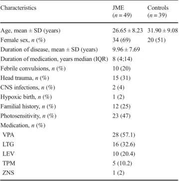
The study comprised 49 patients with JME and 39 healthy subjects in the control group. The mean duration of disease was 9.96 ± 7.69 years. All patients had normal results in their neurologic examinations and imaging studies. When patients were questioned for their medical history, 20% defined past febrile convulsions, 31% had experienced mild head traumas, 4% had CNS bacterial infections, and 2% had hypoxic births. In 26 (53%) of the patients, no risk factors were identified. Familial history of epilepsy was encountered in 25% of pa-tients. Photosensitivity was detected among 47% of papa-tients. Ongoing medications of the patients were as follows: valproic acid (n = 28), lamotrigine (n = 16), levetiracetam (n = 10), topiramate (n = 5), and zonisamide (n = 1) (Table1).
A total of 71 (81%) samples were found to have methyla-tion in the NKCC1 gene, 36 (73%) from patients and 35 (90%) from healthy individuals. Out of the KCC2 samples, 50 (57%) were found to have methylation, 33 (67%) from patients and 17 (44%) from healthy individuals. In patients with JME, methylation of NKCC1 (73%) was lower than in the control group (90%) (p = 0.047, Fisher’s exact test). Methylation of KCC2 in patients with JME (67%) was greater than in the control group (44%) (p = 0.022, Fisher’s exact test) (Table2).
Fig. 1 SLC12A5 gene promoter region methylation in a patient with JME
I
125 . . . . ... • . . . ... • . . . • .•. . . ·1 · ... ... . 100 · · · · · · · · · · · · · · · · · · · · · · · · · · · · · · · · · · · · · · · · · · · · · · · · · · · · ·I
75 ...•...•..•.. . . ... . . ....•... . . .. ·1 ·... . . . . . . . . ... ·•. . . . . . . . . . . . . . . . . . . ... . 50 . . .•...•.... . . ... . . ....•.... . .... ·1 · ... . . . .... . . .... . . ... ... . . . .. . 25 . . . . ... . . . ... . . . ... . . . ·1 .. . .. ... . ... . . . ... . . . .. . 0 ··· "-~ - - - - ' E S T G G T C G G C A T C A G T T C G A T G A T A G A G T C G 5 10 15 20 25Sequence to analyze:
There was no significant difference in duration of epilepsy in patients with and without methylation in the NKCC1 gene (p = 0.448, Mann–Whitney–Wilcoxon test).
The duration of epilepsy in patients with methylation at KCC2 was shorter than in patients without methylation (p = 0.032, Mann–Whitney–Wilcoxon test).
History of febrile convulsion, head trauma, familial history, presence of photosensitivity, and ongoing medications were
not found to be associated with methylation (p > 0.05, Fisher’s exact test) (Table3).
Discussion
This is the first report to examine methylation variations in NKCC1 and KCC2 genes in JME. DNA methylation is the only covalent event known to take place in mammalian DNA and serves as a control mechanism of specific gene expression in adult tissues, whereby the methyl group of S-adenosylmethionine (SAM) is transferred to the cytosine (C) residue preceding a guanine (G) residue in the CpG islands, catalyzed by a family of enzymes called DNA methyltransfer-ases (DNMT), and thus 5-methyl cytosine (5mC) is generated [8,17–19]. Results from studies employing DNA methylation models indicate that DNMT-catalyzed DNA methylation is an important factor in epileptogenesis [7].
Inhibition mediated by ionotropic GABAAreceptors is important in the maintenance of normal brain function and protection against seizures. Leading to the permeability of
Table 1 Baseline demographic characteristics of the study population
Characteristics JME Controls
(n = 49) (n = 39)
Age, mean ± SD (years) 26.65 ± 8.23 31.90 ± 9.08
Female sex,n (%) 34 (69) 20 (51)
Duration of disease, mean ± SD (years) 9.96 ± 7.69
Duration of medication, years median (IQR) 8 (4;14)
Febrile convulsions,n (%) 10 (20) Head trauma,n (%) 15 (31) CNS infections,n (%) 2 (4) Hypoxic birth,n (%) 1 (2) Familial history,n (%) 12 (25) Photosensitivity,n (%) 23 (47) Medication,n (%) VPA 28 (57.1) LTG 16 (32.6) LEV 10 (20.4) TPM 5 (10.2) ZNS 1 (2)
IQR interquartile range, CNS central nervous system, VPA valproic acid, LTG lamotrigine, LEV levetiracetam, TPM topiramate, ZNS zonisamide
Table 2 NKCC1 and KCC2 methylation status in JME and control
groups
JME (n = 49) Controls (n = 39) p value
NKCC1,n (%) Methylation (+) 36 (73) 35 (90) 0.047 Methylation (−) 13 (27) 4 (10) KCC2,n (%) Methylation (+) 33 (67) 17 (44) 0.022 Methylation (−) 16 (33) 22 (56)
Fig. 2 SLC12A2 gene promoter region methylation in a patient with JME
3\ 18\ 4\ 75 · · · · · · · · · · · · · · · · · · · · · · · · · · · · · · · · · · · · · · · · · · · · · · · · · · · · · · · · · · · · · · · 50 · · · · · · · · · · · · · · · · · · · · · · · · · · · · · · · · · · · · 25 · · · • • • · · · · • · · · · · · · · • · · · · · · · · · · · · · · · .• · · • · · · • • · · · · · · · · · · · · · o ...•.. E S T G A G T C T A G T C G A G A T A G T T A G T C G T C G T G A G T T C G 5 10 15 20 25 30 35 Sequence to analyze: GGGAGGGTTGGGYGAGTTTATTTTITTTAGT GT
anion channels through which Cl− is transported into the cell, GABAAreceptors result in hyperpolarization of plas-ma membrane, and thus triggering neuronal inhibition, they help with reducing the seizure activity [20, 21]. The key molecules that act in neuronal Cl− homeostasis are cation–chloride cotransporters (CCCs), Na−K−2Cl c o t r a n s p o r t e r i s o f o r m 1 ( N K C C 1 ) , a n d K− Cl cotransporter isoform 2 (KCC2), which sit at the cell membrane. Recent data indicate the role of CCCs in epi-lepsy, suggesting the critical importance of CCCs in the nervous system, in the regulation of intracellular chloride concentration and therefore control of neuronal function [22,23]. A variety of evidence has indicated a correlation of epileptogenesis with differences in functional expres-sion of NKCC and KCC transporters [23].
Palma et al. demonstrated the association of increased NKCC1 and decreased KCC2 expression with the excitatory effect of GABAAreceptors on cell membranes extracted by temporal lobectomy in patients with epilepsy [24]. Similarly, in the hippocampus and cortex tissues resected from patients with refractory epilepsy, NKCC1 expression was found to be upregulated, whereas KCC2 expression was downregulated [25].
In cortical and hippocampal neurons, NKCC1 mediates Cl− uptake and KCC2 mediates Cl− extrusion [26]. In GABAergic interneurons, NKCC1-mediated elevation of Cl− may weaken the GABA-induced inhibition, leading to increased network activity. Decreased KCC2 expression, on the other hand, gives rise to an elevated susceptibility to sei-zures [20]. Moreover, NKCC1 has been shown to cause intra-cellular Cl−accumulation in hippocampal pyramidal neurons, which may result in facilitation of seizures in the brain and thus attenuation of GABAAreceptor-mediated inhibition [20].
In the present study, we determined significantly lower NKCC1 DNA methylation and significantly higher KCC2 DNA methylation levels in patients with JME compared with the healthy controls. This implies NKCC1 expression may be higher and KCC2 expression may be lower in affected people. According to our results, such a regulation on NKCC1 and KCC2 and the resulting load of [Cl−] concentration is likely to attenuate the effect of GABAAR-mediated inhibition, which in turn contributes to the hyperexcitability mechanism, giving rise to seizures. The reason behind the shorter duration of disease in methylated patients compared with KCC2-unmethylated patients might be the more pronounced methyl-ation during adolescence, which corresponds to the time of onset of disease and a period of GABAergic system matura-tion. In the majority of patients with JME, seizures are well controlled through the use of valproic acid (VPA), with a response rate up to 80%. VPA is a histone deacetylase inhib-itor and a regulator of DNA methylation, and it causes DNA demethylation [9]. Despite these known actions of VPA, we found no differences between the VPA-receiver and non-receiver patients with regard to their methylation status. It is therefore not likely that our results are affected by used medications.
The anticonvulsant effects of CCC inhibitors render their potential use as target molecules in epileptogenesis and anti-epileptic pharmacotreatment. As the intracellular chloride concentration in CNS is adjusted by NKCC1 and KCC2, al-terations to the balance of NKCC1 and KCC2 activity may determine the shift of GABA from exerting a hyperpolarizing effect to a depolarizing effect [23]. Despite the critical role of epigenetic mechanisms in epileptogenesis, there has been less effort to discover drugs targeting specific epigenetic mecha-nisms. Epigenetic modulation, as a field, is bursting with
Table 3 The relation between antiepileptic drugs and methylation
Valproic acid Lamotrigine Levetiracetam Topiramate p value
n (%) n (%) n (%) n (%) (+) (−) (+) (−) (+) (−) (+) (−) NKCC1 Methylation (+) 21 (75) 15 (71) 11 (69) 25 (76) 9 (90) 27 (69) 3 (60) 33 (75) 1.000* 0.733§ Methylation (−) 7 (25) 6 (29.2) 5 (31) 8 (24) 1 (10) 12 (31) 2 (40) 11 (25) 0.253¶ 0.598‡ KCC2 Methylation (+) 19 (68) 14 (67) 9 (56) 24 (73) 7 (70) 26 (67) 2 (40) 31 (70) 1.000* 0.333§ Methylation (−) 9 (32) 7 (33) 7 (44) 9 (27) 3 (30) 13 (33) 3 (60) 13 (30) 1.000¶ 0.313‡ p values were obtained from Fisher’s exact test
*Valproic acid § Lamotrigine ¶ Levetiracetam ‡Topiramate
possibilities for novel drug treatments directed to inhibiting or modulating activities of epigenetic enzymes [7]. The revers-ible potential of epigenetic therapy is in sharp contrast to the persistent nature of genetic mutations on the genome [27]. This substantial difference is likely to offer a robust opportu-nity to develop novel pharmaceutical interventions to treat epilepsy and related brain disorders.
Our study has certain limitations. First, we do not have enough data about the relation of methylation with either dis-ease duration or severity; future studies investigating this sub-ject are needed. Furthermore, DNA methylation is known to occur in a tissue-specific manner and there can be a slight difference in the intensity of methylation between the blood and brain tissues. However, in a previous study on CPA6 promoter methylation, it was reported that blood cells might be used instead of brain tissue [28,29]. Furthermore, the al-most ubiquitous expression of NKCC1, contrary to the re-stricted expression of KCC2, which takes place in CNS neu-rons in particular, should be taken into consideration. Our study is also limited by the lack of quantification of the ulti-mate gene expression amount. Although a good prediction of any differences in gene expression levels by methylation is possible, it does not suffice to qualify as a definite evidence.
Conclusion
In summary, further studies that investigate the potential effect of DNA methylation mechanisms regulating the gene expres-sion on seizure activity and how they change JME network activity, as well as explore the promising therapeutic potential of treatments based on epigenetic pathways, will be helpful. The knowledge gained through such studies may contribute to a better elucidation of mechanisms underlying JME develop-ment, and therapeutic modalities may also benefit from them.
Compliance with ethical standards
The study was approved by the Ethics Committee of Antalya Education and Research Hospital.
Conflict of interest The authors declare that there are no conflicts of
interest.
Publisher’s note Springer Nature remains neutral with regard to
jurisdic-tional claims in published maps and institujurisdic-tional affiliations.
References
1. Camfield CS, Striano P, Camfield PR (2013) Epidemiology of
ju-venile myoclonic epilepsy. Epilepsy Behav 28(Suppl 1):S15–S17. https://doi.org/10.1016/j.yebeh.2012.06.024
2. Dedei Daryan M, Güveli BT, Baslo SA, Mulhan K, Sarı H, Balçık
ZE, Ataklı D (2018) Prevalence and clinical characteristics of
headache in juvenile myoclonic epilepsy: experience from a tertiary
epilepsy center. Neurol Sci 39(3):519–525.https://doi.org/10.1007/
s10072-017-3232-y
3. Baykan B, Wolf P (2017) Juvenile myoclonic epilepsy as a
spec-trum disorder: a focused review. Seizure 49:36–41.https://doi.org/
10.1016/j.seizure.2017.05.011
4. Desai D, Desai S, Jani T (2016) Juvenile myoclonic epilepsy in
rural Western India: not yet a benign syndrome. Epilepsy Res
Treat 2016:1435150.https://doi.org/10.1155/2016/1435150
5. Kobow K, Blümcke I (2018) Epigenetics in epilepsy. Neurosci Lett
667:40–46.https://doi.org/10.1016/j.neulet.2017.01.012
6. International League Against Epilepsy Consortium on Complex
Epilepsies (2018) Genome-wide mega-analysis identifies 16 loci and highlights diverse biological mechanisms in the common
epi-lepsies. Nat Commun 9(1):5269.
https://doi.org/10.1038/s41467-018-07524-z
7. Younus I, Reddy DS (2017) Epigenetic interventions for
epileptogenesis: a new frontier for curing epilepsy. Pharmacol
Ther 177:108–122.https://doi.org/10.1016/j.pharmthera.2017.03.
002
8. Li XQ, Guo YY, De W et al (2012) DNA methylation and
microRNAs in cancer. World J Gastroenterol 18(9):882–888.
https://doi.org/10.3748/wjg.v18.i9.882
9. Pathak S, Miller J, Morris EC, Stewart WCL, Greenberg DA (2018)
DNA methylation of the BRD2 promoter is associated with juvenile
myoclonic epilepsy in Caucasians. Epilepsia 59(5):1011–1019.
https://doi.org/10.1111/epi.14058
10. Hirose C (2014) Mutant GABAA receptor subunits in genetic
(idiopathic) epilepsy. Prog Brain Res 213:55–85.https://doi.org/
10.1016/B978-0-444-63326-2.00003-X
11. Watanabe M, Fukuda A (2015) Development and regulation of
chloride homeostasis in the central nervous system. Front Cell
Neurosci 9:371.https://doi.org/10.3389/fncel.2015.00371
12. Craiu D (2013) What is special about the adolescent (JME) brain?
Epilepsy Behav 28(Suppl 1):S45–S51.https://doi.org/10.1016/j.
yebeh.2012.12.008
13. Hattingen E, Lückerath C, Pellikan S, Vronski D, Roth C (2014)
Frontal and thalamic changes of GABA concentration indicate dys-function of thalamofrontal networks in juvenile myoclonic epilepsy.
Epilepsia 55(7):1030–1037.https://doi.org/10.1111/epi.12656
14. González MI (2016) Regulation of the cell surface expression of
chloride transporters during epileptogenesis. Neurosci Lett 628:
213–218.https://doi.org/10.1016/j.neulet.2016.06.042
15. Kasteleijn-Nolst Trenité DG, Schmitz B, Janz D, Delgado-Escueta
AV, Thomas P et al (2013) Consensus on diagnosis and
manage-ment of JME: From founder’s observations to current trends.
Epilepsy Behav 28(Suppl 1):S87–S90.https://doi.org/10.1016/j.
yebeh.2012.11.051
16. Berg AT, Berkovic SF, Brodie MJ, Buchhalter J, Cross JH, van
Emde Boas W, Engel J, French J, Glauser TA, Mathern GW, Moshé SL, Nordli D, Plouin P, Scheffer IE (2010) Revised termi-nology and concepts for organization of seizures and epilepsies: report of the ILAE Commission on Classification and
Terminology. 2005–2009. Epilepsia 51(4):676–685.https://doi.
org/10.1111/j.1528-1167.2010.02522.x
17. Dalton SR, Bellacosa A (2012) DNA demethylation by TDG.
Epigenomics 4(4):459–467.https://doi.org/10.2217/epi.12.36
18. Song J, Teplova M, Ishibe-Murakami S, Patel DJ (2012)
Structure-based mechanistic insights into DNMT1-mediated maintenance
DNA methylation. Science 335:709–712.https://doi.org/10.1126/
science.1214453
19. Szyf M, Knox DJ, Milutinovic S, Slack AD, Araujo FD (2000)
How does DNA methyltransferase cause oncogenic
transforma-tion? Ann N Y Acad Sci 910:156–174
20. Zhu L, Polley N, Mathews GC, Delpire E (2008) NKCC1 and
Epilepsy Res 79(2–3):201–212. https://doi.org/10.1016/j. eplepsyres.2008.02.005
21. Taskıran E, Bebek N (2015) Drug resistance and resistance
mech-anisms in epilepsy. Epilepsi 21(2):43–53.https://doi.org/10.5505/
epilepsi.2015.50570
22. Blaesse P, Airaksinen MS, Rivera C, Kaila K (2009)
Cation-chloride cotranspoters and neuronal function. Neuron 61:820–
838.https://doi.org/10.1016/j.neuron.2009.03.003
23. Li X, Zhou J, Chen Z, Chen S, Zhu F (2008) Long-term
expressional changes of Na+ K+ Cl cotransporter 1 (NKCC1) and K+ -Cl- co-transporter 2 (KCC2) in CA1 region of hippocampus follow-ing lithium-pilocarpine induced status epilepticus (PISE). Brain Res
1221:141–146.https://doi.org/10.1016/j.brainres.2008.04.047
24. Palma E, Amici M, Sobrero F, Spinelli G, Di Angelantonio S
(2006) Anomalous levels of Cl- transporters in the hippocampal subiculum from temporal lobe epilepsy patients make GABA
ex-citatory. Proc Natl Acad Sci U S A 103(22):8465–8468.https://doi.
org/10.1073/pnas.0602979103
25. Gómez-Lira G, Mendoza-Torreblanca JG, Granados-Rojas L
(2011) Ketogenic diet does not change NKCC1 and KCC2
expression in rat hippocampus. Epilepsy Res 96(1–2):166–171.
https://doi.org/10.1016/j.eplepsyres.2011.05.017
26. Lösher W, Puskarjov M, Kaila K (2013) Cation-chloride
cotransporters NKCC1 and KCC2 as potential targets for novel a n t i e p i l e p t i c a n d a n t i e p i l e p t o g e n i c t r e a t m e n t s .
Neuropharmacology 69:62–74. https://doi.org/10.1016/j.
neuropharm.2012.05.045
27. Jakovcevski M, Akbarian S (2012) Epigenetic mechanisms in
neu-rological disease. Nat Med 18:1194–1204.https://doi.org/10.1038/
nm.2828
28. Belhedi N, Perroud N, Karege F, Vessaz M, Malafosse A, Salzmann
A (2014) Increased CPA6 promoter methylation in focal epilepsy
and in febrile seizures. Epilepsy Res 108:144–148.https://doi.org/
10.1016/j.eplepsyres.2013.10.007
29. Illingworth R, Kerr A, Desousa D et al (2008) A novel CpG island
set identifies tissue-specific methylation at developmental gene
loci. PLoS Biol 6(1):e22.https://doi.org/10.1371/journal.pbio.