Antimicrobial Activity and Chemical Composition of
Senecio sandrasicus on Antibiotic Resistant Staphylococci
Aysel Ugura*, Nurdan Saracb and M. Emin Duruc
aDepartment of Biology, Faculty of Science and Arts, Mugla University, 48121 Kotekli-Mugla, Turkey bMedical Laboratory Program, Vocational School of Health Services, Mugla University,
48700 Marmaris-Mugla, Turkey
cDepartment of Chemistry, Faculty of Science and Arts, Mugla University, 48121 Kotekli-Mugla, Turkey
Received: December 11th, 2008; Accepted: January 28th, 2009
The antimicrobial activity of hexane, chloroform, ethyl acetate and ethanol extracts of the aerial parts of S. sandrasicus P.H.Davis (Asteraceae), endemic to Sandras mountain (Turkey), were determined. The antimicrobial activity of the extracts on microorganisms including multi-resistant staphylococci were evaluated using the disc diffusion method. The strains of multi-resistant staphylococci and the other standart bacteria were inhibited by some extracts. The volatile organic compouds of
S. sandrasicus was determined by Gas Chromatography (GC) and Gas Chromatography-Mass Spectrometry (GC-MS). The
major compounds of hexane extract were aromadendrene oxide 2 (13.3%), spathulenol (12.5%) and β-caryophylene (11.8%), respectively.
Keywords: Antimicrobial activity, Senecio sandrasicus, Staphylococci, GC/MS analysis.
In the last decades, the spread of antibiotic resistance in bacteria, including staphylococci, is increasing and may represent a hazard for human health [1a]. Staphylococci are responsible for a plethora of medical problems, including skin and soft- tissue infections, surgical site infections, endocarditis and hospital acquired bacteraemia [1b]. Some staphylococcal species, such as
Staphylococcus aureus, Staphylococcus epidermidis
and Staphylococcus saprophyticus, are well known for their implications in human health diseases [1c]. Coagulase-positive S. aureus is a major cause of nosocomial infections, food poisoning, osteomyelitis, pyoarthritis, endocarditis, toxic shock syndrome, and a broad spectrum of other disorders [2a,2b]. Coagulase-negative staphylococci (CNS) include S. epidermidis, Staphylococcus haemolyticus,
S. saprophyticus and a number of other species [2c].
In recent years, several species of CNS have been recognized as opportunistic pathogens and have been implicated in human infections and disease, especially in immunocompromised and seriously ill patients [2d].
Among antibiotic resistant staphylococci, multidrug-resistant S. aureus strains are of great public concern since resistances make more difficult the treatment of infections. Moreover, a number of CNS, such as several S. epidermidis strains, are important hospital-acquired infection agents and the 80–90% of these isolates are methicillin-resistant [1a].
The development of resistance by a pathogen to many of the commonly used antibiotics provides an impetus for further attempts to find new antimicrobial agents to combat infections and overcome problems of resistance and side effects of the currently available antimicrobial agents [3a,3b]. Various sources, including medicinal plants [3c] can yield antimicrobials.
The genus Senecio (Asteraceae) is represented by thirty-nine species in Anatolia. S. sandrasicus is endemic to the Sandras mountain (Mugla-Turkey) and it is a East-Mediterranean element [4a]. The aerial parts of the Senecio species, known as Kanarya otu, Kulluce otu in Turkey [4b]. Many species of this
No. 4
579 - 584
genus have been used in traditional medicine and also the many members of this genus are reported to be used in Anatolian folk medicine [4c].
Several species of the Senecio exhibit antimicrobial [5a-5c], antibacterial [6a], antifungal [6b], antiviral [6c], antioxidant [6d], antiinflammatory [6a], antitubercular [6e] and antimalarial [6f] activities. The information concerning the chemical composition of the S. sandrasicus has not been reported earlier. In the literature, there is one study on antimicrobial activity of this plant [5a], whereas the antimicrobial activity of the extracts of S.
sandrasicus, against multi-resistant Staphylococcus
spp., has never before been studied.
The aims of this study were to investigate the antimicrobial activity of various extracts of S.
sandrasicus against multi-resistant Staphylococcus
spp. Also, to investigate the chemical composition of hexane extract in order to determine of volatile organic compounds of S. sandrasicus by GC-MS. In this study, 0.4 μg/disc and 3.6 μg/disc doses of ethanol, hexane, chloroform and ethyl acetate extracts of S. sandrasicus were investigated for their antimicrobial activities. For this purpose, 9 standard test microorganisms (M. luteus NRRL B-4375, B.
subtilis ATCC 6633, S. mutans CNCTC 8/77, S. aureus ATCC 25923, E. aerogenes RSKK 720, P. aeruginosa ATCC 27853, E. coli ATCC 25922, C. albicans ATCC 10239 and C. tropicalis RSKK 665
and 11 multi-resistant strains of various species of
Staphylococcus were used. The antibiotic resistance
patterns of the Staphylococcus spp. were shown in Table 1.
Table 1: Antibiotic resistance patterns of Staphylococcus spp.
Strains Resistance Patterns*
S. xylosus MU 34 P, AK, DA, E, CN, OX, TEC
S. xylosus MU 35 P, DA, E, C, OX, TE
S. xylosus MU 37 P, AK, DA, E, CN, TEC, TE
S. xylosus MU 42 P, AK, DA, CN, OX, TE
S. aureus MU 38 P, AK, DA, CN, ME, TEC, TE, OX
S. aureus MU 40 P, AK, CN, C, ME, OX, TE
S. aureus MU 46 P, AK, DA, E, CN, TE, OX
Staphylococcus sp. MU 28 P, AK, DA, E, CN, TE
S. capitis MU 27 P, AK, DA, E, CN, TE
S. epidermidis MU 30 P, AK, DA, CN, OX, TEC, TE
S. lentus MU 43 P, AK, DA, CN, OX, TE
*Antibiotic resistance patterns of Staphylococci determined according to the recommendations NCCLS.
P: Penicillin-G (10 U); AK: Amikacin (30µg); DA: Clindamycin (2 µg); E: Erythromycin (15 µg); CN: Gentamicin (10 µg); C: Chloramphenicol (30 µg); ME: Methicillin (5 µg); OX: Oxacillin (1 µg); TEC: Teicoplanin (30 µg); TE: Tetracycline (30 µg).
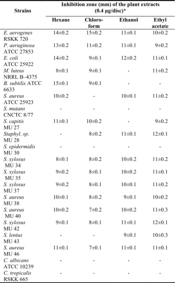
Table 2: Antimicrobial activity of S. sandrasicus extracts (0.4 μg/disc). Inhibition zone (mm) of the plant extracts
(0.4 μg/disc)* Strains Hexane Chloro-form Ethanol Ethyl acetate E. aerogenes RSKK 720 14±0.2 15±0.2 11±0.1 10±0.2 P. aeruginosa ATCC 27853 13±0.2 11±0.2 11±0.1 9±0.2 E. coli ATCC 25922 14±0.2 9±0.1 12±0.2 11±0.1 M. luteus NRRL B–4375 8±0.1 9±0.1 - 11±0.2 B. subtilis ATCC 6633 15±0.1 9±0.1 - - S. aureus ATCC 25923 10±0.2 - 10±0.1 11±0.2 S. mutans CNCTC 8/77 - - - - S. capitis MU 27 11±0.1 10±0.2 - 9±0.2 Staphyl. sp. MU 28 - 8±0.2 11±0.1 12±0.1 S. epidermidis MU 30 - - - - S. xylosus MU 34 8±0.1 8±0.2 10±0.2 11±0.2 S. xylosus MU 35 9±0.2 8±0.1 10±0.2 11±0.1 S. xylosus MU 37 9±0.2 8±0.1 10±0.1 11±0.2 S. aureus MU 38 10±0.1 8±0.2 9±0.1 10±0.2 S. aureus MU 40 10±0.2 7±0.2 10±0.2 11±0.3 S. xylosus MU 42 9±0.1 8±0.1 11±0.1 12±0.1 S. lentus MU 43 - - 9±0.1 10±0.3 S. aureus MU 46 11±0.1 7±0.1 11±0.1 11±0.1 C. albicans ATCC 10239 - - - - C. tropicalis RSKK 665 - - - - *Values represent average ± standard deviation for three replicates.
(-) : No activity.
These extracts demonstrated various antimicrobial activities on tested bacteria. The two different doses of all extracts have not effects on S. mutans, S.
epidermidis MU 30 and yeasts. The inhibition zones
of bacterial strains, sensitive to the 0.4 and 3.6 μg extracts of S. sandrasicus, were in the range of 7-15 and 10-18 mm, respectively. As it can be seen from Table 2 and 3, when the doses of extracts were increased by 9-fold, the antimicrobial activities of extracts were also increased. However, the increase on antimicrobial activities were not as much as the increase in doses of extracts.
The ethanol, hexane, chloroform and ethyl acetate extracts demonstrated antibacterial activities on
Staphylococcus strains which shows resistance to
various antibiotics. All the extracts had no effect on
S. epidermidis MU 30. The hexane extract inhibited
Table 3: Antimicrobial activity of S. sandrasicus extracts (3.6 μg/disc). Inhibition zone (mm) of the plant extracts (3.6
μg/disc)* Strains Hexane Chloro-form Ethanol Ethyl acetate E. aerogenes RSKK 720 17±0.1 18±0.2 15±0.2 13±0.2 P. aeruginosa ATCC 27853 16±0.1 14±0.1 14±0.1 13±0.1 E. coli ATCC 25922 17±0.1 13±0.1 15±0.1 15±0.1 M. luteus NRRL B–4375 12±0.2 13±0.2 - 14±0.2 B. subtilis ATCC 6633 18±0.2 12±0.1 - - S. aureus ATCC 25923 15±0.1 - 14±0.1 14±0.2 S. mutans CNCTC 8/77 - - - - S. capitis MU 27 16±0.1 14±0.1 - 13±0.2 Staphyl. sp. MU 28 - 11±0.2 16±0.3 16±0.1 S. epidermidis MU 30 - - - - S. xylosus MU 34 13±0.1 12±0.2 16±0.2 17±0.2 S. xylosus MU 35 14±0.2 12±0.1 16±0.1 16±0.2 S. xylosus MU 37 13±0.1 12±0.1 14±0.1 14±0.1 S. aureus MU 38 14±0.1 11±0.2 13±0.1 14±0.2 S. aureus MU 40 14±0.1 10±0.2 14±0.3 15±0.1 S. xylosus MU 42 13±0.2 12±0.1 14±0.1 14±0.1 S. lentus MU 43 - - 13±0.1 13±0.1 S. aureus MU 46 15±0.1 10±0.3 15±0.1 15±0.1 C. albicans ATCC 10239 - - - - C. tropicalis RSKK 665 - - - - *Values represent average ± standard deviation for three replicates.
(-) : No activity
strains, except Staphylococcus sp. MU 28, S.
epidermidis MU 30 and S. lentus MU 43. The
chloroform extract exhibited antibacterial activity on multi-resistant Staphylococcus strains, except S.
epidermidis MU 30 and S. lentus MU 43. The ethanol
extract exhibited antibacterial activity on multi-resistant Staphylococcus strains, except S. capitis MU 27 and S. epidermidis MU 30. The 0.4 µg doses of the ethyl acetate extracts inhibited the growth of all Staphylococcus strains, except S. epidermidis MU 30. The four extracts of the S. sandrasicus had antimicrobial activity on the E. coli ATCC 25922, P. aeruginosa ATCC 27853 and E. aerogenes RSKK 720. The chloroform and ethanol extracts showed antibacterial activity on E. aerogenes (cf. [5a]).
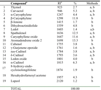
The bioactive hexane extract afforded 19 identifiable compounds by GC and GC-MS methods. Compounds
Table 4: Chemical compositions of S. sandrasicus hexane extract.
Compounda RIa % Methods 1 Thymol 923 2.7 a, b 2 Carvacrol 946 5.3 a, b 3 α-Caryophylene 1247 4.4 a, b 4 β-Caryophylene 1298 11.8 b 5 β-Ionone 1415 1.7 b 6 Dihydroactinidiolide 1559 4.8 b 7 Ledol 1603 1.4 a,b 8 Spathulenol 1636 12.5 a, b 9 Caryophyllene oxide 1647 11.6 a, b 10 Aromadendrene oxide 2 1698 13.3 b 11 Cubenol 1755 3.8 b 12 γ-Gurjenene epoxide 1761 1.6 a, b 13 tau-Cadinol 1784 3.8 a, b 14 δ-Cadinol 1793 3.6 a, b 15 Leden oxide 1801 4.0 b 16 α-Cadinol 1815 6.3 a, b 17 8-hydroxy-endo-cycloisolongifolene 1874 1.9 b 18 Hexahydrofarnesyl acetone 1957 4.3 b 19 Lupeol 2120 1.2 b TOTAL 100.00
a: co-injection with authentic compounds, b: MS,
a : In DB-5 fused silica capillary column.
were identified by comparison with reference substances, NIST 2002, Wiley and locally constructed libraries. The major compounds of hexane extract of this plant were found to be aromadendrene oxide 2 (13.3%), spathulenol (12.5%), β-caryophylene (11.8%), caryophyllene oxide (11.6%), α-cadinol (6.3%) (see Table 4). The hexane extract comprised 69.3% sesquiterpenoids. The antimicrobial activities of aromadendrene oxide 2, spathulenol and caryophyllene oxide are known [7-9]. These are thought to be responsible for the S.
sandrasicus high antimicrobial activity.
In this study, it was observed that methicillin resistant
S. aureus MU 38, MU 40 and oxacillin resistant S. aureus MU 46 were inhibited by all of the extracts.
Oxacillin resistance in both community and hospital acquired strains of staphylococci has emerged as an important and growing resistance threat [10a-10c]. Oxacillin-resistant Staphylococcus aureus (ORSA) was first recognized in the United Kingdom in 1961 after the introduction of methicillin into clinical practice, it has now become a leading cause of nosocomial infections worldwide [10d]. MRSA is currently recognized as a major problem in hospitals and the broader community in the United States and throughout the world [11]. MRSA strains are often resistant to antimicrobials other than β-lactams, thereby limiting the range of therapeutic options and increasing the risk of treatment failure as well as the costs for antimicrobial therapy and hospitalization [12]. There is an established need to develop new
antimicrobial agents to combat these pathogens [5a,13]. In this study, the extracts demonstrated antibacterial activity on some of the CNS, S. xylosus,
S. lentus, S. capitis.
Various extracts of S. sandrasicus have indicated strong activities against S. aureus and CNS, which are resistant to various antibiotics were used in this study. These extracts are possible alternative therapies for infections of the Multidrug-resistant organisms (MDROs) such as Methicillin Resistant
Staphylococcus aureus (MRSA), Vancomycin
Resistant Enterococcus (VRE), extended spectrum beta-lactamases (ESBL), and stably derepressed AmpC enzyme producers among Enterobacteriaceae, nonfermentative Gram-negative bacilli, principally P.
aeruginosa, Acinetobacter spp. and S. maltophilia.
Alternate therapies to accepted antibiotics can prove valuable. The doses of extracts used in this study were active at much lower concentrations than these accepted antibiotics. This in vitro study provides evidence that the plant studied is potentially a rich source of antibacterial agent against the multi-resistant bacteria tested.
Experimental
Plant material: Indigenous S. sandrasicus (an
Asteraceae) were collected at the flowering stage
from Mugla, Turkey. A voucher specimen was identified by Associated Professor Dr. Omer VAROL, Department of Biology, Faculty of Arts and Sciences, Mugla University, and deposited at the Herbarium of Department of Biology at the Mugla University in Turkey under number O.V. 4463.
Obtaining of the crude extracts: The air dried and
powdered aerial parts of S. sandrasicus (200 g) were extracted successively with hexane, chloroform, ethyl acetate and ethanol in a Soxhlet apparatus until the last portion of the extract became colorless. Solvents from the extracts were removed in vacuo using rotary evaporation. Each residue was re-dissolved at 20 mg/mL and 180 mg/mL with hexane, chloroform, ethanol and ethyl acetate, respectively. Crude extracts were maintained at +4ºC prior to use. Crude extracts were investigated for antimicrobial activity. Identity of the compounds from the bioactive hexane extracts was determined using GC and GC/MS.
Microorganisms and condition for cultivation:
Multi-resistant staphylococci (S. aureus MU 38, MU 40, MU 46, Staphylococcus xylosus MU 34, MU 35, MU 37, MU 42, Staphylococcus capitis MU 27,
Staphylococcus epidermidis MU 30, Staphylococcus lentus MU 43, Staphylococcus sp. MU 28) and Bacillus subtilis ATCC 6633, Escherichia coli ATCC
25922, Pseudomonas aeruginosa ATCC 27853,
Staphylococcus aureus ATCC 25923, Streptococcus mutans CNCTC 8/77, Micrococcus luteus NRRL
B-4375, Enterobacter aerogenes RSKK 720, Candida
albicans ATCC 10239, Candida tropicalis RSKK
665 were used as test microorganisms. The strains (MU coded) were obtained from Mugla University Culture Collection. The antibiotic resistance patterns of Staphylococci determined according to the recommendations NCCLS [14].
The above mentioned bacteria, except S. mutans, were cultured in Nutrient Broth (NB) (Difco) S.
mutans were cultured in Brain Heart Infusion Broth
(BHIB) (Difco), C. albicans and C. tropicalis cultured in Sabouraud Dextrose Broth (SDB) (Difco).
P. aeruginosa and the fungi were incubated at
30±0.1°C 18-24 h and 24-48 h, respectively. Other bacteria strains were incubated at 37±0.1°C 24-48 h. Inocula were prepared by adjusting the turbidity of the medium to match the 0.5 Mcfarland Standard Dilutions of this suspension in 0.1 % peptone (w/v) solution in sterile water inoculated on NB, BHIB, SDB to check the viability of the preparation. The cultures of microorganisms were maintained in their appropriate agar slants at 4°C throughout the study and used as stock cultures.
Disc diffusion assay: The antibacterial activity was
evaluated using accepted disc diffusion methodology [15a-15c] using bacterial cell suspension whose concentration was equilibrated to a 0.5 McFarland standard. A 100 μL of each bacterial suspension was spread on a Mueller–Hinton agar plate. Twenty-five µL of each extract containing 0.4 μg and 3.6 μg crude extract were injected in discs of 6 mm in diameter (Schleicher & Schuell). The discs were allowed to dry and then placed on the inoculated agar. The plates were incubated at appropriate temperature and time for microorganisms. Discs of hexane, chloroform, ethanol, ethyl acetate were used as controls. After the prerequisite incubation time a zone of inhibition was measured. The experiment was performed in triplicate and the average values are presented.
Column chromatography (CC): Firstly, CC was
performed to highlight the structure of the active extract. For CC, silica-gel 60 (70-230 mesh) as adsorbent in a column with 2x80 cm measurements
was used and mobile phases were respectively 95:5, 90:10 and 85:15 hexane: acetone systems. The fractions were purified by TLC and subjected to GC and GC-MS analysis.
Gas chromatography (GC): GC analyses of the
extract was performed using a Shimadzu GC-17 AAF, V3, 230V LV series gas chromatograph equipped with a FID and a DB-5 fused silica capillary column (30 m x 0.32 id., film thickness 0.25 µm); the initial oven temperature was held at 100oC
for 5 min, then programmed to 240oC at 3oC/min and
held at this temperature for 30 min; injector temperature and detector temperature was 250 and 270oC, respectively; carrier gas was He at a flow
rate of 1.4 mL/min; sample size, 1.0 µL; split ratio, 50:1. The percentage composition of the hexane extract was determined with Class-GC 10 computer program.
Gas Chromatography – Mass Spectrometry (GC-MS): 50 mg/mL solution of the bioactive hexane
extract was prepared for GC and GC-MS analyses. The analysis of the extract was performed using a Varian Saturn 2100, (E.I Quadrupole) equipped with a DB-5 MS fused silica capillary column (30 m x
0.32 mm id., film thickness 0.25 µm). For GC–MS detection, an electron ionization system with ionization energy of 70 eV was used. Carrier gas was helium at a flow rate of 1.7 mL/min. Injector and MS transfer line temperatures were set at 220oC and
290oC, respectively. The oven temperature was held
at 100oC for 5 min, then increased up to 240oC with
3oC/min increases and held at this temperature for 25
min. Diluted samples (1/100, v/v, in methylene chloride) of 1.0 mL were injected manually in the splitless mode. The relative percentage of the extract constituents was expressed as percentages by peak area normalization.
Identification of components of the extract was computer matching of mass spectra with those of standards (NIST 2002, Wiley library data of GC–MS systems and a locally customized library of 320 spectra), as well as by comparison with the fragmentation patterns of the mass spectra with those reported in the literature [15d] and, whenever possible, by co-injection with authentic compounds.
Acknowledgments - This work was supported by
Mugla University Research Funds.
References
[1] (a) de Mattos EM, Teixeira LA, Alves VM, Rezenda e Resende C, Da Silva Coimbra MV, Da Silva-Carvalho MC, Ferreira-Carvalho BT, Figueiredo AM. (2003) Isolation of methicillin-resistant coagulase-negative staphylococci from patients undergoing continuous ambulatory peritoneal dialysis (CAPD) and comparison of different molecular techniques for discriminating isolates of
Staphylococcus epidermidis. Diagnostic Microbiology and Infectious Diseases, 45, 13–22; (b) Casey AL, Lambert PA, Elliott TSJ.
(2007) Staphylococci. International Journal of Antimicrobial Agents, 27, S23-S32; (c) Yugueros J, Temprano A, Berzal B, Sánchez M,Hernanz C, LuengoJM, Naharro G. (2000) Glyceraldehyde-3-Phosphate Dehydrogenase-Encoding Gene as a Useful Taxonomic Tool for Staphylococcus spp. Journal of Clinical Microbiology, 38, 4351-4355.
[2] (a) Hajjeh RA, Reingold A, Weil A, Shutt K, Schuchat A, Perkins BA. (1999) Toxic shock syndrome in the United States: surveillance update, 1979–1996. Emerging Infectious Diseases, 5, 807–810; (b) Rubin RJ, Harrington CA, Poon A, Dietrich K, Grene JA, Moiduddin A. (1999) The economic impact of Staphylococcus infection in New York City hospitals. Emerging
Infectious Diseases, 5, 9–17; (c) Livermore DM. (2000) Antibiotic resistance in staphylococci. International Journal of Antimicrobial Agents, 16, 3–10; (d) Kloos WE, Bannerman TL. (1995) Staphylococcus and Micrococcus. In: Murray PR, Baron EJ,
Pfaller MA, Tenover FC, Yolken RH, eds., Manual of clinical microbiology. 6th ed, ASM Press, Washington, D.C. USA, pp. 282–298.
[3] (a) Ali-Shtayeh MS, Yaghmour RM-R, Faidi YR, Salem K, Al-Nuri MA. (1998) Antimicrobial activity of 20 plants used in folkloric medicine in the Palestinian area. Journal of Ethnopharmacology, 60, 265- 271; (b) Primo V, Rovera M, Zanon S, Oliva M, Demo M, Daghero J, Sabini L. (2001) Determination of the antibacterial and antiviral activity of the essential oil from
Minthostachys verticillata (Griseb.) Epling. Revista Argentina de Microbiologia, 33, 113-117.(c) Şahin F, Karaman I, Güllüce M,
Oğütçü H, Şengül M, Adıgüzel A, Ozturk S, Kotan R. (2003) Evaluation of Antimicrobial Activities of Satureja hortensis L.
Journal of Ethnopharmacology, 87, 61–65.
[4] (a) Davis PH. (1982) Flora of Turkey and the East Aegean Islands, Vol 7, Edinburgh University Press, Edinburgh; (b) Baytop T. (1997) Turkce Bitki Adlari Sozlugu (2. Baski). Ataturk Kultur, Dil ve Tarih Yuksek Kurumu Turk Dil Kurumu Yayinlari, Ankara; (c) Baytop T. (1999) Turkiye’de Bitkiler ile Tedavi (Gecmiste ve Bugun). Istanbul, Nobel Tip Kitabevleri.
[5] (a) Ugur A, Ertem H, Beyatli Y. (2006) Antibacterial Properties of Senecio sandrasicus on Multidrug-Resistant Stenotrophomonas
maltophilia. Pharmaceutical Biology, 44, 253–257; (b) Bardón A, Borkosky S, Ybarra MI, Montanaro S, Cartagena E. (2007)
Bioactive plants from Argentina and Bolivia. Fitoterapia, 78, 227–231; (c) Loizzo MR, Tundis R, Statti GA, Houghton PJ. (2006) Bioactive extracts from Senecio samnitum Huet. Natural Product Research, 20, 265–269.
[6] (a) Shale TL, Stirk WA, van Staden J. (1999): Screening of medicinal plants used in Lesotho for bacterial and anti-inflammatory activity. Journal of Ethnopharmacology, 67, 347–354; (b) Garcia Navarro VM, Gonzales AC, Fuentes M, Aviles M, Rios MY, Zepeda G, Rojas MG. (2003) Antifungal activities of nine traditional Mexican medicinal plants. Journal of
Ethnopharmacology, 87, 85–88; (c) Fortin H, Vigor C, Lohezic-Le Devehat F, Robin V, Le Bosse B, Boustie J, Amoros M. (2002)
In vitro antiviral activity of thirty-six plants from La Réunion Island. Fitoterapia, 73, 346–350; (d) Conforti F, Loizzo MR, Statti GA, Houghton PJ, Menichini F. (2006) Biological properties of different extracts of two Senecio species. International Journal of
Food Science and Nutrition, 57, 1–8; (e) Hong Q, Minter DE, Franzblau SG, Reinecke MG. (2008) Anti-tuberculosis compounds
from two Bolivian medicinal plants, Senecio mathewsii and Usnea florida, Natural Product Communications, 3, 1377-1384; (f) Waako PJ, Katuura E, Smith P, Folb P. (2007) East African medicinal plants as a source of lead compounds for the development of new antimalarial drugs. Plant Sciences in Development, 45, 102-106.
[7] Nardi U. (1996) Stevia rebaudiana:analisi su un dolcificante naturale. Erboristeria Domani, 6, 58-64.
[8] Ulubelen A, Topcu G, Eris C, Sonmez U, Kartal M, Kurucu S, Bozok-Johansson C. (1994) Terpenoids from Salvia sclarea.
Phytochemistry, 36, 971-974.
[9] Tzakou O, Skaltsa H. (2003) Composition and antibacterial activity of the essential oil of Satureja parnassica subsp. parnassica.
Planta Medica, 69, 282-284.
[10] (a) Karchmer AW. (2000) Nosocomial blood stream infections: Organisms, risk factors and implications. Clinical Infectious
Diseases, 31(Suppl. 4), S139 –S143; (b) Centers for Disease Control and Prevention. (2003) Public health dispatch: Outbreaks of
community-associated methicillin-resistant Staphylococcus aureus skin infections-Los Angeles County, California, 2002–2003.
Morbidity and Mortality Weekly Reports, 52, 88; (c) Centers for Disease Control and Prevention. (2003) Methicillin-resistant Staphylococcus aureus in correctional facilities—Georgia, California, and Texas, 2001–2003. Morbidity and Mortality Weekly Reports, 52, 992–996; (d) Pottumarthy S, Fritsche TR, Jones RN. (2005) Evaluation of alternative disk diffusion methods for
detecting mecA mediated oxacillin resistance in an international collection of staphylococci: Validation report from the SENTRY Antimicrobial Surveillance Program. Diagnostic Microbiology and Infectious Diseases, 51, 57–62.
[11] Diekema DJ, Pfaller MA, Schmitz FJ, Smayevsky J, Bell J, Jones RN, Beach M, Sentry Partcipants Group. (2001) Survey of infections due to Staphylococcus species: frequency of occurrence and antimicrobial susceptibility of isolates collected in the United States, Canada, Latin America, Europe, and the Western Pacific region for the SENTRY Antimicrobial Surveillance Program, 1997–1999. Clinical Infectious Diseases, 32(suppl 2), S114-32.
[12] Bagcigil FA, Moodley A, Baptiste KE, Jensen VF, Guardabassi L. (2007) Occurrence, species distribution, antimicrobial resistance and clonality of methicillin- and erythromycin-resistant staphylococci in the nasal cavity of domestic animals. Veterinary
Microbiology, 121, 307–315.
[13] Pesewu GA, Cutler RR, Humber DP. (2008) Antibacterial activity of plants used in traditional medicines of Ghana with particular reference to MRSA. Journal of Ethnopharmacology, 116, 102–111.
[14] National Committee for Clinical Laboratory Standards (NCCLS). 1999. Performance Standarts for Antimicrobial Susceptibility Testing, Ninth Informational Supplement. NCCLS document M100-S9. NCCLS: Pennsylvania, USA.
[15] (a) Bauer AW, Kirby WM, Sherris JC, Turck M. (1966) Antibiotic susceptibility testing by a standardized single disk method.
American Journal of Clinical Pathology, 45, 493–496; (b) Collins CH, Lyne PM, Grange JM. (1995) Microbiological Methods.
Seventh ed. London, Butterworths; (c) Murray PR, Baron EJ, Pfaller NA. (1995) Manual of Clinic Microbiology, D.C, ASM Press, Washington; (d) Adams RP. (2001) Identification of Essential Oils Components by Gas Chromatography/ Quadrupole Mass Spectroscopy. Allured Publishing Corporation, Illinois, USA.