Доклади на Българската академия на науките Comptes rendus de l’Acad´emie bulgare des Sciences
Tome 73, No 8, 2020
MEDICINE Experimental medicine
APOPTOTIC EFFECTS OF PUNICALAGIN ON PARTIALLY HEPATECTOMIZED RAT LIVER
¨
Oge Artagan#, Filiz Alanyalı∗, Mediha Canbek∗∗
(Submitted by Academician B. Petrunov on June 21, 2019)
Abstract
The antioxidant potential of pomegranate is attributed to its polyphenols, including punicalagin. In this study, we searched the effects of punicalagin on 70% partially hepatectomized rats. Animals were subjected to partial hepatec-tomy (PH), immediately after receiving 15 mg/kg punicalagin. They were dis-sected at different time points (3 h, 6 h) following surgery. NF-kappaB (NFκB), caspase-3 gene expression results were evaluated by RT-PCR and protein ex-pression fold changes were evaluated according to the qPCR method. Apoptotic index results were compared between groups.
Gene expression levels of experimental groups necessarily correlate with protein expression levels. Results of RT-PCR (p < 0.05), qPCR (p < 0.05) and apoptotic index (p < 0.05) which belong to Caspase-3 gene region, have shown that 15 mg/kg of punicalagin induced apoptosis in a time-dependent manner despite the partial hepatectomy (group V–VI). Six hours after PH in group VI, apoptosis reached highest level with the injection of punicalagin. Activation of NFκB, at the end of the 6th hour in group IV, has demonstrated a significant increase compared to the other groups. After PH, 15 mg/kg of punicalagin has time-dependently suppressed regeneration and induced apoptosis. The results of the study showed that apoptosis induction could be associated with the caspase-3 increased activation. The liver, apoptotic index values triggered in group VI at the end of the 6th hour after PH. According to the results, 15 mg/kg intraperitoneally (IP) punicalagin application induced liver apoptosis despite the induction of liver regeneration (by partial hepatectomy).
#
Corresponding author.
This research was supported by Anadolu University Scientific Research Projects Unit (Project Number: 1202F057).
Key words: NFκB, caspase-3, RT-PCR, TUNEL method, Taq-Man pro-tein assay, punicalagin
1. Introduction. The treatment alternatives in curing cancer, which has seriously threatened human life, fail to satisfy expectations. Therefore, protection from cancer has been a vital strategy [1]. Epidemiological studies show that
consumption of fruits and vegetables with high phenolic contents correlates with reduced cardiovascular, cerebrovascular diseases, and reduced risk of cancer [2–4].
Anticancer effects of phenolic contents include antioxidant, anti-inflammatory, and antiproliferative properties [4–6].
Punica granatum L. (Pomegranate) has been traditionally used as medicine for the treatment of various diseases in lots of countries [3]. Pomegranate is a
rich polyphenol source that takes its red colour from hydrolyzable tannins such as anthocyanins, ellagic acid, and punicalagin [5]. Pomegranate has also
anti-oxidant [2], anti-inflammatory [6], anti-arteriosclerotic [7–9], anti-tumour
activ-ity [7], antiproliferative, pro-apoptotic effects [4]. Punicalagin is the major
antiox-idant polyphenol in pomegranate [5, 6, 10].
The liver is one of the organs of the body with the most crucial function. Any functional disorder that may occur in the liver influences all the systems in the body. Various factors such as chemical contaminators, medicines, alcohol, phys-ical damage, liver tumours, viral infections, tumours, and surgphys-ical applications can lead to injuries in the liver tissue. PH in the liver is generally implemented in massive lesions (metastasis, liver tumours, hemangioma, etc.) and liver trans-plantation to any other living being. The liver has the potential to be able to rejuvenate itself entirely against the cell losses after hepatectomy.
Apoptosis begins with the production of the apoptotic signal within the cell which starts to undergo biochemical and morphological modifications. PH is a well-regulated model for the evolution of apoptosis and cell proliferation together during liver regeneration [11]. After PH, mitotic activity rises in the liver tissue.
Most residual hepatocytes enter the cell cycle simultaneously. In the first phase, hepatocytes undergo a transition from resting state (G0) to (G1). The first phase
lasts approximately 4–6 h after PH. DNA synthesis starts around 18 h and peaks at 24 h after PH [1, 11, 12]. The growth of the liver due to the increase in mitotic
ac-tivity continues until the liver lobes reach the dimensions of an adult’s liver lobеs. Between 24 and 30 h after 70% hepatectomy, it has been observed that mitosis has commenced, and the regeneration has been completed in 7–10 days [13–15].
Rejection of 70% of the mass of the liver in rats is considered an optimum ratio in terms of regenerative induction, and it is supposed that a PH to be realized in a lower or upper rate compared to this optimum ratio can inhibit the regeneration [1, 11, 14, 16]. After PH, the remaining hepatocytes synchronously enter
the cell cycle. Those hepatocytes pass to the G1 phase, which has the potential
6 h after hepatectomy. Around 18 h after PH cells commence DNA synthesis and it is completed about 24 h after PH [1].
Induction of liver regeneration with PH (70%) and upon this induction, his-tological and genomic investigation of the apoptotic effect of punicalagin on the rat liver constitutes the basis for the experiment model. Changes in mRNA ex-pressions of Caspase-3 and NFκB genes have been figured out on rat livers at which regeneration has been induced by the method of PH. The effects of proba-ble changes in mRNA levels on liver histology have been researched by the tunnel (terminal deoxynucleotidyl transferase-mediated nick end-labeling) method.
TaqMan Protein Assay allows relative quantitation of specific proteins. The method indicates three main steps: binding, ligation, and amplification. The first step starts with the incubation of protein samples with oligonucleotide probe pair. The protein assay probes are protein-specific antibodies conjugated to oligonu-cleotides with a biotin-streptavidin linkage. The second step indicates ligation of the oligonucleotide probes with the help of DNA ligase. The last step indicates the amplification of oligonucleotides and analyses by real-time PCR with the TaqMan probe. The ligation product is used as a template in the TaqMan real-time PCR assay [17].
2. Material and methods. 2.1. Chemicals. Punicalagin (≥ 98% HPLC) was purchased from Sigma. PureLink RNA Mini Kit, Paris Kit, and TRIzol were obtained from Ambion. High Capacity cDNA Reverse Transcription Kit and TaqMan Gene Expression Master Mix were obtained from Applied Biosys-tems. ApopTag Plus Peroxidase InSitu Apoptosis Detection Kit was obtained from Millipore.
2.2. Animals. Wistar albino male rats weighing 190–250 g were used for the experiment. Animals were housed in polycarbonate cages, kept in an air-conditioned atmosphere at 22◦C with a 12 h light/dark cycle. All animals were
fed the standard commercial diet with tap water. Wistar albino male rats were obtained from the T. R. Ministry of Health, Refik Saydam National Public Health Agency, Experiment Animals Production Laboratory. At the end of 7-day acclima-tization rats were randomly divided into six groups, and each group consisted of seven rats. The experiments were approved by the Institutional Ethical Commit-tee for Animal Care and Use at the Eski¸sehir Osmangazi University, Eski¸sehir, Turkey.
Punicalagin was dissolved (15 mg/kg body weight) in distilled water and applied intraperitoneally immediately after the operation to the partially hepate-ctomized rats. All animals were submitted to a 70% PH and dissected at 3 and 6 h after surgery. Hepatectomized rats were housed in separate cages with free access to water and chow until the dissection time. PH (70%) was performed, according to Higgins and Anderson [18].
2.3. Experimental design.
Group II:Sham-operated group, dissected 6 h after surgery.
Group III: Hepatechtomized and immediately after applied 0.09% NaCl solution (SF). Dissected 3 h after surgery.
Group IV:Hepatectomized and immediately after applied 0.09% NaCl so-lution (SF). Dissected 6 h after surgery.
Group V: Hepatectomized and immediately after applied 15 mg/kg puni-calagin. Dissected 3 h after surgery.
Group VI: Hepatectomized and immediately after applied 15 mg/kg puni-calagin. Dissected 6 h after surgery.
2.4. RNA extraction. Liver tissue pieces were immersed in RNAlater and then frozen. Snap-frozen tissues were homogenized. High-quality total RNA was extracted using Trizol (Applied Biosystems) according to the kit protocol.
2.5. TaqMan gene expression assay. RNA isolation Kit and TaqMan Gene Expression Assay Kit, TaqMan® Gene Expression Master Mix, High
Ca-pacity cDNA Reverse Transcription Kit, RNAlater were obtained from Applied Biosystems. Gene expression assays were performed according to the kit protocol, observing the amplification plots for all experimental groups. Gene expression lev-els between control and experimental groups were determined by using the ∆∆CT
values [17].
2.6. TaqMan protein assay. TaqMan® Protein Assays Open Kit, Taq-Man Protein Expression Core Reagents Kit, TaqTaq-Man® Protein Assays Fast
Mas-ter Mix, All TaqMan®were purchased from Applied Biosystems. Protein Assays
were performed according to the kit protocol. High-quality total RNA was ex-tracted from the liver tissue using Trizol (Applied Biosystems). qPCR assays were performed on a StepOnePlus™ Real-Time PCR System (Applied Biosys-tems). TaqMan® Protein Assays with rat liver tissue samples were performed
using 500 ng/µL protein in each well. Data were analyzed using Protein Assist TM 1.0 (Applied Biosystems, Foster City, CA).
2.7. TUNEL staining (analysis of apoptotic nuclei by immunocyto-chemistry). Apoptotic nuclei of rat liver sections were visualized by in situ nick-end labellings with the use of the ApopTag Plus in situ apoptosis detection kit (Apop Tag Peroxidase Kit) according to the kit protocol.
3. Results and discussion. 3.1. TaqMan® protein assay provides rel-ative protein quantification of the sample. Proteins can typically be detected in reactions containing 500 ng µL−1. The TaqMan Protein Assay has a simpler and
faster way of western blotting process [17].
TaqMan protein assay consists of antibody-protein binding with real-time PCR-based detection of oligonucleotide sequence [19]. The reaction contains three
main steps: 1. Binding of an antibody linked oligonucleotide probes to a spe-cific protein in the sample. 2. Ligation of oligonucleotide probes by DNA ligase. 3. Amplification of the template and analysis by real-time PCR with TaqMan probe [17].
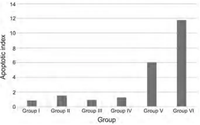
Fig. 1. Apoptotic index values of groups: Asym Sig = 0.001; P < 0.05
3.2. Hepatocellular apoptosis index. In the sections of the liver where TUNEL staining was performed the apoptotic index was derived by proportioning the number of apoptotic cells in the randomly selected five-section areas to the number of cells having normal physiology. Changes in the apoptotic index between the experimental groups were determined. According to results, it was found out that the highest apoptotic index value was detected in group VI, and the lowest apoptotic index was detected in group I and III. After PH, it has been observed that exposure of liver cells to punicalagin has induced apoptosis. Group VI has shown a significant difference compared to group V (P < 0.05). In group VI, it has been seen that implementation of 15 mg/kg [2] punicalagin just after PH has
induced apoptosis (Fig. 1).
3.3. RT-PCR. Analyzing the results of caspase-3 gene expressions, differ-ences between groups have been found statistically significant. In group VI, caspase-3 gene expression has differed significantly when compared to group V and group IV. In groups V and VI, caspase-3 activity has displayed a significant increase in a time-dependent manner (Fig. 2).
Fig. 2. Caspase-3 gene expression graph of groups (n=7) 14 12 x 10 Q) "O .S 8 u '= 0 6 a.
I
0 a. <l:: 4 2 0-
•
•
•
Group I Group II Group Ill Group IV Group V Group VI Group 18 Caspaise-3 16 14 C: ·
..
~ 12 [ 10 ><I
., 8 ., "' 6 " (!) 4 2I
0-
•
•
Groopl Group II Gro~p Ill Group IV Group V Group VI
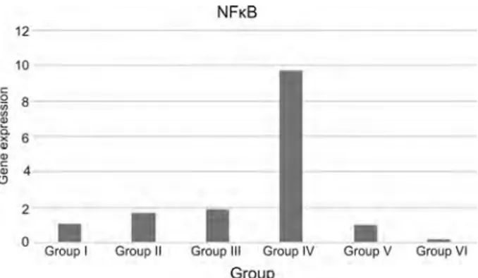
Fig. 3. NFκB gene expression graph of groups (n = 7)
As a result of the comparison of NFκB gene expression between the groups, in group III, there was an increase but the level of NFκB in group IV was quite high. NFκB gene expression level has a significant rise in group IV. In group V and VI, punicalagin application after PH has suppressed NFκB gene expression level time-dependently (Fig. 3).
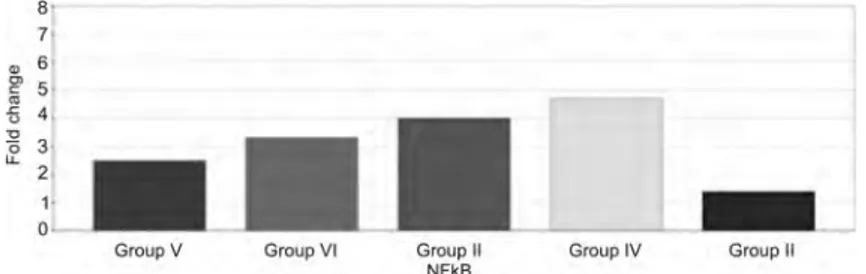
3.4. qPCR.Caspase-3 protein expression has indicated an increase in group V compared to groups I, II, III, and IV. Caspase-3 protein expression has reached its peak value at the end of the 6th hour (group VI). As a result of application of IP punicalagin after PH, caspase-3 protein expression has increased reasonably at the end of the 3rd hour; however, at the end of the 6th hour, a considerable increase was observed (Fig. 4). NFκB protein expression in group IV has differed signif-icantly compared to the other experimental groups. In group III, NFκB protein expression has started to increase at the end of the 3rd hour, and in group IV that value reached its maximum. In group V, NFκB protein expression was suppressed. In group VI, NFκB protein expression was further suppressed in a time-dependent manner (Fig. 5).
4. Conclusion. In this study, the apoptotic effects of punicalagin on rat liver after PH have been investigated in vivo. We used the TaqMan protein assay (a new method for relative protein quantification) and the TaqMan gene expression
Fig. 4. Caspase-3 protein expression graph of groups (n = 7). Group I, accepted as a reference 12 10 C: 0 8 ·;;; ~ ~ 6 ., ., C: 4 " (.!) 2 0
•
Group I >20.0 20.0 17.5 " 15.0 g, "' 12.5 £ 10.0 'O 0 7.5 u. 0.0 Group V•
Group II Group VI NFKBI
Group Ill Group IV
Group Group II Caspase-3
•
Group V Group VI Group IV Group IIFig. 5. NFκB protein expression graph of groups (n = 7). Group I, accepted as a reference
assay. The goal is to induce apoptosis with punicalagin injection on the rat liver, where regeneration was induced experimentally by PH.
We investigated whether punicalagin triggered apoptosis in the hepatocytes of hepatectomized liver tissue. Results demonstrated that the punicalagin appli-cation triggered apoptosis in hepatectomized rat liver in a short period of time (3 and 6 h). Punicalagin-induced apoptosis was associated with repression of the activity of NFκB and up-regulation of caspase-3 activity.
Caspase-3 is the essential killer and a sign of early apoptosis [20]. Caspase-9
and caspase-8 cleave procaspase-3 and generate the active caspase-3 that serves as the crucial executioner of apoptotic cell death. Caspase-3 activates the other caspases and breaks the cytoskeleton [21]. The activity of caspase-3 also reached
the maximum level. TaqMan Protein analysis showed that NFκB protein expres-sion level increased 6 h after PH without punicalagin injection. The increase in NFκB was followed by a decrease of caspase-3 6 h after PH in SF injected groups. While PH increased the NFκB activation, punicalagin induction triggered caspase-3 activation. Results revealed that punicalagin caused a significant induction of apoptosis in the hepatectomized liver section.
TUNEL-positive staining results showed typical morphological features of apoptotic nuclei in the punicalagin applied on hepatectomized rat liver. The identification of stained apoptotic bodies was confirmed by specific morphological features, including nuclear fragmentation, cytoplasmic shrinkage, and dissociation from other cells in the section [22]. The results of in situ end-labelling showed the
appearance of apoptotic bodies in the regenerating liver, DNA fragmentation was observed as early as 3 h after PH with punicalagin. Apoptotic bodies in the regenerating liver sections increased significantly at the end of the 6th hour after PH.
Results revealed that caspase-3 activity was seen as a maximum level whereas NFκB activity was suppressed in group VI.
Finally, according to the results, punicalagin induced apoptosis and depressed cell proliferation despite the induction of regeneration (by partial hepatectomy) in the liver tissue. The apoptotic effect comparison of punicalagin against cancer cell lines and healthy cell lines will provide new aspects to develop novel approaches.
8 7 " 6 0 ) 5 C ., -fi 4 "0 3 0 u. 2 1• 0
-Group V Group VI Group II Group IV Group II
Statistical analysis. All data were evaluated using non-parametric tests (Kruskal–Wallis and Kolmogorov–Smirnov). SPSS 20.0 (SPSS: Statistical Pack-age for the Social Sciences) was used for assessing the significance of differences between groups. Differences were considered significant P < 0.05.
REFERENCES
[1] Chagas C. E. A., A. Vieira, T. P. Ong, F. S. Moreno (2009) Farnesol inhibits cell proliferation and induces apoptosis after partial hepatectomy in rats, Acta Cirurgica Brasileira, 24(5), 377–382.
[2] Hertog M. G. L., P. M. Sweetnam, A. M. Fehily, P. C. Elwood, D. Krom-bout (1997) Antioxidant flavonols and ischemic heart disease in a Welsh population of men: the Caerphilly Study, Am. J. Clin. Nutr., 65, 1489–1494.
[3] Kulkarni P. K., S. M. Aradhya, S. Divakar (2004) Isolation and identification of a radical scavenging antioxidant-punicalagin from pith and carpellary membrane of pomegranate fruit, Food Chem., 87, 551–557.
[4] Seeram N. P., L. S. Adams, S. M. Henning, Y. Niu, Y. Zhang (2005) In vitro antiproliferative, apoptotic and antioxidant activities of punicalagin, ellagic acid and a total pomegranate tannin extract are enhanced in combination with other polyphenols as found in pomegranate juice, J. Nutrit. Biochem., 16(6), 360–367. [5] Bell C., S. Hawthorne (2007) Ellagic acid, pomegranate and prostate cancer,
J. Pharm. Pharmacol., 60, 139–144.
[6] Adams L. S., N. P. Seeram, B. B. Aggarwal, Y. Takada, D. Heber (2006) Pomegranate juice, total pomegranate ellagitannins, and punicalagin suppress in-flammatory cell signaling in colon cancer cells, J. Agricult. Food Chem., 54, 980– 985.
[7] Aviram M., L. Dornfeld (2001) Pomegranate juice consumption inhibits serum angiotensin converting enzyme activity and reduces systolic blood pressure, Atherosclerosis, 158, 195–198.
[8] Kaplan M., M. Aviram (2001) Retention of oxidized LDL by extracellular matrix proteoglycans leads to its uptake by macrophages: An alternative approach to study lipoproteins cellular uptake, Arterioscler. Thromb. Vasc. Biol., 21, 386–393. [9] Nigris F., S. Williams-Ignarro, L. O. Lerman, E. Crimi, C. Botti et al.
(2005) Beneficial effects of pomegranate juice on oxidation-sensitive genes and en-dothelial nitric oxide synthase activity at sites of perturbed shear stress, Proc. Nat. Acad. Sci. USA, 102, 4896–4901.
[10] Gil M. I., F. A. Tom´as-Barber´an, B. Hess-Pierce, D. M. Holcroft, A. A. Kader (2000) Antioxidant activity of pomegranate juice and its relationship with phenolic composition and processing, J. Agric. Food Chem., 10, 4581–4589. [11] ¨Ozeki A., I. Tsukamoto (1999) Retinoic acid repressed the expression of c-fos
and c-jun and induced apoptosis in regenerating rat liver after partial hepatectomy, Biochim. Biophys. Acta, 1450(3), 308–319.
[12] Fausto N. (2000) Liver regeneration, J. Hepatol., 32(1), 19–31.
[13] Ehrenfried J. A., T. C. Ko, E. A. Thompson, B. M. Evers (1997) Cell cycle-mediated regulation of hepatic regeneration, Surgery, 122(5), 927–935.
[14] Palmes D., H. U. Spiegel (2004) Animal models of liver regeneration, Biomate-rials, 25, 1601–1611.
[15] Taub R. (2004) Liver Regeneration: From Myth to Mechanism, Nat. Rev. Mol. Cell Biol., 5, 836–847.
[16] Borowiak M., A. N. Garratt, T. W¨ustefeld, M. Strehle, C. Trautwein et al. (2004) Met provides essential signals for liver regeneration, Proc. Nat. Acad. Sci. USA, 101, 10608–10613.
[17] Swartzman E., M. Shannon, P. Lieu, S. M. Chen, C. Mooney et al. (2010) Expanding applications of protein analysis using proximity ligation and qPCR, Methods, 50, 23–26.
[18] Higgins G. M., R. M. Anderson (1931) Experimental pathology of the liver. I. Restoration of the liver of the white rat following partial surgical removal, Arch. Pathol., 12, 186–202.
[19] Gullberg M., S. M. G´ustafsd ´ottir, E. Schallmeiner, J. Jarvius, M. Bjarneg˚ard et al. (2004) Cytokine detection by antibody-based proximity lig-ation, Proc. Nat. Acad. Sci. USA, 101(22), 8420–8424.
[20] Pfister C., H. Pfrommer, M. S. Tatagiba, F. Roser (2013) Detection and quantification of farnesol-induced apoptosis in difficult primary cell cultures by TaqMan protein assay, Apoptosis, 18, 452–466 .
[21] Khan N., F. Afaq, H. Mukhtar (2007) Apoptosis by dietary factors: the suicide solution for delaying cancer growth, Carcinogenesis, 28, 233–239.
[22] Suzuki T., I. Tsukamoto (2004) Apoptosis induced by 5-(N,N-hexamethylene)-amiloride in regenerating liver after partial hepatectomy, Eur. J. Pharmacol., 503, 1–7.
Medical Service and Techniques Department Vocational School of Health Service
Mu˘gla Sıtkı Ko¸cman University 48700 Marmaris, Mu˘gla, Turkey e-mail: [email protected]
∗Department of Biology
Faculty of Science Anadolu University 26470 Eski¸sehir, Turkey e-mail: fsusuz@eski¸sehir.edu.tr
∗∗Department of Biology
Faculty of Science Eski¸sehir Osmangazi University
26480 Eski¸sehir, Turkey e-mail: [email protected]