Cumhuriyet Science Journal
CSJ
e-ISSN: 2587-246X
ISSN: 2587-2680 Cumhuriyet Sci. J., Vol.40-4 (2019) 830-837
* Corresponding author. Email address: [email protected]
http://dergipark.gov.tr/csj ©2019 Faculty of Science, Sivas Cumhuriyet University
On fluorescent sensing of metal ions using water extracts of Salvia officinalis
Idris SARGIN1,*
1Department of Biochemistry, Faculty of Science, Selcuk University, 42075 Konya, Turkey
Received: 02.07.2019; Accepted: 14.11.2019 http://dx.doi.org/10.17776/csj.585503
Abstract. Sensing of metal ions using fluorometric tools has wide applications in chemical, biological and environmental analysis. Plant phytochemicals, like flavonoids, exhibit intense fluorescence upon excitation by UV light. Leaves sage (Salvia officinalis), which is rich in polyphenolic and flavonoids compounds, were extracted using Soxhlet and microwave-assisted extractors. The extraction methods led to variations in the phytochemical composition of the extracts, which in turn affected their interaction with metal ions. Despite the variations in the composition, both of the extracts gave high fluorescence emissions when excited at 365 nm. Variations in fluorescence emissions of the extracts were studied in upon addition of each metal ion; i.e., Li+,
Na+, K+, Cs+, Be2+, Mg2+, Sr2+, Ba2+, Al3+, Tl3+, Ge4+, Sn4+, Pb2+, Sb3+, Bi3+, Se4+, Cu2+, Ag+, Zn2+, Cd2+, Ti4+,
Cr3+, Cr6+, Mo6+, W6+, Mn2+, Fe3+, Ni2+, Co2+ and Pd2+. When they were added into the Soxhlet extract, some
ions (Cr3+, Pb2+, Co2+) induced intense fluorescence and some (Ge4+, Mg2+, K+, Na+) ions quenched the
fluorescence emission. As for microwave-assisted extract, the addition of Sr2+, Mg2+ and Co2+ ions enhanced
the fluorescence emission of the extract, but Fe3+, Be2+ and Cs+ lowered the fluorescence intensity. However,
the results of the study should be considered as introductory and further selectivity and sensitivity studies should be done for each extract if they are used for sensing of metal ions. Yet, this study demonstrated that sage extracts has a potential for fluorescent sensing of certain metal ions.
Keywords:Sage; Flavonoid; Metal ion sensing; Soxhlet extraction; Microwave irradiation.
Salvia officinalis'in su özütlerini kullanarak metal iyonlarının floresanla
algılanması
Özet. Metal iyonlarının florometrik araçlar kullanarak algılanması kimyasal, biyolojik ve çevresel analizlerde
geniş uygulamalara sahiptir. Flavonoidler gibi bitki fitokimyasalları, UV ışığı ile uyarıldıklarında yoğun floresans ışıma yaparlar. Polifenolik bileşikler ve flavonoidler bakımından zengin olan adaçayı (Salvia
officinalis) yaprakları Soxhlet ve mikrodalga ekstraktörler kullanılarak özütlendi. Ekstraksiyon yöntemleri,
özütlerin fitokimyasal kompozisyonunda değişikliklere neden olmuştur, bu da metal iyonlarıyla etkileşimlerini etkilemiştir. Kompozisyondaki değişikliklere rağmen, özütlerin her ikisi de 365 nm'de uyarıldığında yüksek floresans emisyonu vermiştir. Özütlerin floresans emisyonundaki değişmeler her bir iyonun ilave edilmesiyle çalışıldı; Li+, Na+, K+, Cs+, Be2+, Mg2+, Sr2+, Ba2+, Al3+, Tl3+, Ge4+, Sn4+, Pb2+, Sb3+, Bi3+, Se4+, Cu2+, Ag+, Zn2+,
Cd2+, Ti4+, Cr3+, Cr6+, Mo6+, W6+, Mn2+, Fe3+, Ni2+, Co2+ ve Pd2+. Soxhlet özütüne ilave edildiğinde, bazı iyonlar
(Cr3+, Pb2+, Co2+) yoğun floresansa neden olmuş, bazı (Ge4+, Mg2+, K+, Na+) iyonlar ise floresans emisyonunu
söndürmüştür. Mikrodalga destekli özütte, Sr2+, Mg2+ ve Co2+'nın ilavesi, özütün floresans emisyonunu arttırdı,
ancak Fe3+, Be2+ ve Cs+ floresan şiddetini düşürdü. Bununla birlikte, çalışmanın sonuçları giriş niteliğinde
olarak değerlendirilmeli ve metal iyonlarını algılamak için kullanılacaksa her özüt için seçicilik ve duyarlılık çalışmaları yapılmalıdır. Yine de, bu çalışma adaçayı özütlerinin belirli metal iyonlarının floresan algılama potansiyelinin olduğunu göstermiştir.
1.
INTRODUCTIONExtracts from plants have been subject of intensive studies because of their wide range of
pharmacological activities. Sage (Salvia
officinalis) is a medicinal and herbal plant [1]. Its biological activities are attributed to its phenolic compounds including carnosic acid, carnosol, rosmarinic acid, diterpenes, triterpenes and flavonoids [2, 3].
Apart from therapeutic activities and physiological importance [4], flavonoids, one group of the active ingredients in S. officinalis extracts, also display important characteristics [5]. These polyphenolic phytochemicals emit brilliant fluorescence when excited by UV light [6].
Detection of metal ions using fluorescence spectrometry is a simple and powerful technique in analytical chemistry. Designing fluorescent and water-soluble chemosensors for sensing metal ions in aqueous environments is of importance. Water-soluble and natural fluorescent chemosensors are needed for detection of metal ions by fluorescence spectrometry [7, 8].
Natural compounds as fluorescent probes for metal ions have attracted considerable attention in the past few years. For example, a plant alkaloid berberine was isolated from the stems of Mahonia
leschenaultti and then used for detection of Ag+ ion
[9]. In one study natural Isorhamnetin from Ginkgo leaves was applied for determination of Cu2+ in
samples from rivers, lakes, vegetables and fruits [10]. In another study, a simple and green analytical procedure based on chlorophyll a was developed by Gao and et al., (2006) [11]. The authors reported the extraction and purification of chlorophyll a from the leaves of pea and use of chlorophyll a fluorometric detection of Hg2+ ion. A
more recent study by Ahmad et al., (2018) reported that flavonoid containing methanolic extract of Corchorus depressus could be used as a spectrofluorometric assay for the detection of Benzo[a]pyrene [12]. Flavonoids also display affinity for metal ions by forming metal–flavonoids complexes. Flavonoids are able to form fluorescent chelates with a variety of metal ions [13].
This study aimed to test whether it is possible to
develop a simple, cheap and fast
spectrofluorometric chemosensor based on the water extract of common sage (S. officinalis) for detection of metal ions. In the study, two extraction
procedures were used to see the effect of the extraction method on the composition of the S. officinalis extracts; the Soxhlet extraction and microwave-assisted extraction. Both extracts exhibited high fluorescence emission when excited by UV light despite the variations in their compositions. The change in the fluorescence intensity of the extracts was tested for 30 metal ions; Li+, Na+, K+, Cs+, Be2+, Mg2+, Sr2+, Ba2+, Al3+,
Tl3+, Ge4+, Sn4+, Pb2+, Sb3+, Bi3+, Se4+, Cu2+, Ag+,
Zn2+, Cd2+, Ti4+, Cr3+, Cr6+, Mo6+, W6+, Mn2+, Fe3+,
Ni2+, Co2+, Pd2+.
2.
MATERIALS AND METHODS2.1. Preparation of S. officinalis extracts
Commercially available, dry sage (Salvia
officinalis) was obtained from a local supplier. Dry aerial parts of S. officinalis were ground to powder using a commercial blender. Two extraction procedures were followed; Soxhlet extraction [14] and microwave-assisted extraction [15]. In Soxhlet extraction, 5.0 g of S. officinalis in a cellulose thimble was placed in the extractor and a flaks with 300 mL of ultrapure water (ELGA, PURELAB Option-Q) was fitted to the assembly. The extraction bed was heated at 80°C for 24 h. In microwave-assisted extraction, 5.0 g of S. officinalis powder in 300 mL of ultrapure water was microwave-irradiated at 400 W for 30 min in a MARS CEM microwave oven. The Soxhlet and microwave extracts were filtered using a filter paper (Whatman, No: 42), transferred into the glass flasks sealed with aluminium foil and stored at 4°C in dark (To protect from direct sunlight, the vessels were covered with aluminium foil). The pH of the Soxhlet and microwave extracts was measured as 5.13 and 5.67.
2.2. Flavonoid and phenolic content of the Soxhlet and microwave-assisted extracts of S. officinalis
Analysis of the flavonoid and phenolic content of the Soxhlet and the microwave extracts was done using high-performance liquid chromatography (Shimadzu HPLC-DAD) [5, 16]. The HPLC working conditions were as follows: Detector: DAD detector, max = 278 nm; auto sampler: SIL– 10AD vp; system controller: SCL-10Avp; pump: LC-10ADvp; degasser: DGU-14A; column oven: CTO-10Avp; column: Agilent Eclipse XDB-C18, 250x4.60 mm, 5 µ; mobile phase: A: 3% acetic acid, B: methanol; flow rate: 0.8 mL min−1; column temperature: 300 °C; injection volume: 20 µL.
Prior to the HPLC analysis, 10.0 mL of the extracts from the stock solutions were heated at 60 °C to dryness, and then the dried residue was dissolved in ultrapure water (ELGA, PURELAB Option-Q) to give a final concentration of 20 mg mL−1.
2.3. Fluorescence properties of the S. officinalis extracts
The extracts were excited at 365 nm wavelength and the corresponding fluorescence intensity was recoded on Perkin Elmer LS 55 Fluorescence Spectrometer. Fluorescence intensity of the extracts in relation to excitation wavelength (λex)
was measured in range of 315‒395 nm. Fluorescence emission (λem) of the extracts as
function of dilution was also studied at λex = 365
nm. Consecutive dilutions from the extracts were done with ultrapure water from 100 to 0.2 %.
2.4. Preparation of metal ion solutions
Thirty metal ion solutions were prepared from the solutions of metal ions for Merck AAS standard. Standard metal solution (1000 mg L−1) was diluted to 1 mg L−1 with ultrapure water. The metal ions that were studied are as follows; Li+, Na+, K+, Cs+,
Be2+, Mg2+, Sr2+, Ba2+, Al3+, Tl3+, Ge4+, Sn4+, Pb2+,
Sb3+, Bi3+, Se4+, Cu2+, Ag+, Zn2+, Cd2+, Ti4+, Cr3+,
Cr6+, Mo6+, W6+, Mn2+, Fe3+, Ni2+, Co2+, Pd2+.
2.5. Fluorescence emission of S. officinalis extracts in the presence of metal ions
Metal ion solution (2.0 mL, 1 mg L−1) was added into 2.0 mL of the Soxhlet extract or the microwave-assisted extract, and the final solution was shaken for one minute and rested for 10 min. Then, the fluorescence emission spectrum of the final solution was recorded at λex = 365 nm. The
same procedure was applied to each metal solution.
3.
RESULTS AND DISCUSSION3.1. Variation in the flavonoid and phenolic content of S. officinalis extracts
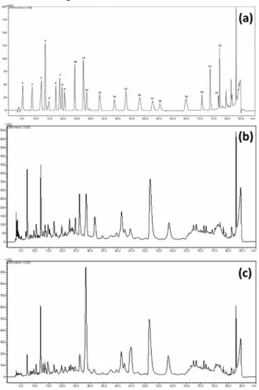
The standard chromatogram and the
chromatograms of the Soxhlet and microwave extracts are presented in Fig. 1. Table 1 lists the results of the HPLC analysis. The analysis revealed that both extracts had high content of catechin and rosmarinic acid. However, microwave irradiation led to higher results with regard to these compounds. But there were variations in phenolic and flavonoid contents of the extracts. For example, two phenolic compounds (benzoic acid
and o-coum acid) were not detected in the microwave extract. Cinnamic acid was detected in the microwave extract but not in the Soxhlet extract. As for flavonoids, two flavones (apigenin and luteolin) and one flavanone (hesperidin) were detected in the extracts. Apigenin and luteolin contents of both extracts were very close to one another but hesperidin content of the extract from the microwave irradiation was higher by two folds than that of the Soxhlet extract. The chemical structure of apigenin, luteolin and hesperidin are presented in Fig. 2.
Fig. 1. The chromatogram of the standards (a) and
the chromatograms of the Soxhlet (b) and microwave (c) extracts (1: gallic acid, 2: protocatechuic acid, 3: catechin, 4: p-hydroxy benzoic acid, 5: chlorogenic acid, 6: caffeic acid, 7: epicatechin, 8: syringic acid, 9: vanillin, 10: p-coum acid, 11: ferulic acid, 12: sinapinic acid, 13: benzoic acid, 14: o-coum acid, 15: rutin 16: hesperidin, 17: rosmarinic acid, 18: eriodictiol, 19: cinnamic acid, 20: quercetin, 21: luteolin, 22: kaempferol, 23:apigenin).
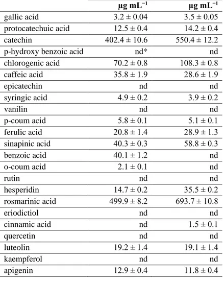
Table 1. The flavonoid and phenolic content of the Soxhlet and
microwave-assisted S. officinalis extracts. (*nd: Not detected. (±) refers to standard deviations. Three repetitions were done.).
Soxhlet extract Microwave extract
µg mL−1 µg mL−1
gallic acid 3.2 ± 0.04 3.5 ± 0.05
protocatechuic acid 12.5 ± 0.4 14.2 ± 0.4
catechin 402.4 ± 10.6 550.4 ± 12.2
p-hydroxy benzoic acid nd* nd
chlorogenic acid 70.2 ± 0.8 108.3 ± 0.8 caffeic acid 35.8 ± 1.9 28.6 ± 1.9 epicatechin nd nd syringic acid 4.9 ± 0.2 3.9 ± 0.2 vanilin nd nd p-coum acid 5.8 ± 0.1 5.1 ± 0.1 ferulic acid 20.8 ± 1.4 28.9 ± 1.3 sinapinic acid 40.3 ± 0.3 58.8 ± 0.3 benzoic acid 40.1 ± 1.2 nd o-coum acid 2.1 ± 0.1 nd rutin nd nd hesperidin 14.7 ± 0.2 35.5 ± 0.2 rosmarinic acid 499.9 ± 8.2 693.7 ± 10.8 eriodictiol nd nd cinnamic acid nd 1.5 ± 0.1 quercetin nd nd luteolin 19.2 ± 1.4 19.1 ± 1.4 kaempferol nd nd apigenin 12.9 ± 0.4 11.8 ± 0.4
Fig. 2. Chemical structures of flavonoids identified in the Soxhlet and microwave-assisted extracts of common
Previous literature studies have clearly demonstrated that flavonoids and phenolic compounds can coordinate metal ions and form stable complexes with metal cations. Biological activity of metal–flavonoids complexes, therefore, has been widely studied for their free radical scavenging activity [13]. In one study it was
demonstrated that flavonoids (kaempferol,
quercetin, myricetin, luteolin, catechin and naringenin) are capable of interacting of metal ions such as Cu2+ and Fe3+ ions through chelation [17].
Electron donating moieties (usually carbonyl and hydroxyl) of flavonoids are involved in formation of complexes with metal species. However, their interaction with metal ions are affected by the number and location of coordinating or chelating sites in flavonoid molecule [18]. Thus, due to this property, formation of flavonoid metal complexes has been widely utilized in spectrophotometric and spectrofluorometric studies [19, 20]. As depicted in Fig. 2, apigenin, luteolin and hesperidin have different number of hydroxyl functionalities in
their structures, which may affect their interaction with metal ions.
3.2. Dependency of fluorescence emission of S.
officinalis extracts on the extract concentration and excitation wavelength
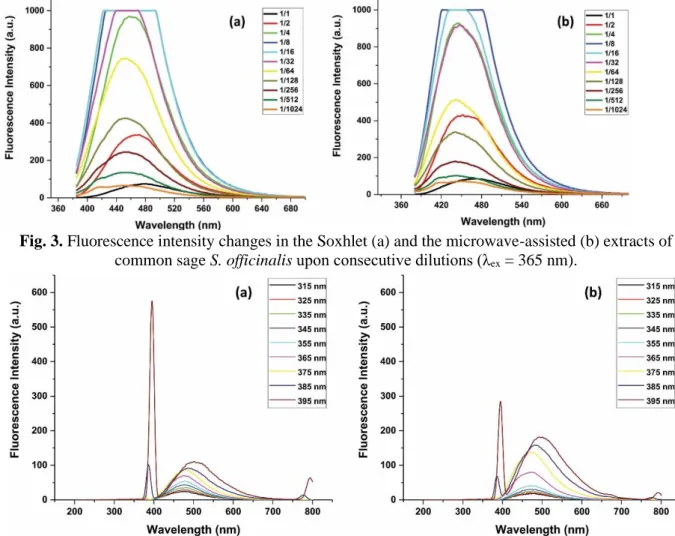
The measurements revealed that dilution of the
extract solutions led to enhancement in
fluorescence emission intensity to a certain point. Further dilution, on the other hand, decreased the fluorescence intensity (Fig. 3). This behaviour was observed for both Soxhlet extract and the microwave-assisted extract. Dilution of the extracts led to blue shift in the spectra. Emitted fluorescence of the extracts was also affected by change in the excitation wavelength (Fig. 4). At higher wavelength a red shift was recorded for both of the extracts. Excitation of S. officinalis extracts at 395 nm led to the highest emission peak at 480 nm for the Soxhlet extract and 472 nm for microwave-assisted extract.
Fig. 3. Fluorescence intensity changes in the Soxhlet (a) and the microwave-assisted (b) extracts of
common sage S. officinalis upon consecutive dilutions (λex = 365 nm).
Fig. 4. Fluorescence emission spectra of the Soxhlet (a) and microwave-assisted (b) extracts of common
3.3. Fluorescence emission response of S.
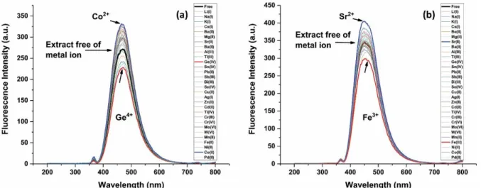
officinalis extracts to various metal ions Fluorescence emission spectra of the Soxhlet and the microwave-assisted extracts in the presence of metal ions are presented in Fig. 5. A column graph according to maximum fluorescence emission peaks of the extracts upon addition of various metal ions is shown in Fig. 6. Co2+ ion led to increase in
the fluorescence intensity of the Soxhlet extract. Ge4+ ions, on the other hand, had an opposite effect
and lowered the fluorescence of the extract. A completely different fluorescence emission spectrum was obtained for the microwave-assisted extract. Addition of Sr2+ ions to the
microwave-assisted extract enhanced the fluorescence emission of the extract. Fe3+ ions in the extract
quenched the fluorescence.
Fig. 5. Fluorescence emission changes in the Soxhlet (a) and microwave-assisted (b) extracts of common
sage S. officinalis upon addition of different metal ions (final concentration: 0.5 mg L−1). Equal volumes of the extracts and metal solutions were mixed, shaken and rested for 10 min (λex = 365 nm).
Fig. 6. Maximum fluorescence emission of the Soxhlet (a) and microwave-assisted (b) extracts of
common sage S. officinalis upon addition of different metal ions (final concentration: 0.5 mg L−1). Equal volumes of the extracts and metal solutions were mixed, shaken and rested for 10 min (λex = 365 nm).
The variations observed in the fluorescence emission of the extracts upon addition of the metal ions can be attributed to the interaction of metal ions with apigenin, luteolin and hesperidin content of the extracts. These observations are in line with the earlier literature reports. In one study on apigenin and luteolin by Favora et al., (2007), it was demonstrated that chelation of apigenin and
luteolin with Al3+ ions led to intense fluorescence
emission [21]. The authors concluded that complexation with metal cations could transform poorly fluorescent molecular systems into efficient fluorophores. A study by Perez-Ruiz et al., (1999) reported that spectrofluorometric determination of hesperidin in the presence of Al3+ [22]. The authors
fluorescent complex between hesperidin molecule and Al3+ ion. Yet, in some studies contradictory
results were also reported. For example, in a recent study the authors concluded that presence of Fe3+,
Cu2+, Mg2+, Mn2+, Zn2+ and Ca2+ ions did not have
obvious effect on the interaction of apigenin with bovine serum albumin [23].
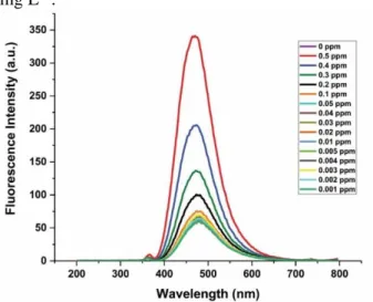
3.4. Turn-on fluorescence sensing of Co2+
using the Soxhlet extract of S. officinalis
The microwave extract exhibited the significant fluorescence intensity at presence of Co2+ ion.
Therefore, the fluorescence response of the Soxhlet extract to Co2+ was studied further as a model ion
(Fig. 7). In the experiments, the final concentration of the extract was adjusted to 50%, Co2+ ion
solutions were added into the extract solutions, shaken for one minute and rested for 10 min. The lowest concentration of Co2+ ion that gave obvious
fluorescence emission intensity was found to be 0.2 mg L−1.
Fig. 7. Fluorescence emission spectra of the
Soxhlet extract of S. officinalis as a function of Co2+ concentration.
4.
CONCLUSIONThis study revealed that the flavonoid and phenolic content of S. officinalis extract is highly dependent on the extraction method and parameters that are followed during an extraction procedure. Upon excitation by UV light, the Soxhlet and the microwave-assisted extracts emitted brilliant fluorescence, which can be attributed to the presence flavonoids (apigenin, luteolin and hesperidin). The extraction method greatly affected the content of the extracts by giving different HPLC profiles. Numerous previous studies have demonstrated clearly that flavonoid and phenolic molecules from plant extracts are capable of
interacting with metal ions through chelation or complex formation. This study, however, aimed to find out whether it was possible to use plant extracts with high flavonoid contents for fluorescent sensing of metal ions. As the chemical contents of plant extracts are highly dependent on the solvent type and extraction procedure, in the study two different methods were followed for water extraction of S. officinalis. Due to the variation of their composition, the extracts showed different interaction with 30 metal ions including alkaline, alkaline earth and transition metal ions. The result of the study with Co2+ was encouraging;
the Soxhlet extract was sensitive to Co2+ ion
concentration of 0.2 mg L−1. However, further studies are still needed to clarify the phenomena. The extracts can be used as simple, fast and low-cost turn-on fluorescence method for detection of, for example, Co2+ and Sr2+ ions; or for a turn-off
fluorescence method for signalling of Ge4+ and Fe3+
ions; yet, this study should be considered as an introductory study. Therefore, preliminary studies were not done to optimize the conditions for each ion. Especially, in case of Co2+, Sr2+, Ge4+ and Fe3+
ions optimization conditions have to be
investigated in a more detailed manner if sage extracts are used in any analytical metal sensing study. In future studies phytochemicals of sage plant can be extracted using different methods and the extracts can be tested in fluorescent sensing applications of metal ions.
REFERENCES
[1] Ghorbani A., Esmaeilizadeh M.,
Pharmacological properties of Salvia officinalis and its components, J. Tradit. Complem. Med., 7(2017) 433-440.
[2] Durling N.E., Catchpole O.J., Grey J.B., Webby R.F., Mitchell K.A., Foo L.Y., Perry N.B., Extraction of phenolics and essential oil from dried sage (Salvia officinalis) using ethanol– water mixtures, Food. Chem., 101(2007) 1417-1424.
[3] Ollanketo M., Peltoketo A., Hartonen K., Hiltunen R., Riekkola M.-L., Extraction of sage (Salvia officinalis L.) by pressurized hot water and conventional methods: Antioxidant activity of the extracts, Eur. Food. Res. Technol., 215(2002) 158-163.
[4] de Rijke E., Out P., Niessen W.M.A., Ariese F., Gooijer C., Brinkman U.A.T., Analytical
separation and detection methods for flavonoids, J. Chromatogr. A, 1112(2006) 31-63.
[5] Lu Y., Foo L.Y., Flavonoid and phenolic
glycosides from Salvia officinalis,
Phytochemistry, 55(2000) 263-267.
[6] Havsteen B.H., The biochemistry and medical significance of the flavonoids, Pharmacol. Therapeut., 96(2002) 67-202.
[7] Formica M., Fusi V., Giorgi L., Micheloni M., New fluorescent chemosensors for metal ions in solution, Coordin. Chem. Rev., 256(2012) 170-192.
[8] Jeong Y., Yoon J., Recent progress on fluorescent chemosensors for metal ions, Inorg. Chim. Acta, 381(2012) 2-14.
[9] Affrose A., Parveen S.D.S., Kumar B.S., Pitchumani K., Selective sensing of silver ion using berberine, a naturally occurring plant alkaloid, Sensor. Actuat. B-Chem., 206(2015) 170-175.
[10] Yang S., Jiang W., Zhao F., Xu L., Xu Y., Gao B., Sun H., Du L., Tang Y., Cao F., A highly sensitive and selective fluorescent sensor for detection of copper ions based on natural Isorhamnetin from ginkgo leaves, Sensor. Actuat. B-Chem., 236(2016) 386-391.
[11] Gao S., Tan G., Yuan H., Xiao D., Choi M.M.F., A Simple fluorometric method using Chlorophyll A for determination of Hg2+ ion,
Microchim. Acta, 153(2006) 159-162.
[12] Ahmad W., Rana N.F., Riaz S., Ahmad N.M., Hameed M., Naeem A., Tahir R., Chemical sensing of Benzo [a] pyrene using Corchorus depressus fluorescent flavonoids, Nat. Prod. Res., 32(2018) 968-971.
[13] Malešev D., Kuntić V., Investigation of metal-flavonoid chelates and the determination of flavonoids via metal-flavonoid complexing reactions, J. Serbian Chem. Soc., 72(2007) 921-939.
[14] Veličković D., Milenović D., Ristić M., Veljković V., Kinetics of ultrasonic extraction of extractive substances from garden (Salvia officinalis L.) and glutinous (Salvia glutinosa L.) sage. Ultrason. Sonochem., 13(2006) 150-156.
[15] Putnik P., Kovačević D.B., Penić M., Fegeš M.,
Dragović-Uzelac V., Microwave-assisted
extraction (MAE) of dalmatian sage leaves for the optimal yield of polyphenols: HPLC-DAD identification and quantification, Food Anal. Method., 2016;9:2385-94.
[16] Kontogianni VG, Tomic G, Nikolic I, Nerantzaki AA, Sayyad N, Stosic-Grujicic S, Stojanovic I., Gerothanassis I.P., Tzakos A.G.,
Phytochemical profile of Rosmarinus
officinalis and Salvia officinalis extracts and correlation to their antioxidant and anti-proliferative activity. Food Chem. 136(2013) 120-129.
[17] Fernandez M.T., Mira M.L., Florencio M.H., Jennings K.R., Iron and copper chelation by flavonoids: An electrospray mass spectrometry study, J. Inorg. Biochem., 92(2002) 105-111. [18] Kasprzak M.M., Erxleben A., Ochocki J.,
Properties and applications of flavonoid metal complexes, RSC Adv., 5(2015) 45853-45877. [19] Samsonowicz M., Regulska E., Spectroscopic
study of molecular structure, antioxidant activity and biological effects of metal hydroxyflavonol complexes, Spectrochim. Acta A, 173(2017) 757-771.
[20] Özbek N., Alp H., Çelik G., Ak T., Çağılcı O.C., Yaylı N., Ocak U., Ocak M., A simple
spectrofluorimetric method for iron
determination with a chalcone-based Schiff base, J. Fluoresc. 27(2017) 635-641.
[21] Favaro G., Clementi C., Romani A.,
Vickackaite V., Acidichromism and
ionochromism of luteolin and apigenin, the main components of the naturally occurring yellow weld: A spectrophotometric and fluorimetric study, J. Fluoresc., 17(2007) 707-714.
[22] Perez-Ruiz T., Martinez-Lozano C., Tomas V., Fenoll J., Spectrofluorometric determination of hesperidin by manual and flow-injection methods, Fresenius J. Anal. Chem., 364(1999) 279-283.
[23] Bi S., Yan L., Pang B., Wang Y., Investigation of three flavonoids binding to bovine serum
albumin using molecular fluorescence