Summary
The aim of this study was to determine coagulation profi les of spontaneous premature calves with respiratory distress syndrome. The study involved spontaneous premature calves with respiratory distress syndrome (n= 20) and clinically healthy newborn calves (n= 10). Blood samples were collected from all calves within the period between 2nd and 12th h of delivery to determine platelets (PLT), activated partial thromboplastin time (APTT), prothrombin time (PT), thrombin time (TT), serum fibrin/fibrinogen degradation products (FDPs), and fibrinogen. Premature calves had prolonged APTT (P<0.001) and PT (P<0.01), decreased fibrinogen concentration (P<0.001) and decreased platelet counts (P<0.001) when compared to controls. An insignificant prolondation of TT was observed in 8 premature calves and two of which had also FDPs when compared to control calves. Abnormal, but insignifi cant fi ndings of FDPs were noted in six premature calves with respiratory distress syndrome when compared to healthy calves. As a result, premature calves with respiratory distress syndrome had abnormal coagulation profi le and seven of these died despite intensive therapy. The veterinarian should take these results in consideration when dealing with premature calves with respiratory distress syndrome and constitute an appropriate treatment. Additionally, these findings may be of value in forming bases for future studies.
Keywords: Premature calf, Coagulation profile, Respiratory distress syndrome
Respiratorik Distres Sendromlu Spontan Premature Buzağılarda
Koagulasyon Profi linin Değerlendirilmesi
Özet
Bu çalışma, respiratorik distres sendromlu spontan premature buzağılarda koagülasyon profilini belirlemek amacıyla yapıldı. Bu çalışmada, respiratorik distres sendromlu spontan premature (n=20) ve klinik olarak sağlıklı yenidoğan buzağılar (n=10) kullanıldı. Trombosit (PLT), aktive edilmiş parsiyel tromboplastin zamanı (APTT), protrombin zamanı (PT), trombin zamanı (TT), fibrin/fibrinojen yıkım ürünleri (FDPs) ve fibrinojen düzeylerini belirlemek için doğumdan itibaren 2 ila 12. saatleri arasında tüm buzağılardan kan örnekleri toplandı. Sağlıklı yenidoğan buzağılarla karşılaştırıldığında, premature buzağılarda APTT (P<0.001) ve PT’de (P<0.01) uzama, fibrinojen konsantrasyonunda (P<0.001) düşme ve trombosit sayısı azalma (P<0.001) tespit edildi. Premature buzağıların 8’inde TT’da istatistiksel olarak önemsiz bir uzama ve aynı zamanda bu buzağıların 2’sinde de anormal FDPs bulgusu kaydedildi. Prematüre buzağıların 6’sında istatistiksel olarak önemsiz, fakat anormal FDPs bulgusu tespit edildi. Sonuç olarak, respiratorik distres sendromlu premature buzağılarda anormal koagülasyon profi li belirlendi ve bu buzağıların 7’si yoğun tedaviye rağmen öldü. Veteriner Hekimler respiratorik distres sendromlu premature buzağıların tedavisinde bu sonuçları göz önünde tutarak uygun bir tedavi yöntemi geliştirebilirler. Ayrıca, bu bulgular yapılacak çalışmalara temel oluşturabilir.
Anahtar sözcükler:Premature buzağı, Koagulasyon profili, Respiratorik distres sendromu
The Evaluation of Coagulation Profi les in Spontaneous Premature
Calves with Respiratory Distress Syndrome
Kemal IRMAK *
Kürşat TURGUT **
* Kafkas Üniversitesi Veteriner Fakültesi, İç Hastalıkları Anabilim Dalı, TR-36100 Kars - TÜRKİYE ** Selçuk Üniversitesi Veteriner Fakültesi, İç Hastalıkları Anabilim Dalı, TR-42003 Konya - TÜRKİYE
Makale Kodu (Article Code): KVFD-2010-2729
INTRODUCTION
The rate of prematurely born calves has gradually cattle farms in recent years. The underlying causes have increased and thus causing great economical losses in been reported as physical, genetic, environmental and
İleti şim (Correspondence)
+90 474 2426807/1249
[email protected]endocrinologic factors and infectious diseases 1-3.
Neonatal health problems associated with Disseminated Intravascular Coagulation (DIC) are shock, idiopathic respiratory distress syndrome, and various bacterial infections, especially gram-negative sepsis 4. Studies
have shown that respiratory acidosis is mainly developed in spontaneous premature calves, but both respiratory and metabolic acidosis does also occur 3,5,6. Respiratoric
insufficiency of calves may be due to improper development of lung and accordingly haemostasis factors may not adequately be synthesized. In such cases asphyxia develops and severe acidosis occurs, this in turn results in consumption coagulopathy. Consumption coagulopathy in calves may not only be a result of severe respiratoric insufficiency, but also be due to severe illness process and their complications 5. Acidosis induces DIC, because
it inhibits Anti thrombin III (AT III) and heparin 7,8 and
also triggers endothelial sloughing with the attendant activation of factor XII to XIIa and/or XI to XIa, and/or platelet release, with a later activation of the procoagulant system. Less commonly alkalosis may also trigger DIC, but the mechanism is unclear 9. Anoxia augments DIC,
as it causes acidosis and endothelial damage. Stasis may also lead to DIC because stasis prevents the removal of activated clotting enzymes and interferes with local AT III supply, and causes local anoxemia 7,10.
Oxygen radical injury has been thought to be one of the common mechanisms of several neonatal diseases in premature calves. Bronchopulmonary dysplasia and intraventricular hemorrhage of prematurity have been associated with excessive production of oxygen free radicals 11-13. This mechanism may also play role in the
development of DIC in premature subjects. However, there is a scarcity of literature on coagulation profile in premature calves.
This study was designed to evaluate the some coagulation parameters of spontaneous premature calves with respiratory distress syndrome.
MATERIAL and METHODS
In this study, 20 spontaneous premature calves with respiratory distress (Premature group), referred to the Clinic of Internal Medicine, Faculty of Veterinary Medicine, University of Selcuk, Konya, and 10 clinically healthy newborn calves (Control group) belonging to the faculty farm were used. All premature and control calves were Swiss Holstein breed of both sexes. The diseased calves were included into the study based on the case defi nition detailed below.
A premature calf with respiratory distress syndrome was defined as calf with decreased gestational age (from
gestation day 260 till birth), low birth weight, presence of short silky hair coat, general weakness or fl oppiness, inability to stand, weak or no suckling refl ex, hyperemic gums and incomplete tooth eruption, and soft claw 14-17,
and also with acute dyspnea (elevated respiratory rate, labored breathing and abnormal lung sounds on auscultation) 18.
Premature calves with respiratory distress were not able to receive colostrum because of general weakness, inability to stand, absence of suckling refl ex and respiratory distress. They were 2 to 12 h old. Blood samples were collected from both premature and control calves within the period between 2nd and 12th h of delivery.
An intensive therapy including fl uid and electrolyte, antibiotics, inotrope, nonsteroidal anti-infl ammatory drugs, oxygen therapy, warm therapy, colostrums via stomach tube was applied to the premature calves.
Jugular vein blood samples were collected once prior to treatment from all calves into tubes containing K-EDTA and sodium citrate.
Haematological Examinations: Platelet count was
determined on the K-EDTA whole blood sample using an automatic cell counter (Medonic, Ca530).
Coagulation Testing: 9 ml of venous blood sample
for the determination of APTT, PT, TT, fibrinogen and FDPs concentrations were collected into collection tube including 1 ml of 3.8% sodium citrate and centrifuged at 1.500 g for 15 min. The plasma samples were separated from blood within 30 min of blood collection and measurements were done within 12-24 h.
APTT (STA-CK Prest® , Kit Cat. No: 00597) and PT (STA®
-Neoplastine® Cl Plus , Kit Cat. No: 00597) were measured
by STA® analyser. TT (ThromboquikTM; Product no: 35515)
and fibrinogen (Fibriquik®; Product No: 35529) were
measured by Thromblyser-XR (Organon Teknika). FDPs were determined by Latex agglutination test method using FDP plasma kit (Kit Cat. No: 00540; Organon Teknika Corp; Durham, NC, USA). The scoring of FDPs was established as 1= <5 μg/ml, 2= 5-20 μg/ml, 3= >20 μg/ml.
Statistical Analysis
FDPs determined for both groups were compared with Chi Square test. Two samples student t test was used for other analyses. P<0.05 was considered statistically significant (SPSS 8.0.0 for Windows; Statistical Package of Social Science, SPSS Inc. USA).
RESULTS
The clinical examination of the spontaneous pre-mature calves with gestational age of 259-265 days
revealed low body weight (32-40 kg), general weakness, soft claw, inability to stand, hyperemic gums and in-complete tooth eruption, weak or no suckling reflex, respiratory distress and short silky hair coat. Seven of the spontaneous premature calves with respiratory distress syndrome died in spite of therapy, but no necropsy was perfomed. The rest of the calves (n=13) recovered. Spontaneous premature calves did not naturally receive colostrum due to weak or no suckling refl ex.
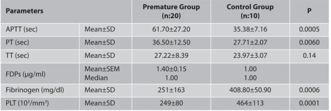
The mean APTT, PT, TT, fibrinogen, and PLT counts and mean and median FDPs concentrations of both premature and control groups are given in Table 1. The results revealed a significant prolongation of APTT (P<0.001) and PT (P<0.01) and a marked decrease in fibrinogen concentration (P<0.001) and platelet counts (P<0.001) in premature group when compared with control group. However, FDPs concentrations and TT prolongation in premature group did not signifi cantly increase when compared to the control group.
DISCUSSION
The results of this study revealed that aff ected calves had clinical signs consistent with signs previously reported for premature calves with respiratory distress 14-18 and that
abnormal coagulation values were evident.
Parameters of haemostasis may be abnormal during DIC; however, currently there is no single test consistently or specifically provides a definitive diagnosis 19. The laboratory
diagnosis of DIC is usually based on the prolonged APTT, PT, TT, thrombocytopenia, hypofibrinogenemia, FDPs, schistocytes in blood smears, and decreased concentrations of coagulation factors (usually factors V, VIII) and antithrombin III. Excessive coagulation is refl ected by reduced plasma concentrations of platelets, coagulant and anticoagulant proteins, and increased concentrations of coagulant by-products. Fibrinolysis is indicated by elevated FDPs or reduced concentrations of fibrinolytic
and antifibrinolytic proteins 19,20. The results of these tests
will also vary according to the severity and fulminant nature of the process and the time of sampling 21,22.
Diagnosis of DIC is assumed when at least three of fi ve tests included on a coagulation profile are abnormal 23.
In one study, the tests that were correlated most highly with the diagnosis of DIC in humans were thrombocyte numbers, PT, FDPs, and AT III levels 24. In dogs, it has been
suggested that platelet count and AT III concentrations are the most sensitive indicators of DIC 25. In our study,
at least three coagulation profile tests were abnormal in all premature calves. The most common of these were prolonged APTT (13 cases), PT (14 cases), TT (8 cases) FDPs (6 Cases), thrombocytopenia (3 cases), and hypo-fibrinogenemia (11 cases). Furthermore, in our study, prolonged APTT and PT, thrombocytopenia and hypo-fibrinogenemia were found to be statistically significant in the premature group when compared with control group.
APTT test is used to evaluate the intrinsic and common
pathways. The PT is a measure of the extrinsic and common pathways of coagulation. The most common cause of prolonged APTT and PT is increased consumption of clotting factors during DIC. In this study, the pro-longed APTT and PT were found to be common coagulation profile abnormalities in spontaneous premature calves. Similar results were reported earlier 5, where calves
developed respiratory distress syndrome showed a slightly prolonged prothrombin time and partial thromboplastin time as well as a decreased AT III activity. It may be assumed that in calves with respiratory distress syndrome - in analogy to pulmonary immaturity - the blood clotting mechanism is not yet fully developed. In healthy prematures and surviving asphyctic calves haemostasis remains largely stable during the first day of life, whereas plasma fibrinogen concentration increases. In the calves not surviving, the examination period post natum prothrombin time and partial thromboplastin time became significantly longer. Only in these severely asphyctic calves the presence of
Table 1. The mean±SD of APTT, PT, TT, fibrinogen concentrations and PLT counts, mean±SEM and median FDPs
concentrations in both premature and control groups and their statistical significance
Tablo 1. Premature ve kontrol grubu buzağılarında ortalama APTT, PT, TT, fibrinogen konsantrasyonları ve
trombosit sayısı ile ortalama ve median FDPs konsantrasyonları ve bu değerlerin istatistiksel önemlilikleri
Premature Group Control Group
Parameters P
(n:20) (n:10)
APTT (sec) Mean±SD 61.70±27.20 35.38±7.16 0.0005
PT (sec) Mean±SD 36.50±12.50 27.71±2.07 0.0060 TT (sec) Mean±SD 27.22±8.39 23.97±3.07 0.14 Mean±SEM 1.40±0.15 1.00 FDPs (μg/ml) Median 1.00 1.00 Fibrinogen (mg/dl) Mean±SD 251±163 408.80±50.90 0.0006 PLT (103/mm3) Mean±SD 249±80 464±113 0.0001 FDP graded 1= <5 (μg/ml), 2= 5-20 (μg/ml), 3= >20 (μg/ml)
a consumption coagulopathy seems likely. A secondary reactive fibrinolysis was not observed 5.
Thrombin time is a measure of the rate of fibrinogen to fibrin conversion. It is prolonged when fibrinogen is less than 60 mg/dl, fibrinogen is nonfunctional, or FDPs are present that interfere with fibrin polymerization. Since hypofibrinogenemia is rare in large animals, a prolonged thrombin time most likely would indicate the presence of FDPs 22. In our study, prolongation of TT was
not statistically significant, but it was prolonged when compared to control calves. Prolonged TT was observed in eight spontaneous premature calves and two of which had also FDPs. However, the data obtained were not in favour of the report in which TT prolongation due to consumption coagulopathy in calves with respiratory distress where reactive hyperfibrinolysis was evident 5.
Increased serum concentration of FDPs refl ects the proteolytic action of plasmin on fibrin and/or fibrinogen at a rate that exceeds the clearance capacity of the mononuclear phagocyte system. Measurable serum FDPs generally indicate increased fibrinolysis response to excessive activation of coagulation (i.e. DIC); however, severe inflammatory processes, hemorrhagic disorders, or postoperative states that cause extensive intra-vascular fibrin deposition may elevate serum FDPs significantly 22. In the present study FDPs concentration
in premature group did not significantly increase when compared to the control group. However, abnormal finding of FDPs along with other parameters in six spontaneous premature calves may be suggestive of DIC.
Hypofibrinogemia may result from impaired hepatic synthesis, increased consumption during DIC, degradation during primary hyperfibrinolysis, or uncompensated loss during massive hemorrhage 22. Fibrinogen concentration
was found to be decreased in premature group when compared to the control group (P<0.001). In our study, hypofibrinogenemia was found to be common coagulation profile abnormality in spontaneous pre-mature calves. A previous study carried out in prepre-mature calves also shown a decreased average plasma fibrinogen concentration than animals delivered in due time 5.
Plasma fibrinogen increase in non asphyctic calves and neonatal babies may suggest the commencement of fibrinogen synthesis 5,26.
In this study, platelet counts in premature group were less than the control group (P<0.001). Thrombo-cytopenia (a platelet count less than 150.000/l) is caused by one of three basic mechanisms: decreased or ineff ective production of platelets, platelet sequestration, or shortened platelet life span. Excessive consumption of platelets to fulfill their normal role in haemostasis occurs during DIC. The platelet count is a useful test for monitoring the rate and severity of consumption or destruction. Other components of the haemostatic
system should be evaluated (e.g., PT, APTT, FDPs), since thrombocytopenia may be only part of a disseminated coagulopathy 22. Thrombocytopenia together with
prolonged APTT, PT, TT, increased FDPs and hypo-fibrinogenemia may suggest that DIC might have developed in these calves.
The results of our study indicated that some coagulation parameter abnormalities were common in spontaneous premature calves with high mortality. These results are parallel with the studies of DIC in small animals where severe hemostatic dysfunction is associated with high mortality rate and DIC is a significant risk factor for mortality 27-29. The veterinarian should take
these results in consideration when dealing with pre-mature calves with respiratory distress syndrom to constitute an appropriate treatment protocol.
REFERENCES
1. Jensen R, Pier AC, Kaltenbach CC, Murdoch WJ, Becerra VM, Mills KW, Robinson JL: Evaluation of histopathologic and physiologic changes in cows having premature births after consuming ponderosa pine needles. Am J Vet Res, 50 (2): 285-289, 1989.
2. Murray RD: Pathophysiology of abortion in cattle. Bovine Pract, 25,
110-114, 1990.
3. Ok M, Birdane FM: Prematüre buzağılarda kan asit-baz dengesi, bazı kan gazları ve elektrolit düzeyleri. Vet Bil Derg, 16 (1): 147-150, 2000. 4. Mangurten HH: Hemorrhagic disease due to vitamin K defi ciency in a premature infant: A syndrome which may resemble disseminated intravasculer coagulation (DIC). Clin Pediatr, 12 (6): 372-375, 1973. 5. Aurich JE, Grunert E, Zaremba W: Veraenderungen im Blutgerinnungspotential frühgeborener Kaelber mit und ohne Atemnotsyndrome. Tierarztl Prax, 17, 27-33, 1989.
6. Sarıalıoğlu F, Yurdakök M, Kutluk MT, Çalıkoğlu AS: Çocuk Hastalıklarında Tanı ve Tedavi. 1. Cilt, 1. Baskı. pp. 644-647. Barış Kitapevi/ Appleton & Lange, 1993.
7. Slappendel RJ: Disseminated intravascular coagulation. Vet Clin North
Am: Small Anim Pract, 18 (1): 169-184, 1988.
8. Gökçe E, Irmak K: Dissemine intravaskuler koagulasyon (DIC). Kafkas
Univ Vet Fak Derg, 13 (2): 215-222, 2007.
9. Bick RL: Disseminated intravasculer coagulation: Objective clinical and laboratory diagnosis, treatment, and assesment of therapeutic response. Semin Thromb Hemost, 22 (1): 69-88, 1996.
10. Turgut K: Hemostazis: Koagulasyon ve Trombosit Bozuklukları. In, Turgut K (Ed): Veteriner Klinik Laboratuvar Teşhiş. 2. Baskı. s. 124-165. Bahçıvanlar Basım Sanayi AŞ, 2000.
11. Hittner HM, Godio LB, Rudolph AJ, Adams JM, Garcia-Prats JA, Friedman Z, Kautz JA, Monaco WA: Retrolental fibroplasias: Efficacy of vitamin E in a double-blind clinical study of preterm infants. N Engl J Med, 305, 1365-1371, 1981.
12. Johnson L, Quinn GE, Abbasi S,Gerdes J, Bowen FW, Bhutani V: Severe retinopathy of prematurity in infants with birth weights less than 1250 grams: incidence and outcome of treatment with pharmacologic serum levels of vitamin E in addition cryotherapy from 1985 to 1991. J
Pediatr, 127, 632-639, 1995.
13. Sullivan JL: Iron, plasma antioxidants, and the oxygen radical disease of prematurity. Am J Dis Child, 142, 1341-1344, 1998.
14. Koterba A, Madigan JE: Manifestations of Disease in the Neonate. In, Smith BP (Ed): Large Animal Internal Medicine. pp. 316-319. The CV Mosby Company, Philadelphia. 1990.
biochemical parameters in premature calves. Indian Vet J, 77, 859-861, 2000.
16. Miller RB: Bovine abortion. In, Marrow DA (Ed): Current Therapy in Theriogenology. pp. 291-300. WB Saunders Co, Philadelphia, 1986. 17. Roberts SJ: Diseases and accidents of the gestation periods (Abortion). In, Veterinary Obstetrics and Genital Diseases (Theriogenology). Third ed., pp. 123-125. Edwards Brothers, Inc. Ann Arbor, Michhigan, USA, 1986.
18. Wilson WD, Lofstedt J: Alterations in respiratoric functions. In, Smith BP (Ed): Large Animal Internal Medicine. pp. 46-99. The CV Morby Co, USA, 1996.
19. Morris DD: Disease of hematopoietic and hemolymphatic systems. In, Smith BP (Ed): Large Animal Internal Medicine. pp. 1077-1080. The CV Morby Co, USA, 1990.
20. Jain NC: Coagulation and Its Disorders. Essentials of Veterinary Haematology. pp. 82-104. Lea & Febiger, Philadelphia, 1993.
21. Blood DC, Radostits OM, Henderson JA: Veterinary Medicine. Sixth ed., pp. 304-305. Bailliere Tindall, London, 1983.
22. Morris DD: Alterations in the clotting profi le. In, Smith BP (Ed): Large
Animal Internal Medicine. pp. 445-452. The CV Mosby Co, Missouri, USA, 1990.
23. Ritt MG, Rogers KS, Thomas JS: Nefrotic syndrome resulting in thromboembolic disease and Disseminated Intravascular Coagulation in a dog. J Am Anim Hosp Assoc, 33, 385-391, 1997.
24. Bovill EG: Laboratory diagnosis of diseminated intravascular coagulation. Semin Hematol (Suppl), 31, 35-39, 1994.
25. Keyes ML, Rush JE, Knowles KE: Pulmonary thromboembolism in dogs. J Vet Emerg Crit Care, 3, 23-32, 1993.
26. Boyer C, Ménaché D, Beaufils F, Mathieu H: Haemostatic disorders and respiratory distress in the newborn. Intensive Care Med, 3, 273-278, 1977.
27. Drazner IH: Clinical implications of disseminated intravascular coagulation. Compend Contin Educ Pract Vet, 4, 974-981, 1982.
28. Hammer AS, Couto CG, Swardson C: Hemostatic abnormalities in dogs with hemangiosarcoma. J Vet Intern Med, 5, 11-14, 1991.
29. King LG: Postoperative complications and prognostic indicators in dogs and cats with septic peritonitis: 23 cases (1989-1992). J Am Vet Med