See discussions, stats, and author profiles for this publication at: https://www.researchgate.net/publication/272278607
First report of Conidiobolus coronatus in Turkey
Article in Mycotaxon -Ithaca Ny- · January 2011DOI: 10.5248/115.121 CITATION 1 READS 36 3 authors, including:
Some of the authors of this publication are also working on these related projects:
The Isolation of Entomopathogenic Fungi to be used in Biological Control with Two-spotted Spider MiteView project
Fındık kurdu [Curculio nucum l. (col.: curculionidae)]’na Karşı Kimyasal Mücadelede Kullanılan Farklı Pülverizatör Tiplerinin Bazı Faydalı ve Zararlı Böceklere Etkileri
View project Cafer Eken
Aydın Adnan Menderes University 53 PUBLICATIONS 364 CITATIONS
SEE PROFILE
Kibar Ak
Black Sea Agricultural Research Institute 22 PUBLICATIONS 66 CITATIONS
SEE PROFILE
All content following this page was uploaded by Kibar Ak on 07 September 2018. The user has requested enhancement of the downloaded file.
MYCOTAXON
Volume 115, pp. 121–124 January–March 2011
doi: 10.5248/115.121
First report of Conidiobolus coronatus in Turkey
Cafer Eken
1,2*, Şaban Güçlü
2& Kibar Ak
31Graduate School of Natural and Applied Sciences, Ardahan University, Ardahan,75000,Turkey 2Department of Plant Protection, Faculty of Agriculture, Atatürk University, Erzurum,25240, Turkey 3Black Sea Agricultural Research Institute, Gelemen, Samsun,55001, Turkey
*Correspondence to: [email protected]
Abstract — Conidiobolus coronatus (Entomophthorales, Zygomycota) was isolated from infected specimens representing an Issus sp. (Issidae, Hemiptera) collected from the Trabzon province of Turkey. The species, which represents a new record for the Turkish mycoflora, is described briefly and illustrated.
Key words — insect, entomopathogenic fungi, hazelnut
Introduction
Members of the widespread order Entomophthorales (Zygomycota) are
predominantly pathogens of insects and mites (Pell et al. 2001). Species of
Conidiobolus, most notably the ubiquitous C. coronatus, are recorded as
widespread soil saprophytes utilising a variety of substrates, including plant
detritus, living plants, different dead arthropods and the fruiting bodies
of other fungi in various regions of the world (MacLeod & Müller-Kögler
1973, Keller 1987, Sajap et al. 1997, Dromph et al. 2001, Laxman et al. 2005,
Manning et al. 2007, Comerio et al. 2008). Conidiobolus coronatus, mainly
tropical strains, is known to cause disease in both insects and humans (Ribes et
al. 2000, Prabhu & Patel 2004); the disease has been named
rhinophyco-mycosis, rhinophycomycosis entomophthorae, rhinoentomophthororhinophyco-mycosis,
and conidiobolomycosis (King 1979, Ochoa et al. 1996, Yang et al. 2010).
Material & methodsDead insects collected in 2008 from a hazelnut orchard in in the Black Sea region of Turkey were cultured for entomopathogenic fungi. After the cadavers were washed in a solution of 2% sodium hypochlorite for 1 min, they were dried on filter paper. After transfer to Petri dishes containing 20 ml of PDA, the cadavers were incubated at 25°C for 1 week with high humidity (80 ± 10% rh). Colonies of filamentous fungi
122
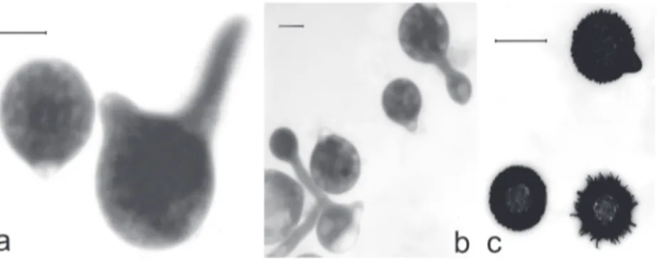
... Eken, Güçlü & AkFigure 1. Conidiobolus coronatus: a— primary conidium and germinating primary conidium; b— primary conidia and secondary conidia produced on germ tube arising from primary conidia; c— villose conidia may be the equivalent of resting spores in this species. Bar = 20 μm.
emerging from each cadaver and identifiable as the genus Conidiobolus were transferred to PDA and identified to species using the relevant literature (Emmons & Bridges 1961, Prasertphon 1963, MacLeod & Müller-Kögler 1973, King 1979, Keller 1987, Humber 1997, Hatting et al. 1999, Toledo et al. 2007, Comerio et al. 2008). After identification, all isolates were deposited in the fungal collection of Department of Plant Protection, Faculty of Agriculture, Atatürk University, Erzurum-Turkey.
Results
The description and illustration of Conidiobolus coronatus given below
are based on the Turkish collections of the material. This is the first report of
C. coronatus from Turkey.
Conidiobolus coronatus
(Costantin) A. Batko, Entomophaga,Mem. Hors. Ser. 2: 129. 1964. Fig. 1
Colonies (PDA) expanding, hyaline, soon with irregular radial blooms; at
25°C reaching 85 mm diam. after 2 days; primary conidia (Fig. 1a) globose,
with more prominent basal papilla tapering toward obtuse apex; variable in
size, (23.2–)32.1(–46.8) × (18.1–)24.6(–35.1) μm; basal papilla prominent with
pointed apex, forcibly discharged; secondary conidia (Fig. 1b) forming
singly and forcibly discharged or (more commonly) producing many forcibly
discharged secondary microconidia on short germ tubes arising from primary
conidia; conidiophores simple, unbranched; villose conidia (resting spores;
Fig. 1c) resembling primary conidia but covered with villose appendages
(unique to this species), (19.9–)25.8(–34.8) μm diam.
Specimen examined: On cadavers of Issus sp. (Issidae, Hemiptera). TURKEY: Trabzon
province, Of district, Corylus avellana L. (Corylaceae) orchards, 40°51’30’’N,
Discussion
Conidiobolus coronatus, originally isolated from a culture of Agaricus
campestris L. (possibly derived from a dead insect hidden between the lamellae),
was described in 1897 as Boudierella coronata Costantin (MacLeod &
Müller-Kögler 1973). Since then it has been isolated from numerous and diverse
sources (MacLeod & Müller-Kögler 1973, King 1979, Keller 1987, Sajap et al.
1997, Dromph et al. 2001, Laxman et al. 2005, Manning et al. 2007, Comerio
et al. 2008). Utilization of the species as a biological control is limited by its
potential to cause human disease (King 1979, Ochoa et al. 1996, Ribes et al.
2000, Prabhu & Patel 2004).
Diameters of both the primary and villose conidia measured in this study
fell within the ranges observed by Emmons & Bridges (1961; primary conidia:
36–44 μm), Prasertphon (1963; primary: 25–61 μm; villose: 8–42 μm), Keller
(1987; primary: 37–74 μm; villose: 16–42 μm), Hatting et al. (1999; primary:
41.5–66 × 30–48 μm), Toledo et al. (2007; primary: 17.6–39.5 × 24.7–29.6 μm;
villose: 19.8–24.7 μm), and Comerio et al. (2008; primary: 30–38 μm).
Acknowledgments
We are grateful to Dr. Celal Tuncer and Dr. Yusuf Yanar for reviewing the manuscript.
Literature cited
Comerio RM, Andorno AV, Botto EN. 2008. Conidiobolus coronatus isolation from a pest aphid of chives (Allium schoenoprasum L.). Revista Iberoamericana de Micología 25: 193–195. Dromph KM, Eilenberg J. Esbjerg P. 2001. Natural occurrence of entomophthoralean fungi
pathogenic to collembolans. Journal of Invertebrate Pathology 78: 226–231. doi: 10.1006/ jipa.2002.5077
Emmons CW, Bridges CH. 1961. Entomophthora coronata, the etiologic agent of a phycomycosis of horses. Mycologia 53: 307–312. doi: 10.2307/3756277
Hatting JL, Humber RA, Poprawski TJ, Miller RM. 1999. A survey of fungal pathogens of aphids from South Africa, with special reference to cereal aphids. Biological Control 16: 1–12. doi: 10.1006/bcon.1999.0731
Humber RA. 1997. Fungi: identification. 153–185, in LA Lacey (ed.), Manual of Techniques in Insect Pathology. London, Academic Press.
Keller S. 1987. Arthropod-pathogenic Entomophthorales of Switzerland. I. Conidiobolus,
Entomophaga and Entomophthora. Sydowia 40: 122–167.
King DS. 1979. Systematics of fungi causing entomophthoramycosis. Mycologia 71: 731–745. doi: 10.2307/3759185
Laxman RS, Sonawane AP, More SV, Rao BS, Rele MV, Jogdand VV, Deshpande VV, Rao MB. 2005. Optimization and scale up of production of alkaline protease from Conidiobolus coronatus. Process Biochemistry 40: 3152–3158. doi: 10.1016/j.procbio.2005.04.005
MacLeod DM, Müller-Kögler E. 1973. Entomogenous fungi: Entomophthora species with pear-shaped to almost spherical conidia (Entomophthorales: Entomophthoraceae). Mycologia 65: 823–893. doi: 10.2307/3758521
124
... Eken, Güçlü & AkManning RJ, Waters SD, Callaghan AA. 2007. Saprotrophy of Conidiobolus and Basidiobolus in leaf litter. Mycological Research 111: 1437–1449. doi: 10.1016/j.mycres.2007.08.019
Ochoa LF, Dubque CS, Velez A. 1996. Rhinoentomophthoromycosis. Report of two cases. Journal of Laryngology and Otology 110: 1154–1156. doi: 10.1017/S002221510013600X
Pell JK, Eilenberg J, Hajek AE, Steinkraus DC. 2001. Biology, ecology and pest management potential of Entomophthorales. 71–153, in TM Butt et al. (eds.), Fungi as Biocontrol Agents. Wallingford, CABI Publishing. doi: 10.1079/9780851993560.0071
Prabhu RM, Patel R. 2004. Mucormycosis and entomophthoramycosis: a review of the clinical manifestations, diagnosis and treatment. Clinical Microbiology and Infection 10 (Suppl 1): 31–47. doi: 10.1111/j.1470-9465.2004.00843.x
Prasertphon S. 1963. Conidial formation in Entomophthora coronata (Costantin) Kevorkian. Journal Insect Pathology 5: 318–335.
Ribes JA, Vanover-Sams CL, Baker DJ. 2000. Zygomycetes in human disease. Clinical Microbiology Reviews 13: 236–301. doi: 10.1128/CMR.13.2.236-301.2000
Sajap AS, Atim AB, Husim H, Wahab YA. 1997. Isolation of Conidiobolus coronatus (Zygomycetes:
Entomophthorales) from soil and its effect on Coptotermes curvignathus (Isoptera: Rhinotermitidae). Sociobiology 30: 257–262.
Toledo AV, Remes Lenicov AMM de, López Lastra CC. 2007. Primer registro de Conidiobolus
coronatus (Zygomycetes: Entomophthorales) en crías experimentales de dos especies plaga del
maíz: Delphacodes kuscheli y D. haywardi (Hemiptera: Delphacidae) en la Argentina. Boletín de la Sociedad Argentina de Botánica 42: 169–174.
Yang X, Li Y, Zhou X, Wang Y, Geng S, Liu H, Yang Q, Lu X, Hiruma M, Sugita T, Ikeda S, Ogawa H. 2010. Rhinofacial conidiobolomycosis caused by Conidiobolus coronatus in a Chinese rice farmer. Mycoses 53: 369–373. doi: 10.1111/j.1439-0507.2009.01716.x
View publication stats View publication stats