Molecular modelling, spectroscopic characterization and
nonlinear optical analysis on N -Acetyl-DL-methionine
N. G¨unaya, ¨O. Tamerb,∗, D. Avcıb, E. Tarcanc, and Y. Atalayb
aBeykent University, School of Health Sciences, Department of Audiology, Istanbul, Turkey. bSakarya University, Faculty of Arts and Sciences, Department of Physics, Sakarya, Turkey. cKocaeli University, Faculty of Arts and Sciences, Department of Physics, Kocaeli, Turkey.
Received 2 April 2020; accepted 4 May 2020
In this present methodical study, besed on the density functional theory (DFT), the first-principles calculations have been employed suc-cessfully to study the structural and electronic properties of N-acetyl-DL-methionine (C7H13NO3S) which is a derivative of DL-methionine
which is also known as DL-2-amino-4-methyl-thiobutanoic acid. Optimized molecular structure, vibrational frequencies, and also13C and
1
H NMR chemical shift values of the title compound are provided in a detailed manner by using B3LYP and HSEH1PBE functionals by applying 6-311++G(d,p) basis set for calculations using Gaussian 09W program. The comparison of the calculated values with the experi-mental values provides important information about the title compound. Besides the electronic properties (UV-Vis calculations) of the title compound, such as HOMO-LUMO energy values and energy gap, absorption wavelengths, oscillator strengths, were performed basing on the optimized structure in the gas phase. Moreover, the molecular electrostatic potential surface, dipole moment, nonlinear optical properties, linear polarizabilities, and first hyperpolarizabilities and chemical parameters have also been studied.
Keywords: N-Acetyl-DL-methionine; IR; NMR; DFT; nonlinear optic. PACS: 42.65.-k; 61.66.Hq; 67.30.er; 71.15.Mb; 95.30.Ky.
DOI: https://doi.org/10.31349/RevMexFis.66.749
1.
Introduction
Methionine is an amino acid that contains an α-amino group (protonated NH+3 ), an α-carboxylic acid group (deproto-nated COO−), and an S-methyl thioether side chain. Just like other amino acids, methionine is an organic compound used in the biosynthesis of proteins. It is also a non-polar, aliphatic amino acid and does not synthesize by the human body, so it must be obtained from foods. Methionine has the potential to the field of medicine to be used as, for example, anti-inflammatory, pain-reliever, and it stimulates the forma-tion of cartilage tissue cells to create more cartilaginous tis-sue, is also used as an additive in animal feedstuffs.
All amino acids (except glycine) can exist in two iso-meric forms, which are called D-(dextrorotatory) and L-(levorotatory) forms, analogous to left-handed and right-handed configurations, because of forming two different enantiomers (also called enantiomers stereoisomers and op-tical isomers which are mirror images to each other) around the central α-carbon atom. Methionine is one of the essential sulfur-containing amino acids, and it occurs naturally as L-methionine. A racemic DL- methionine mixture that is com-posed of equal amounts of D-methionine and L-methionine forms of the same compound is not optically active. Syn-thetic animal feed grade DL-methionine is a white crystalline powder with a faint characteristic odor and sweet, slightly bitter taste [1]. N-Acetyl DL-methionine (NAM) is one of the derivatives of DL-methionine, and, in this content, den-sity functional theory investigations were performed on the structural, electronic, and spectroscopic properties of NAM molecule.
2.
Calculation Details
In this study, the Gaussian 09W software package [2] was used to perform quantum chemical calculations, the geome-try, the normal modes, HOMO-LUMO energy gaps, and the molecular electrostatic potential (MEP) surface of the title compound were visualized by GaussView 5.0 [3]. All cal-culations were performed in the gas phase at the DFT hy-brid methods: the B3LYP (Becke’s three-parameter exact exchange functional (B3) [4,5] combined with the gradient-corrected correlational functional of Lee, Yang and Parr (LYP) [6] hybrid method is also called the first-generation method) and the HSEH1PBE (it is deciphered as the Heyd, Scuseria, Ernzerhof (HSE) [7,8] hybrid combined with Perdew, Burke and Ernzerhof’s exchange and correlation functions (PBE) [9], which is also known as the HSE06 ap-proach, is a second-generation method) with 6-311++G(d,p) basis set. The selection of DFT levels can be validated by a comparison between the experimental and computed UV-Vis spectrum. The excitation energies for the title molecule calculated by B3LYP and HSEH1PBE levels in conjunction with 6-311++G(d,p) basis set were compared with the ex-perimental emission spectrum. These levels were found to predict energies accurately, with differences between the ex-perimental and computed excitations below the usually ac-cepted threshold of 0.3 eV [10]. Additionally, considering that the HOMO-LUMO energy gap obtained by experimen-tal and DFT levels are very close to each other, it will be easily understood that the selection of DFT levels is cor-rect. The 1H and 13C NMR isotropic shielding are
calcu-lated with the GIAO (the Gauge Independent Atomic Or-bital) [11-15] CSGT (the Continuous Set of Gauge Trans-formations) [15-17] and IGAIM (the Individual Gauges for Atoms In Molecules) [16] methods, using corresponding TMS shielding calculated at the B3LYP/6-311++G(d,p) and HSEh1PBE/6-311++G(d,p) levels. First of all, the molec-ular structure of TMS was optimized by the selected lev-els of FDT. After that, 1H and 13C NMR isotropic chemi-cal shifts for TMS were computed by using GIAO, CSGT, and IGAIM method in conjunction with the same levels of DFT. The average1H and13C NMR chemical shifts for each method and DFT level were used as reference values for TMS [18,19]. The electronic properties, HOMO-LUMO energies, absorption wavelengths, and oscillator strengths are calcu-lated using the B3LYP method of the time-dependent DFT (TD-DFT) (20-24) using the optimized parameters obtained from the B3LYP/6-311++G(d,p) method.
3.
Results and discussion
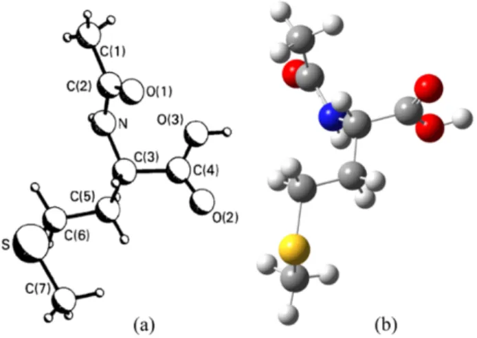
3.1. Molecular StructureIn 1985, the crystal structure of NAM has been reported by Ponnuswamy and Trotter [25] and five years later, re-determined it and its calcium adduct viz., Ca(N -acetyl-DL-methionine)2. H2O by Kim and Eriks [26]. Moovendaran and Natarajan reported structure and some other properties by recording X-ray, IR, UV-Vis-NIR, TGA/DTA, and pho-toluminescence spectra [27]. According to the best of our knowledge, in the literature, there are no available spectro-scopic and quantum chemical calculation results for this com-pound and, therefore, we calculated for the molecular struc-ture for the first time. For the compound, we have employed a 6-311++G(d,p) basis set at B3LYP and HSEH1PBE levels without symmetry constrain, to provide integrity and com-pared computed geometry parameters through experimental bond lengths and bond angles, which are taken from Refs. [25,26]. The atom numbering scheme for the molecule is shown in Fig. 1a) [25].
The optimized molecular geometry of the title molecule has been obtained in the gas phase at B3LYP/6-311++G(d,p) level using is shown in Fig. 1 (b). NAM contains a carboxyl group (-C4(O2)O3H), an N -acetyl group (-NHC2O1C1H3), and a side chain (-C5H2SC6H3C7H2) attached to a C3 atom (see Fig. 1). According to the available reported studies in the literature, the calculated lattice parameters are gathered in Table I, and for these analyses confirm that the crystal belongs to the monoclinic crystal system with space group P 21/c with Z = 4 number of formula units in the unit cell.
FIGURE 1. a) The experimental structure and b) the optimized molecular structure obtained by the B3LYP method for NAM.
Table II gives the unscaled theoretical geometry parame-ters and also experimental X-ray data of the title compound with the estimated standard deviations in parentheses. It is observed that calculated values of bond lengths and bond angles through both methods B3LYP and HSEH1PBE are closer to each other.
In consideration of the bond lengths, as shown in Table, most of the calculated values are higher than experimental ones but this can be explained by the differences in the solid-state and liquid or gas phases. The experimental values were performed for the solid phase, while the theoretical calcula-tions were for the isolated molecule in the gas phase. The observed differences of bond parameters between the calcu-lated and experimental values being most probably due to the subsistence of the crystal field along with the intermolecu-lar interactions have connected the molecules, which occur in the crystal.
It can be observed that the C1-C2, C3-C4, C3-C5, and C5-C6 carbon-carbon bond lengths on the molecule are smaller than typical C-C single (1.54 ˚A) bond, and the C2-N bond lengths in the peptide group of the title compound are much shorter than the normal C-N single bond that is referred to 1.49 ˚A, which confirms this bond to have some characteristics of a double or conjugated bond [28]. The C3-N carbon-nitrogen bond length is intermediate between C-N typical single bond characteristics, as presented in related compounds previously studied [29].
C-C bond lengths in the range of ∼ 1.509 − 1.546 ˚A are also close to the experimental bond lengths in the range of ∼ 1.483 − 1.531 ˚A, varying with a little difference. In the
TABLEI. The unit cell parameters, angel, volume and density for NAM.
a ( ˚A) b ( ˚A) c ( ˚A) β (◦C) V ( ˚A3) Density (g cm−3) References
5.882 (1) 9.285 (1) 21.934 (3) 124.88 (5) 982.7 (2) 1.292 (25)
5.8849 (6) 9.290 (1) 18.024 (2) 92.51 (1) 984.39 (21) 1.30 (26)
TABLEII. Experimental and calculated bond lengths ( ˚A), bond angles (◦) and torsion angles (◦) for NAM.
Parameters Experimental Theoretical
Bond lengths ( ˚A) [25] [26] B3LYP HSEH1PBE
C1-C2 1.489 (2) 1.483 (4) 1.518 1.509 C2-N 1.333 (2) 1.330 (3) 1.379 1.372 C2-O1 1.236 (2) 1.238 (3) 1.219 1.215 C3-C4 1.523 (2) 1.520 (3) 1.531 1.522 C3-C5 1.531 (2) 1.522 (3) 1.546 1.536 C3-N 1.445 (2) 1.447 (3) 1.454 1.443 C4-O2 1.207 (2) 1.205 (3) 1.204 1.201 C4-O3 1.300 (2) 1.300 (3) 1.353 1.343 C5-C6 1.518 (3) 1.526 (4) 1.530 1.522 C6-S 1.796 (2) 1.794 (3) 1.833 1.814 C7-S 1.786 (3) 1.764 (5) 1.827 1.809 Bond angles (◦) C1-C2-N 117.2 (1) 117.5 (3) 117.5 117.2 C1-C2-O1 122.6 (1) 122.5 (2) 122.2 122.4 N-C2-O1 120.2 (1) 120.0 (2) 120.2 120.4 C4-C3-C5 108.8 (1) 110.1 (2) 109.4 109.1 C4-C3-N 112.7 (1) 112.8 (2) 112.8 113.0 C5-C3-N 110.1 (1) 108.8 (2) 111.2 111.0 C3-C4-O2 122.4 (1) 122.5 (2) 124.8 124.7 C3-C4-O3 113.4 (1) 113.3 (2) 111.9 111.9 O2-C4-O3 124.2 (1) 124.2 (2) 123.3 123.4 C3-C5-C6 114.3 (1) 114.4 (2) 112.3 112.1 C5-C6-S 115.6 (1) 115.6 (2) 114.2 114.1 C2-N-C3 121.0 (1) 121.3 (2) 128.0 127.4 C6-S-C7 101.4 (1) 102.0 (2) 100.7 100.4 Torsion Angles (◦) O2-C4-C3-C5 - 80.3 -101.0 -99.0 O3-C4-C3-C5 - -97.1 77.5 79.3 O2-C4-C3-N - -157.2 134.7 136.9 C4-C3-C5-C6 - -172.4 167.3 168.3 C4-C3-N-C2 - 64.8 -90.6 -89.6 C5-C3-N-C2 - -173.4 146.1 147.5 C3-N-C2-O1 - 1.9 170.1 170.0 C3-N-C2-C1 - -177.1 -11.3 -11.4 N-C3-C4-O3 (ψ2) -25.5 (2) 25.4 -46.8 -44.7 N-C3-C5-C6 (χ1) -63.1 (2) 63.5 -67.5 -66.5 C3-C5-C6-S (χ2) -59.2 (2) 58.9 177.7 177.5 C5-C6-S-C7 (χ3) -59.3 (2) 60.4 -77.3 -75.6
title compound, the carbon sulfur bond lengths C6-S and C7-S are 1.833 ˚A and 1.827 ˚A with B3LYP and 1.814 ˚A and 1.809 ˚A with HSEH1PBE while the reported values are in the range ∼ 1.764 − 1.796 ˚A [25,26]. As discussed by John-son et al., the various DFT methods predict bond lengths, which are systematically too long (30). The results of the
calculated bond lengths of NAM molecule using DFT meth-ods have provided proof of these findings. According to Ta-ble I, the computed C2-N bond length (1.379 and 1.372 ˚A) is a bit longer than from experimental bond lengths (1.333 and 1.330 ˚A).
TABLEIII. Comparison of experimental and calculated vibration frequencies for NAM.
Assignments Experimental Theoretical
(27) B3LYP HSEH1PBE Scaled freq.a Ib IR Scaled freq.a IIRb υ(N-H)as 3343 3435 21.2700 3469 23.5340 υ(CH2)s 2968 2928 24.1035 2943 22.7840 υ(CH2-S)as 2920 2919 1.1755 2936 0.7793 υ(C=O) 2461 1740 326.5540 1776 330.5009 υ(C=O)s 1693 1681 495.0484 1718 515.5060 γ(N-H) 1618 1442 5.5508 1445 41.9708 β(C-N-H) 1560 1421 44.2275 1421 55.1498 υ(C-C) 1381 1297 68.8192 1303 47.8395 β(CH3) 1248 1284 185.7300 1287 84.4035 τ (CH2) 1190 1164 28.7162 1173 30.6745 υ(C-C-N) 1115 1082 5.3365 1099 3.8105 ρ(C-O-H) 1115 1082 5.3365 1099 3.8105 υ(C-N) 1042 1029 14.6397 1044 9.2707 υ(C-C-N) 1015 998 15.3698 961 5.4230 ρ(CH3) 961 934 2.8278 936 2.9764 υ(C-C) 895 798 0.7839 812 0.4921 ρ(CH2) 743 723 4.5378 723 5.5597 ω(N-H) 743 622 130.2450 623 133.9825 β(C-C) 590 567 25.7306 573 23.2463
υ: Stretching; β: in plane bending; γ: out?of plane bending; τ : twisting; ρ: rocking; ω: wagging.
aScaled frequencies are in unit of cm−1. bI
IRinfrared inten. are in unit of km mol−1.
The shortening of the bond length of C2-O1 (∼1.237 ˚A) and C4-O2 (∼1.206 ˚A) could be assigned a double bond char-acter, whereas the corresponding values for the title com-pound are 1.219 ˚A 1.215 ˚A (B3LYP) and 1.204 ˚A, 1.201 ˚A (HSEH1PBE).
Computed C1-C2-N, C1-C2-O1, and N-C2-O1 bond lengths of the side chain have the same values (respectively, 117◦, 122◦, and 120◦) with the experimental results. About the X-ray data, the bond angle C2-N-C3 is ∼ 121.2◦, whereas it is computationally 128.0◦ and 127.4◦ with B3LYP and HSEH1PBE methods, respectively, which provide the pos-sibility of the presence of hydrogen bond. According to liter-ature [25,26] only two hydrogen bonds are formed between neighboring molecules N-H· · · O2 and O3-H· · · O1.
On the other hand, the carbon-carbon-oxygen bond an-gles in the carboxyl group lie in the range 111.9-124.8◦, (DFT) and 113.3-122.5◦(XRD). The bond angles are C4-C3-C5=109.3◦, C4-C3-N=112.9◦, and C5-C3-N=111.1◦(DFT), and this asymmetry gives the interaction between the car-boxyl group and the neighboring peptide group and side chain.
3.2. Vibrational wavenumbers
Vibrational frequency calculations for the ground spin state of the NAM were calculated with the same level of theory calculations based on the optimized geometries. The experi-mental IR spectra were compared with the calculated spectra (Fig. 2), and also theoretical and experimental absorption fre-quencies and assignments of the title compound are listed in Table III. The vibrational band assignments have been made using the GaussView molecular visualization program [3].
According to the literature [31], there are disagreements in the calculated harmonic vibrational frequencies when they are compared to the experimental fundamental vibrational frequencies, and these discrepancies can partially be at-tributed to two errors: the neglect of anharmonicity and the inaccurate description of the electron-electron interaction. A scaling factor is commonly used to correct the calculated value to match the experimental observable. Therefore, we used 0.96 [32] scale factors for the computed frequencies of the NAM. All the experimental results were confirmed by Moovendaran and Natarajan [27].
FIGURE2. Experimental and calculated IR spectra for NAM. The asymmetric stretching mode v(N-H) is located 3343 cm−1 experimentally (3435 and 3469 cm−1 at DFT), while the weak stretching mode v(C=O) were around 2461 cm−1 experimentally (1740 and 1776 cm−1 at DFT) after we applied the scale factor. The calculated results by frequency analysis indicate deviations from experimental val-ues due to the intramolecular hydrogen bond between N and O. The bands corresponding to both out of the plane and in-plane C-H deformations are calculated in the region 1164-1287 cm−1. The bending vibrations for γ(N-H) and β(C-N-H) groups in IR spectra are observed in the region 1618 and 1560 cm−1, respectively, while the calculated vibration fre-quencies of them are ∼1444 cm−1 and 1421 cm−1, respec-tively. On the other hand, the bands at 1115, 1042, and 1015 cm−1 are assigned to the v(C-N) vibration, and the theoreti-cally computed values are observed in the wave region 1099-961 cm−1. The theoretically computed values of ρ(CH3) vi-bration mode at ∼935 cm−1show a good agreement with the experimental value at 961 cm−1. The IR band at 1693 cm−1 in the experimental IR spectrum of the title compound were designated to v(C=O) symmetric stretching fundamentals of C4 and O2 atoms, and this band appeared at 1681 cm−1 (B3LYP) and 1718 cm−1(HSEH1PBE) in the DFT calcula-tions. The v(CH2-S) stretching mode is another typical mode for NAM, and this vibration experimentally appeared at 2920 cm−1. Theoretically, this band could be assignable to: at 2919 cm−1 for the B3LYP method and 2936 cm−1 for the HSEH1PBE method.
3.3. NMR studies
The structural parameters are obtained with the B3LYP and HSEH1PBE functional and 6-311++G(d,p) basis set, and they are used to predict 13C and 1H NMR chemical shifts with the recommended GIAO, CSGT, and IGAIM ap-proaches and Tetramethylsilane (TMS) is used for refer-ence. The isotropic magnetic shielding values were used to calculate the isotropic chemical shifts (δ) with respect to TMS (δXiso = σisoT M S− σisoX ). The appropriate value of the 1H/13C isotropic chemical shifts for TMS (σT M S
iso ) was found
to be 184.0807/31.9723 ppm for GIAO, 182.3844/30.7302
ppm for IGAIM, and 182.3611/30.7287 ppm for CSGT by B3LYP/6-311++G(d,p). Those obtained by HSEH1PBE level were also found to be 188.3372/31.8158ppm for GIAO, 186.7863/30.5605 ppm for IGAIM, and 186.7641/30.5590 ppm for CSGT.
The experimental and theoretical values for13C and1H NMR chemical shifts of the title molecule are given in Ta-ble IV. The correlation values between the experimental and calculated chemical shifts in Table IV shows that all com-putations are in good agreement with the experimental data except the proton of the amino group in1H NMR. The chemi-cal shifts of the hydrogen atoms of the CH3 groups are 2.040 ppm (C1), 1.850 ppm (C7), and the hydrogen atom of the CH2groups are 1.93 (C5), 1.82 ppm (C5), 2.48 ppm (C6) ex-perimentally. In our present investigation, the chemical shifts of hydrogen atoms in the CH3 groups are calculated in the range 1.036- 2.270 ppm, and in the CH2 groups are in the range 1.324 - 2.713 ppm. The chemical shift value of the H atom bonded to C3 is calculated in the range 3.668 - 2.949 ppm and observed at 4.284 ppm.
There are seven carbon atoms in the title molecule, and the13C NMR spectrum of the NAM exhibits seven signals. The signal at 173.36 ppm is assigned to the C4 carbon atom of the carboxyl group computed as in the range 178.167 -173.800 ppm. As would be expected, 13C NMR chemical shifts for C4 and C2 atoms are calculated as higher than the other C atoms due to the electronegativity properties of O and N atoms. As can be seen from Table IV, the theoretical13C chemical shift results for NAM are generally closer to the ex-perimental13C shift data.
3.4. Non-linear optical (NLO) analysis
The electric dipole (hyper) polarizabilities of a molecule are a measure of the ability to respond to an electric field (E) [33] and determine the strength of molecular interactions (long-range intermolecular induction and dispersion forces), the cross sections of different scattering, collision processes [34] and the nonlinear optical properties of the system [35].
In the presence of the electric field, the expectation value of the electric dipole moment is the sum of a permanent dipole moment and the contribution induced by the field:
hµi = µ0+ αE +
1 2βE
2+ · · · (1) In this expression, α is the linear polarizability, and β is the first hyperpolarizability (else, sometimes, second-order or quadratic hyperpolarizability).
The relation between the three Cartesian components of the dipole moment vector (~µ) and the three Cartesian com-ponents of the electric field vector ( ~E) requires nine pro-portionality factors, therefore α is a second rank tensor that can be described by a 3 × 3 matrix with the 9 components. The second interaction involves evaluating the effect on the dipole moment of combinations of Cartesian components of the field; therefore, β is the third rank tensor that can be
TABLEIV. Experimental and theoretical13C and1H isotropic chemical shifts for NAM.
Atom Experimental Theoretical
GIAO IGAIM CSGT
13
C B3LYP HSEH1PBE B3LYP HSEH1PBE B3LYP HSEH1PBE
C4 173.36 178.167 176.345 175.443 173.832 175.411 173.800 C2 169.38 174.759 172.713 172.615 170.775 172.589 170.749 C3 50.86 63.419 60.409 61.876 58.939 61.852 58.915 C6 30.65 39.776 36.948 39.541 36.708 39.527 36.694 C5 29.63 39.296 36.922 38.774 36.526 38.761 36.512 C1 22.26 22.431 22.061 20.300 19.889 20.297 19.885 C7 14.46 21.013 19.251 21.147 19.419 21.148 19.419 R2 0.9968 0.9980 0.9960 0.9974 0.9960 0.9974 1 H H (O3) - 5.641 5.557 4.739 4.659 4.741 4.661 H (N) 8.16 4.587 4.502 4.055 3.979 4.047 3.971 H (C3) 4.284 3.688 3.701 2.962 2.998 2.949 2.985 H (C6) 2.48 2.713 2.707 2.300 2.278 2.292 2.269 H (C6) 2.48 2.356 2.309 1.645 1.605 1.639 1.599 H (C7) 1.850 2.270 2.230 1.919 1.862 1.919 1.861 H (C7) 1.850 1.973 1.888 1.639 1.537 1.639 1.537 H (C1) 2.040 1.881 1.841 1.203 1.168 1.198 1.163 H (C7) 1.850 1.853 1.858 1.270 1.240 1.271 1.242 H (C1) 2.040 1.852 1.826 1.119 1.091 1.115 1.087 H (C1) 2.040 1.851 1.835 1.065 1.039 1.062 1.036 H (C5) 1.93 1.829 1.807 1.392 1.379 1.384 1.370 H (C5) 1.82 1.809 1.780 1.353 1.331 1.345 1.324 R2 0.8924 0.8807 0.8445 0.8396 0.8445 0.8400
scribed by a 3 × 3 × 3 matrix with the 27 components. Ac-cording to Kleinman symmetry, α can be reduced into 6 com-ponents (αxy = αyx, αxz = αzx, αzy = αyz) and β can
be reduced into 10 components (βxyy = βyxy = βyyx =
βyyz = βyzy = βzyz...) [36]. In the lower tetrahedral point
group, the Gaussian 09W output file printout of the α matrix is αxx, αxy, αyy, αxz, αyz, αzz, and β matrix is βxxx, βxxy,
βxyy, βyyy, βxxz, βxyz, βyyz, βxzz, βyzz, βzzz.
For the title molecule, we use the following expressions to calculate. The dipole moment,
µ =qµ2
x+ µ2y+ µ2z. (2)
The isotropic polarizability, hαi = 1
3(αxx+ αyy+ αzz). (3)
The anisotropy polarizability,
∆α = ( 1 2 · (αxx+ αyy)2+ (αyy+ αzz)2 + (αzz+ αxx)2+ 6 ¡ α2 xy+ α2xz+ α2yz ¢¸)1/2 (4)
And the magnitude of the first order hyperpolarizability tensor (hβi) is defined in terms of three axes as:
hβi =£(βx)2+ (βy)2+ (βz)2 ¤1/2
(5) where, in general, components equation of β can be calcu-lated using the following equation:
βi= βiii+1 3 X i6=j β2
ijj+ βjij2 + β2jji
1/2
(6)
The complete equation for the β in terms of Cartesian components leads to:
hβi =h¡βxxx+ βxyy+ βxzz ¢2
+¡βyyy+ βxxy+ βyzz ¢2
+¡βzzz+ βxxz+ βyyz ¢2i1/2
(7) In this study at DFT with 6-311++G(d,p) basis set, based on the finite field (FF) approach, parameters such as the total
TABLEV. Total static dipole moment (µ), the mean polarizability (hαi), the anisotropy of the polarizability (∆α) and the mean first-order hyperpolarizability (hβi) for NAM (units in esu).
Property B3LYP HSEH1PBE pNA Urea
µx -1.184 -1.101 µy 1.601 1.637 µz 0.314 0.414 µ/ Debye 2.016 2.015 6.2 [40] 4.56 [41] αxx 20.5 20.1 αyy 17.6 17.2 αzz 17.1 16.8 hαi/10−24 18.4 18.1 17.0 [40] ∆α/10−24 3.15 3.10 βx 1.58 1.64 βy 1.33 1.15 βz -0.69 -0.62 hβi/10−30 2.17 2.10 16.9 [42,43]
static dipole moment (µ), the component of the dipole mo-ment (µi(i = x, y, z)), the isotropic polarizability (hαi),
the anisotropy of the polarizability (∆α), the first order hy-perpolarizability (hβi) are calculated by using B3LYP and HSEH1PBE functionals for NAM and collected in Table V. Also, Table V collects these parameters for two reference molecules, i.e., urea and para-nitroaniline (pNA). Urea and pNA are well-known prototypical donor-acceptor organic standards with large hyperpolarizability [37-42]. The first-order hyperpolarizability values for NAM are higher than urea and pNA molecules.
All the tensors values of the Gaussian 09W program out-put file are reported in the atomic system of the unit (a.u.) and therefore the calculated values are converted into elec-trostatic units (esu) (for hαi and ∆α1 a.u. = 0.1482 × 10⁻²⁴ esu and for hβi 1 a.u. = 8.6393 × 10⁻³³esu).
3.5. HOMO-LUMO gap (HLG) analysis
The terms HOMO and LUMO are shorthand for the high-est occupied and lowhigh-est unoccupied molecular orbitals, re-spectively. HOMO is the orbital of the highest energy for a molecule that contains electrons, and the empty molecu-lar orbital that is closet in energy to the HOMO is called the LUMO. The HOMO and LUMO are the frontier orbitals: the frontier is the site of much of the reactive and spectroscopic activity of the species.
The DFT calculations that are the main working horse for ground-state properties such as atomic structures and binding energies significantly underestimate band gaps in particular the Kohn-Sham eigen energy. In prior studies, band struc-tures predicted by the DFT method are reported to match ex-perimental observations [43].
Ionization energy is the energy required to remove an electron from a neutral atom. The first ionization energy can also be defined as the energy to remove one electron from a
neutral atom, to make an ion with a positive charge. Electron affinity is to do with the energy required to add an electron to a neutral atom to become a negative ion. To evaluate the relative reducibility of the title compound, ionization poten-tial and electron affinity were calculated from differences of total energies of the neutral (ENEUTRAL) and ionic systems
(ECATION, EANION) obtained from optimized molecular
con-figurations using the following equations. The ionization potential,
IP = ECATION− EANION. (8)
The electron affinity,
EA = ENEUTRAL− EANION. (9)
The restricted calculation was used in computing the total energy for the neutral ground state, whereas the unrestricted model was used for cations and anions.
According to Koopmans’ theorem, the first ionization en-ergy directly relates to removing an electron from the orbital is given by the negative value of the energy of the orbital (IP = −EHOMO) if the wave function relaxations are
ig-nored, as calculated within the Hartree-Fock theorem [44]. In the literature, this theorem has become generally accepted.
Based on the HOMO and LUMO energy for the title molecule, we use the following expressions to calculate.
The electronegativity:
χ = (IP + EA)/2. (10)
The chemical potential:
µ = −χ = −(IP − EA)/2, (11) [45].
TABLEVI. Frontier orbital energies and calculated physico-chemical properties for NAM.
Property B3LYP HSEH1PBE
HOMO energy EHOMO(eV) -6.364 -6.180
LUMO energy ELUMO(eV) -0.899 -1.006
HOMO-LUMO energy gap ∆E (eV) 5.464 5.174
Ionization potential IP (eV) 8.405 8.389
Electron affinity EA (eV) -0.225 -0.255
Electronegativity χ (eV) 4.090 4.067
Chemical potential µ (eV) -4.090 -4.067
Global hardness η (eV) 4.315 4.322
Global softness S (eV−1) 0.232 0.231
Electrophilicity ω (eV) 1.938 1.913
FIGURE3. The FMOs, energies (in eV), and HOMO-LUMO energy gaps of NAM calculated at B3LYP.
The hardness:
η = (IP − EA)/2 (12)
[46-48 ]. The softness:
S = 1/η. (13)
The electrophilicity (in terms of chemical potential and hardness):
ω = µ2/2η. (14)
The single point energies and frontier molecular orbitals (FMOs) were determined according to quantum chemical calculations, and in these calculations, molecular orbital 51 (the HOMO) is described by as much as 363 molecular or-bital coefficients, each of the thirteen hydrogen atoms con-tributing 7, the three oxygen, seven carbon, and nitrogen
atoms contributing 22 and sulfur contributing 30 basis func-tions. The quantum chemical parameters of the molecule are summarized in Table VI.
The HOMO which is the outermost orbital contain-ing electrons and its vicinal orbital, play an important role in electron donor, while the LUMO, which is the inner-most orbital containing free places to accept electrons and its vicinal orbital, play an important role in electron ac-ceptor. The energy value of HOMO was computed as -6.364 eV (B3LYP), -6.180 eV (HSEH1PBE) and LUMO as -0.899 eV (B3LYP), -1.006 eV (HSEH1PBE). The HOMO-LUMO energy gap, which reflects optical and electric properties and chemical activity of the molecule was cal-culated 5.464 eV (B3LYP) and 5.174 eV (HSEH1PBE); both the values were strongly supported the previous work (5.2 eV [27]). To understand the bonding scheme and fa-cilitate discussion of the transitions of the title compound, contour plots of FMOs, HOMO-1 to LUMO+1, are pre-sented in Fig. 3 for the molecule in the gas phase. The red and green colors of the molecular orbital’s plot show the
FIGURE4. DOS spectrum of NAM using B3LYP/6-311++G(d,p) level in the range of -20 to 15 eV.
positive and negative phases, respectively. In the NAM, the HOMO represents the charge density localized over the side chain (-C5H2SC6H3C7H2) unit attached to a C3 atom; by contrast, the LUMO is located over the carboxyl group unit.
In order to obtain the total density of the state (DOS) curve of NAM as shown in Fig. 4, the output which extracts data from Gaussian .log file was analyzed and plotted as a graph using the Gauss Sum 3.0 program [49].
3.6. Electronic transitions
B3LYP/6-311++G(d,p) level using the TDDFT approach method on the previously optimized ground-state geometry
of NAM was employed to simulate the optical properties of the molecule for the gas phase. The computed vertical ex-cited singlet states, transition energies (E), wavelength (λ), and oscillator strengths (f ) in the gas phase are tabulated in Table VII.
As it was clear from TDDFT/B3LYP calculation, the theoretical absorption bands in the UV region are corre-spond to two absorption maximum at λ = 257.21 with E = 4.8204 eV and f = 0.0725 and λ = 384.61 nm with E = 3.2237 eV and f = 0.0092 for gas phase at 6-311++G(d,p) basis set. Also, the maximum absorption wavelength corresponds to the electronic transition from the HOMO to LUMO with 100%. In the visible region, we found no calculated vertical transitions and seems to be with the same character as the experimental absorption spectra in this region. According to the experimental spectrum, the UV transparency cutoff occurs around 235 nm, and there is no absorption, and the crystal is colorless in the entire UV-Vis regions [27].
3.7. Molecular electrostatic potential
The electrostatic potential V (~r) at any point ~r for a molecu-lar system having an electronic density function ρ(~r) is given by: V (~r) =X A ZA | ~RA− ~r| − Z ρ(~r)d~r0 |~r0− ~r|, (15)
TABLEVII. Absorption wavelengths (λ), excitation energies (E), oscillator strengths (f ) and orbital configurations spectral transitions for NAM calculated at TDDFT (B3LYP/6-311++G(d,p)) (H=HOMO L=LUMO, H-2=HOMO-2, etc).
Excited Transition Spin
States Character Contribution Multiplicity Symmetry E (eV) λ(nm) f
S1 H → L 100% Singlet A 2.2552 549.76 0.0005 S2 H-4 → L 8% Singlet A 3.2237 384.61 0.0092 H-2 → L 66% H-1 → L 25% S3 H-4 → L 4% Singlet A 3.3471 370.43 0.0002 H-2 → L 21% H-1 → L 75% S4 H-4 → L 76% Singlet A 4.1504 298.73 0.0014 H-3 → L 11% H-2 → L 12% S5 H-3 → L 2% Singlet A 4.7631 260.3 0.0015 H → L+1 72% H → L+3 21% H → L+4 3% S6 H-4→ L 10% Singlet A 4.8204 257.21 0.0725 H-3 → L 86% Rev. Mex. F´ıs. 66 (6) 749–760
FIGURE5. MEP surface of NAM using B3LYP/6-311++G(d,p) level of theory.
in which ZAis the charge on nucleus A, located at ~RA. In
this expression, the two terms represent the nuclear and elec-tronic contributions to the potential, respectively.
The electrostatic potential is a physical observable, which can be determined experimentally by diffraction methods (50) and computationally. The Gaussian program calculates the potential at the points that located on the molecule’s size on the ρ(~r) isodensity surface and then according to poten-tial assigns colors to these points. In this way, MEP sur-face maps are surveyed. As is well known, MEP sursur-faces are useful diagrams that illustrate the charge distributions of molecules three-dimensionally. These maps allow us to visualize the charge distributions, charge related properties, size, and shape of molecules. Moreover, MEP provides inter-preting and predicting relative reactivities sites for the elec-trophilic and nucleophilic attack, studies of the biological recognition process, hydrogen bonding interactions, molec-ular cluster, and crystal behavior, and the correlation and pre-diction of a wide range of macroscopic properties [51-55].
Figure 5 presents the MEP surface of the molecule under investigation is composed by using B3LYP/6-311++G(d,p) level of theory and a value of 0.0004 a.u. for the isodensity surface. On the map, the most negative (the lowest) and
pos-itive (the highest) electrostatic potentials are colored red and blue, respectively, and intermediate potentials are colored in-termediate color spectrum.
Furthermore, the MEP map provides a visual method to understand the relationship between the structure and activ-ity of the molecule. It is used to identify electrophilic and nucleophilic sites of a molecule. It can be seen from Fig. 5, the obtained electrostatic potential is composed of the pos-itive electrostatic potential around the O1 atom in the pep-tide group, which is the most reactive site for nucleophilic at-tack. The negative electrostatic potential is mainly localized around the N-H in the peptide group region and O3-H in the carboxyl group, which is the possible site for electrophilic at-tack. The remaining green areas of the molecule (C-C bonds) are surrounded by close to zero electrostatic potential.
4.
Conclusions
This study analyzes computational studies on N-acetyl-DL-methionine. We have calculated the geometric parameters, vibrational frequencies,13C and1H NMR values, the nonlin-ear optical properties, frontier molecular orbitals, UV-Vis cal-culations, and molecular electrostatic potential surface of the monomer form of the N-acetyl-DL-methionine using DFT methods with 6-311++G(d,p) basis set. In particular, a good agreement between experimental and calculated vibrational frequencies has been observed. DFT/HSEH1PBE method has shown a better fit to experimental ones than DFT/B3LYP in evaluating vibrational frequencies. The nonlinear optical behavior of the title molecule was investigated by the deter-mination of the dipole moment, the polarizability, and the first-order hyperpolarizability. The HOMO-LUMO energy gap results reveal that the NAM is the highest kinetic sta-bility and can be termed as a hard molecule with the largest HOMO-LUMO gap of average 5.319 eV. The molecular elec-trostatic potential surface has also been drawn to explain the activity of the title molecule. In general, we can easily state that the theoretical results obtained from our studies for the mentioned compound are in good agreement with the exper-imental data. Hence, we anticipate that the present quantum chemical study will be very useful in the understanding of the structure, activity, and dynamics of the molecule. We hope that our results for N-acetyl-DL-methionine would provide more information for further investigation in the future.
1. T. Willke, Methionine production-a critical review, Appl. Mi-crobiol. Biotechnol. 98 (2014) 9893, https://doi.org/ 10.1007/s00253-014-6156-y.
2. M. J. Frisch, H. P. Hratchian, and A. B. Nielsen, Gaussian 09: Programmer’s Reference, (Gaussian, 2009).
3. R. Dennington, T. Keith, and J. Millam, GaussView, Version 5, (Semichem Inc., Shawnee Mission, KS, 2009).
4. A. D. Becke, Density-functional exchange-energy approxi-mation with correct asymptotic behavior, Phys. Rev. A 38 (1988) 3098,https://doi.org/10.1103/PhysRevA. 38.3098.
5. A. D. Becke, Density functional thermochemistry. IV. A new dynamical correlation functional and implications for exact ex-change mixing, J. Chem. Phys. 104 (1996) 1040, https: //doi.org/10.1063/1.470829.
6. C. Lee, W. Yang, and R. G. Parr, Development of the Colle-Salvetti correlation-energy formula into a functional of the elec-tron density, Phys. Rev. B 37 (1988) 785, https://doi. org/10.1103/PhysRevB.37.785.
7. J. Heyd, G. E. Scuseria, and M. Ernzerhof, Hybrid func-tionals based on a screened Coulomb potential, J. Chem. Phys. 118 (2003) 8207,https://doi.org/10.1063/1. 1564060.
8. J. Heyd and G. E. Scuseria, Assessment and validation of a screened Coulomb hybrid density functional, J. Chem. Phys. 120 (2004) 7274,https://doi.org/10.1063/1. 1668634.
9. J. P. Perdew, K. Burke, and M. Ernzerhof, Gener-alized gradient approximation made simple, Phys. Rev. Lett. 77 (1996) 3865, https://doi.org/10.1103/ physrevlett.77.3865.
10. M. E. Ochoa, P. Labra-V´azquez, N. Farf´an, and R. San-tillan, Designed synthesis and crystallization of isomorphic molecular gyroscopes with cell-like bilayer self-assemblies, Cryst. Growth Des. 18 (2018) 2795, https://doi.org/ 10.1021/acs.cgd.7b01542.
11. F. London, Th´eorie quantique des courants interatomiques dans les combinaisons aromatiques, J. Phys. Radium 8 (1937) 397, https://doi.org/10.1051/jphysrad: 01937008010039700.
12. R. McWeeny, Perturbation theory for the Fock-Dirac density matrix, Phys. Rev. 126 (1962) 1028,https://doi.org/ 10.1103/PhysRev.126.1028.
13. R. Ditchfield, Self-consistent perturbation theory of diamag-netism: I. A gauge-invariant LCAO method for N.M.R. chem-ical shifts, Mol. Phys. 27 (1974) 789,https://doi.org/ 10.1080/00268977400100711.
14. K. Wolinski, J. F. Hinton, and P. Pulay, Efficient implementa-tion of the gauge-independent atomic orbital method for NMR chemical shift calculations, J. Am. Chem. Soc. 112 (1990) 8251,
https://doi.org/10.1021/ja00179a005.
15. J. R. Cheeseman, G. W. Trucks, T. A. Keith, and M. J. Frisch, A comparison of models for calculating nuclear magnetic res-onance shielding tensors, J. Chem. Phys. 104 (1996) 5497, https://doi.org/10.1063/1.471789.
16. T. A. Keith and R. F. W. Bader, Calculation of mag-netic response properties using atoms in molecules, Chem. Phys. Lett. 194 (1992) 1, https://doi.org/10.1016/ 0009-2614(92)85733-Q.
17. T. A. Keith and R. F. W. Bader, Calculation of magnetic re-sponse properties using a continuous set of gauge transforma-tions, Chem. Phys. Lett. 210 (1993) 223, https://doi. org/10.1016/0009-2614(93)89127-4.
18. P. Labra-V´azquez, A. Z. Lugo-Aranda, M. Maldonado-Dom´ınguez, R. Arcos-Ramos, M. Carre ´on-Castro, R. Santill´an, and N. Farf´an, On the molecular structure of (E)-3-(9H-fluoren-2-yl)-1-(pyridin-2-yl) prop-2-en-1-one, theoretical calculations and SXRD studies, J. Mol. Struct. 1101 (2015) 116,https:
//doi.org/10.1016/j.molstruc.2015.08.023.
19. T. Pawlak, D. Czajkowska-Szczykowska, I. Jastrzebska, R. Santillan, B. Seroka, J. Maj, J. W. Morzycki, P. Labra-V´azquez,
N. Farf´an, G. D. Bujacz, and M. J. Potrzebowski, Influ-ence of Hydrogen/Fluorine Substitution on Structure, Ther-mal Phase Transitions, and Internal Molecular Motion of Aro-matic Residues in the Crystal Lattice of Steroidal Rotors, Cryst. Growth Des. 20 (2020) 2202, https://doi.org/ 10.1021/acs.cgd.9b01179.
20. E. Runge and E. K. U. Gross, Density-functional theory for time-dependent systems, Phys. Rev. Lett. 52 (1984) 997, https://doi.org/10.1103/PhysRevLett.52. 997.
21. R. Bauernschmitt and R. Ahlrichs, Treatment of elec-tronic excitations within the adiabatic approximation of time dependent density functional theory, Chem. Phys. Lett. 256 (1996) 454, https://doi.org/10.1016/ 0009-2614(96)00440-X.
22. E. K. U. Gross, J. F. Dobson, and M. Petersilka, Density Functional Theory II: Relativistic and Time Dependent Exten-sions, edited by R. F. Nalewajski (Springer, Berlin, Heidel-berg, 1996), pp. 81-172, https://doi.org/10.1007/ BFb0016643.
23. K. Burke and E. K. U. Gross, Density Functionals: Theory and Applications, edited by D. Joubert (Springer, Berlin, Heidel-berg, 1998), pp. 116-146,https://doi.org/10.1007/ BFb0106735.
24. M. E. Casida, C. Jamorski, K. C. Casida, and D. R. Salahub, Molecular excitation energies to high-lying bound states from time-dependent density-functional response theory: Character-ization and correction of the time-dependent local density ap-proximation ionization threshold, J. Chem. Phys. 108 (1998) 4439,https://doi.org/10.1063/1.475855. 25. M. N. Ponnuswamy and J. Trotter, N-Acetyl-DL-methionine,
C7H13NO3S, Acta Cryst. C 41 (1985) 917,https://doi. org/10.1107/S0108270185005996.
26. E. E. Kim, A. Sicignano, and K. Eriks, Binding of calcium to amino acids: the crystal structure of pentaaquobis(hydroxy-(L)-prolinato)calcium, Ca(C5H8O3N)2.5H2O, J. Am. Chem.
Soc. 107 (1985) 6042, https://doi.org/10.1021/ ja00307a036.
27. K. Moovendaran and S. Natarajan, Growth of bulk single crys-tal of N-acetyl DL-methionine and its spectral characterization, Spectrochim. Acta A 135 (2015) 317,https://doi.org/ 10.1016/j.saa.2014.07.012.
28. W. He, G. Zhou, J. Li, and A. Tian, Molecular design of analogues of 2,6-diamino-3,5-dinitropyrazine-1-oxide, J. Mol. Struct. 668 (2004) 201,https://doi.org/10.1016/j. theochem.2003.10.058.
29. N. Dege, N. S¸eny¨uz, H. Bat?, N. G¨unay, D. Avc?, ¨
O. Tamer, and Y. Atalay, The synthesis, characteri-zation and theoretical study on nicotinic acid [1-(2,3-dihydroxyphenyl)methylidene]hydrazide, Spectrochim. Acta A 120 (2014) 323, https://doi.org/10.1016/j.saa. 2013.10.030.
30. B. G. Johnson, P. M. W. Gill, and J. A. Pople, The performance of a family of density functional methods, J. Chem. Phys. 98 (1993) 5612,https://doi.org/10.1063/1.464906.
31. A. P. Scott and L. Radom, Harmonic vibrational frequencies: an evaluation of Hartree-Fock, Møller-Plesset, quadratic configu-ration interaction, density functional theory, and semiempiri-cal ssemiempiri-cale factors, J. Phys. Chem. 100 (1996) 16502, https: //doi.org/10.1021/jp960976r.
32. H. Pir, N. G¨unay, D. Avcı, and Y. Atalay, Molecular struc-ture, vibrational spectra, NLO and NBO analysis of bis(8-oxy-1-methylquinolinium)hydroiodide, Spectrochim. Acta A 96 (2012) 916, https://doi.org/10.1016/j.saa. 2012.07.044.
33. C. R. Zhang, H. S. Chen, and, G. H. Wang, Structure and prop-erties of semiconductor microclusters GanPn(n=1-4): A first principle study, Chem. Res. Chin. Univ. 20 (2004) 640. 34. Y. Sun, X. Chen, L. Sun, X. Guo, and W. Lu, Nanoring
struc-ture and optical properties of Ga8As8, Chem. Phys. Lett. 381 (2003) 397, https://doi.org/10.1016/j.cplett. 2003.09.115.
35. O. Christiansen, J. Gauss, and J. F. Stanton, Frequency-dependent polarizabilities and first hyperpolarizabilities of CO and H2O from coupled cluster calculations, Chem. Phys. Lett. 305 (1999) 147, https://doi.org/10.1016/ S0009-2614(99)00358-9.
36. D. A. Kleinman, Nonlinear dielectric polarization in optical media, Phys. Rev. 126 (1962) 1977, https://doi.org/ 10.1103/PhysRev.126.1977.
37. D. A. Dixon and N. Matsuzawa, Density functional study of the structures and nonlinear optical properties of urea, J. Phys. Chem. 98 (1994) 3967, https://doi.org/10.1021/ j100066a011.
38. A. J. Garza, G. E. Scuseria, S. B. Khan, and A. M. Asiri, Assessment of long-range corrected functionals for the pre-diction of non-linear optical properties of organic materials, Chem. Phys. Lett. 575 (2013) 122,https://doi.org/10. 1016/j.cplett.2013.04.081.
39. L. T. Cheng, W. Tam, S. H. Stevenson, G. R. Meredith, G. Rikken, and S. R. Marder, Experimental investigations of or-ganic molecular nonlinear optical polarizabilities. 1. Meth-ods and results on benzene and stilbene derivatives, J. Phys. Chem. 95 (1991) 10631, https://doi.org/10.1021/ j100179a026.
40. W. D. Kumler and G. M. Fohlen, The Dipole Moment and Structure of Urea and Thiourea1, J. Am. Chem. Soc. 64 (1942) 1944,https://doi.org/10.1021/ja01260a054. 41. C. C. Teng and A. F. Garito, Dispersion of the nonlinear
second-order optical susceptibility of organic systems, Phys. Rev. B 28 (1983) 6766,https://doi.org/10.1103/PhysRevB. 28.6766.
42. M. St¨ahelin, D. M. Burland, and J. E. Rice, Solvent depen-dence of the second order hyperpolarizability in p-nitroaniline, Chem. Phys. Lett. 191 (1992) 245,https://doi.org/10. 1016/0009-2614(92)85295-L.
43. R. W. Godby, M. Schl¨uter, and L. J. Sham, Self-energy op-erators and exchange-correlation potentials in semiconductors, Phys. Rev. B 37 (1988) 10159, https://doi.org/10. 1103/PhysRevB.37.10159.
44. T. Koopmans, ¨Uber die Zuordnung von Wellenfunktionen und Eigenwerten zu den einzelnen Elektronen eines Atoms, Physica 1 (1934) 104, https://doi.org/10.1016/ S0031-8914(34)90011-2.
45. R. S. Mulliken, A new electroaffinity scale; together with data on valence states and on valence ionization potentials and elec-tron affinities, J. Chem. Phys. 2 (1934) 782,https://doi. org/10.1063/1.1749394.
46. R. G. Pearson, Hard and soft acids and bases, J. Am. Chem. Soc. 85 (1963) 3533, https://doi.org/10. 1021/ja00905a001.
47. R. G. Pearson, Hard and soft acids and bases, HSAB, part 1: Fundamental principles, J. Chem. Educ. 45 (1968) 581, https://doi.org/10.1021/ed045p581.
48. R. G. Pearson, Maximum chemical and physical hardness, J. Chem. Educ. 76 (1999) 267, https://doi.org/10. 1021/ed076p267.
49. N. M. O’Boyle, A. L. Tenderholt, and K. M. Langner, cclib: A library for package-independent computational chemistry al-gorithms, J. Comput. Chem. 29 (2008) 839,https://doi. org/10.1002/jcc.20823.
50. P. Politzer and D. G. Truhlar, Chemical applications of atomic and molecular electrostatic potentials: reactivity, structure, scattering, and energetics of organic, inorganic, and biolog-ical systems (Springer, New York, 2013), pp. 1-6, https:
//doi.org/10.1007/978-1-4757-9634-6.
51. E. Scrocco and J. Tomasi, New concepts II, edited by A. Davison and M. J. S. Dewar (Springer, Berlin, Heidelberg, 1973), pp. 95-170, https://dx.doi.org/10.1007/ 3-540-06399-4 6.
52. E. Scrocco and J. Tomasi, Advances in quantum chem-istry, edited by P. L¨owdin (Acedemic Press, New York, 1978), pp.115-193,https://doi.org/10.1016/ S0065-3276(08)60236-1.
53. P. Politzer and K. C. Daiker, The force concept in chemistry, edited by B. M. Deb (Van Nostrand Reinhold, New York, 1981), pp. 294-387.
54. P. Politzer, P. R. Laurence, and K. Jayasuriya, Molecular elec-trostatic potentials: An effective tool for the elucidation of bio-chemical phenomena, Environ. Health Perspect. 61 (1985) 191,
https://doi.org/10.1289/ehp.8561191.
55. P. Politzer and J. S. Murray, The fundamental nature and role of the electrostatic potential in atoms and molecules, Theor. Chem. Acc. 108 (2002) 134, https://doi.org/ 10.1007/s00214-002-0363-9.