REGULAR ARTICLE
A novel homozygous nonsense mutation in
CAST associated
with PLACK syndrome
Şehime Gülsün Temel1,2&B. Karakaş3&Ü. Şeker4&B. Turkgenç5&Ö. Zorlu4&H. Sarıcaoğlu4&Ç. Oğur6&Ö. Kütük7& D. P. Kelsell8&M. C. Yakıcıer9
Received: 18 September 2018 / Accepted: 8 July 2019
# Springer-Verlag GmbH Germany, part of Springer Nature 2019 Abstract
Peeling skin syndrome is a heterogeneous group of rare disorders. Peeling skin, leukonychia, acral punctate keratoses, cheilitis and knuckle pads (PLACK syndrome, OMIM616295) is a newly described form of PSS with an autosomal recessive mode of inheritance. We report a 5.5-year-old boy with features of PLACK syndrome. Additionally, he had mild cerebral atrophy and mild muscle involvements. Whole exome sequencing was performed in genomic DNA of this individual and subsequent analysis revealed a homozygous c.544G > T (p.Glu182*) nonsense mutation in the CAST gene encoding calpastatin. Sanger sequencing confirmed this variant and demonstrated that his affected aunt was also homozygous. Real-time qRT-PCR and immunoblot analysis showed reduced calpastatin expression in skin fibroblasts derived from both affected individuals compared to hetero-zygous family members. In vitro calpastatin activity assays also showed decreased activity in affected individuals. This study further supports a key role for calpastatin in the tight regulation of proteolytic pathways within the skin.
Keywords Peeling skin syndrome . PLACK syndrome . Whole exome sequencing . CAST gene . Calpastatin
Introduction
Peeling skin syndrome (PSS; OMIM 270300) is characterized by continuous shedding of the stratum corneum of the epider-mis accompanied by diverse clinical findings including ery-thema, vesicular lesions, or other ectodermal features (Kurban and Azar1969; Levy and Goldsmith1982; Hashimoto et al.
2000; Judge et al.2004). It is classified into two clinical forms; either acral PSS (APSS; OMIM 609796) or generalized PSS (GPSS; OMIM 270300) (Bowden 2011). APSS involves mainly skin fragility of the palmar, plantar and dorsal part of hands and feet. Previously, mutations in genes encoding for transglutaminase (TGM5) and cysteine protease inhibitor cystatin A (CSTA; OMIM 184600), which are known to have Part of this research was presented as a poster presentation at the
American Academy of Dermatology Annual Meeting 2018, San Diego and the abstract was published in the Journal of American Academy Dermatology (JAAD) (2018): Volume 79, Issue 3, Supplement 1, Page AB16, DOI:https://doi.org/10.1016/j.jaad.2018.05.108.
* Şehime Gülsün Temel
[email protected]; [email protected]
1 Faculty of Medicine, Department of Medical Genetics, University of
Uludag, Bursa, Turkey
2
Faculty of Medicine, Department of Histology & Embryology, University of Uludag, Bursa, Turkey
3
Molecular Biology, Genetics and Bioengineering Program, Sabancı University, Istanbul, Turkey
4 Faculty of Medicine, Department of Dermatology, University of
Uludag, Bursa, Turkey
5 Acıbadem Genetic Diagnosis Center, University of Acıbadem,
Istanbul, Turkey
6
Bahçeci Genetic Diagnosis Center, Altunizade, Istanbul, Turkey
7
Faculty of Medicine, Department of Medical Genetics, Baskent University, Adana, Turkey
8
Centre for Cell Biology and Cutaneous Research, The Blizard Institute, Barts and The London School of Medicine and Dentistry, Queen Mary University of London, London E1 2AT, UK
9 Faculty of Arts and Sciences, Department of Molecular Biology and
Genetics, University of Acibadem, Istanbul, Turkey / Published online: 7 August 2019
key roles in cross-linking of cornified cell envelope proteins in epidermis, have been associated with APSS (Cassidy et al.
2005; Kharfi et al.2009; Krunic et al.2013). GPSS can pres-ent with severe pruritis, atopy, patchy feeling of pres-entire skin, food allergies, repeated episodes of angioedema, urticaria and asthma, which can be further divided into inflammatory or non-inflammatory GPSS. Mutations in corneodesmosin (CDSN; OMIM 602593) and CHST8 (OMIM 610190), encoding N-acetylgalactosamine-4-O-sulfotransferase have been associated with inflammatory GPSS (Oji et al.2010; Israeli et al.2011; Cabral et al.2012).
Recently, a new recessive form of GPSS with peeling skin, leukonychia, acral punctate keratoses, cheilitis and knuckle pads (termed PLACK syndrome) has been described and is associated with the loss of function mutations in the CAST gene (OMIM 114090; Refseq NM_001042440.4) encoding calpastatin (Lin et al.2015; Alkhalifah et al.2017). To date, homozygous mutations of c.607dupA (p.Ile203Asnfs∗8), c.424A>T (p.Lys142∗) and c.1750delG (p.Val584Trpfs∗37) (Lin et al.2015) and, recently, a homozygous 4-base insertion c.461dupGCAT (p.Ser154Cysfs*6) (Alkhalifah et al.2017) have been reported in the CAST gene leading to loss of func-tion of calpastatin. The aim of this study is to investigate a family displaying features of PLACK syndrome.
Materials and methods
Cases and clinical data
The proband is a 5.5-year old boy who initially present-ed with skin fragility to the Department of Dermatology Uludag University, Faculty of Medicine, Bursa, Turkey. Clinical assessment identified fragile skin, wooly hair, sparse eyelashes and brows, palmoplantar punctate ker-atoderma, follicular hyperkeratosis, knuckle pads and cheilitis. Cerebral atrophy and mild muscle involvement were observed on magnetic resonance imaging (MRI) and nerve conduction velocity (NCV)/electromyography (EMG), respectively. Full neurologic assessment by a pediatric neurologist revealed just a slight activation of the deep tendon reflexes. Family members were recruit-ed to the study and their personal mrecruit-edical history was reviewed and physically examined.
This revealed that the proband’s aunt had similar clinical cutaneous features but in milder form with in addition nail dystrophy. Though not evaluated by MRI and NCV/EMG due to pregnancy, his aunt’s neurological assessment was nor-mal. Pedigree analysis revealed that the condition was segre-gating as an autosomal recessive trait. Written informed con-sent for the study was obtained from all participants and blood was collected from the two affected and five unaffected family members.
The study and data collection were in accordance with the principles of the Declaration of Helsinki and the study had the approval of the local ethical committee.
Whole exome sequencing and sanger sequencing
Genomic DNA was isolated from blood samples using Wizard® Genomic DNA Purification Kit, Promega. Whole exome sequencing (WES) and analysis were performed on 3 samples (proband, father and mother) at the core facility of Advanced Genomics and Bioinformatics Research Center (IGBAM, TUBITAK, Gebze, Turkey) using Illumina technology.
DNA samples were prepared for massively parallel se-quencing by using an Illumina TruSeq Sample Preparation kit. Exonic regions were captured by a NimbleGen SeqCap EZ Human Exome Library v3.0 Kit. Illumina TruSeq PE Cluster Kit v3-cBot-HS was used for paired-end cluster generation and a TruSeq SBS Kit v3-HS re-agent kit was used for sequencing the post-capture librar-ies. Initial clustering was performed on an Illumina cBot machine. Paired-end sequencing was done on an Illumina HiSeq 2500 system with a read length of PE 2 × 10⁴. All procedures were carried out according to the manufac-turer’s instructions. Base calling and image analysis were done using Illumina’s Real Time Analysis software ver-sion 1.13 with default parameters.
Raw sequencing data were aligned to the hg19 refer-ence human genome using BWA (Li and Durbin 2009) with standard parameters in paired-end (PE) mode. SAMtools (Li et al. 2009) was then used to remove PCR duplicates. To calculate the coverage of targeted ex-ome regions, BEDtools (Quinlan and Hall 2010) was used. To perform local realignment around indels, Genome Analysis Toolkit v1.6 (GATK) (DePristo et al.
2011) IndelRealigner was used. Then, SNPs and small indels were called by using GATK UnifiedGenotyper. SnpEff (Cingolani et al. 2012) was used for functional annotation of variants such as gene/exonic region, minor allele frequency, segmental duplications and effect of var-iants. HomSI (Görmez et al.2014) was used for homozy-gosity mapping analysis. Homozygous candidate variants were sifted using FMFilter (Akgün et al.2016).
The homozygous rare/novel sequence variants identi-fied in CAST and VPS13C were veriidenti-fied by Sanger se-quencing in family members using the GenomeLab™ GeXP Genetic Analysis System (Beckman Coulter, Germany). DNASTAR sequence analysis software and sub-components were used for sequence assembly and analysis of Sanger data (DNASTAR Inc., USA). CAST sequencing primers, forward 5′ TTTGGTAGCAAGGG A AT T G G a n d r e v e r s e 5′ AGATCAGGCTCTGG AAAGCA; and VPS13C sequencing primers, forward 5′
TTAGGGAACAGCAGAAACTCA a nd reverse 5′ AACATCCCACTTGATTACGC oligonucleotide primers, were used for PCR amplification following DNA sequencing.
Histogical and immunohistochemical analysis
Hematoxyline-eosine staining was applied to the paraffin-embedded skin tissue sections of the proband (III:7). Immunohistochemical staining was performed with a calpastatin antibody (1:100, sc-376547, Santa Cruz Biotechnology, Inc., Santa Cruz, CA) in the paraffin-embedded skin tissue sections of the proband (III:7) and the control of a skin age and sex matched tissue, available in the Pathology Department of Near East University.
Transmission electron microscopy analysis
Briefly, skin tissue was fixed in 2.5% glutaraldehyde solution in PBS (phosphate-buffered saline) (1×, pH 7.4), pH 7.4, for 4 h and post-fixed for 1 h in 1% osmium tetroxide in 0.1 M 1X-PBS. After washing in 1× PBS, they were dehydrated through ethanol series, treated with propylene oxide and em-bedded in Araldite/Epon812 (Cat. no. 13940, EMS, Hatfield, PA, USA). After heat polymerization, sections were cut using a microtome. Semi-thin sections were stained with methylene blue-azure II and examined using a light microscope (Leica Microsystems, Germany) with a DC490 digital camera (Leica Microsystems, Germany). Ultrathin sections (Leica ultracut R, Germany) were double-stained with uranyl acetate and lead citrate (Leica EM AC20, Germany). These sections were ex-amined in a JEOL-JEM 1400 (Jeol USA Inc) electron
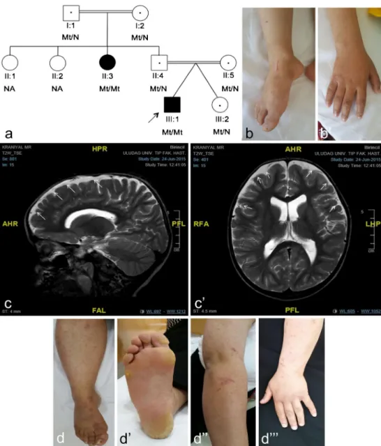
Fig. 1 The PLACK family and clinical features. a Pedigree of PLACK family. Parents are first cousins. b skin fragility and b” knuckle pads. c Axial and c’ sagittal T2-weighted images re-vealed mild cerebral atrophy and evidence of peripheral CSF areas secondary to atrophy by MRI (indicated by white arrows). d Showing the paternal aunt’s (II-3) nail dystrophy, d’ follicular hy-perkeratosis d” and eroded bul-lous lesion residuals d”’ in her arm
microscope and photographed by CCD camera (Gatan Inc., Pleasanton, CA, USA).
Cell culture of human skin fibroblasts
Human skin biopsies were obtained from both affected pa-tients and, initially, washed with 1× PBS and incubated in 37 °C for 60 min. The samples were washed once with 1× PBS, diced and incubated with collagenase type CLS IV (Millipore, Germany) in 37 °C for 120 min under rotation. Samples were then washed in DMEM plus 5% FBS and cen-trifuged 1200 rpm for 10 min. Cell pellets were then cultured in a 12-well plate in DMEM supplemented with 10% (v/v) fetal bovine serum (FBS; PAN, Cat. no. P30-3302), antibiotics (penicillin/streptomycin; Biological Industries, Cat. no. 03-031-1B) and 1% (2 mM)L-glutamine (Biological Industries, Cat. no. 03-020-1B).
Real-time qPCR
Total RNA was extracted from cultured fibroblasts using RNeasy kit (Qiagen, USA). To quantify mRNA expression of CAST gene, qRT-PCR was carried out using following p r i m e r s ( C A S T C o n t F : c a t g a t t t c t g c t g g t g g a g , CASTContR:ccctatgggtttcgaagagtc primer pair that amplify u p s t r e a m o f t h e v a r i a n t r e g i o n , a n d CASTcE182XF:GGACCAGAAGTTTCAGATCCAA, CASTcE182XR:TCCCTGCTGACTGAGCTTTT primer pair that amplify the region that includes c.544G>T (p.Glu182*)) and 1-step QuantiTect SYBR Green qRT-PCR Kit (Qiagen, USA) according to the manufacturer’s standard protocol on Light Cycler 480 instrument (Roche, S w i t z e r l a n d ) . T h e h o u s e k e e p i n g g e n e G A P D H (Hs_GAPDH_1_SG QuantiTect Primer Assay, NM_002046, Qiagen) was used for normalization, and relative gene expres-sion levels were calculated using the 2−ΔΔCtmethod. Results are shown as fold expression (mean ± SEM, n = 3; bars, SE; *P < .05, **P < .01). Statistical significance was analyzed by using Student’s t-tail test on GraphPad Prism 6.2 software.
Immunoblotting
Total cell lysates from fibroblasts were prepared in 1% CHAPS buffer [5 mM MgCl2, 140 mM NaCl, 1 mM EDTA,
1 mM EGTA, 1% CHAPS, 20 mM Tris-HCl (pH 7.5) and protease inhibitors (completeULTRA, Roche)]. Total cell ly-sates were separated on 12% SDS-PAGE gels. After SDS PAGE, proteins were transferred onto PVDF membranes (Millipore Darmstadt, Germany) and then blocked with 5% dried milk in PBS-Tween 20. Membranes were incubated with primary and secondary antibodies (GE Healthcare) in a buffer containing 10% milk diluent-blocking concentrate (KPL, Sera Care, USA), detected with Luminata Crescendo Western HRP Table
1 Novel and rare homozygous variants in the exome file o f the proband Chr G ene T ranscript ID V ariant type V ariant dbSNP ID Mi nor allele frequency (MAF ) Protein p redictions OMIM fenotype Pathogenic cla ss gnomAD exomes gnomAD genomes SIF T Mut ati on tas ter Provean ch r1 KHDRBS1 NM_0 065 59. 3 S yn ony mou s c. 12 66G>A (p.Se r42 2=) rs780 046 000 0. 000 016 NA –– – NA VUS ch r1 OLFML2B NM_0 154 41. 2 M isse ns e c.1 136 C>T (p. Se r3 79Le u ) rs145 215 702 0. 000 18 NA D P N N A VUS ch r5 TM CC1 NM_ 001 017 395 .4 Syn ony mou s c.18 15C>T (p .Ser 605 =) rs 775 227 252 0. 000 024 NA –– – NA VUS ch r5 CAST NM_ 001 284 213 .3 No nse n se c.54 4G>T (p .Glu 182 *) NA NA NA – DC – # 616 295 Pa thog en ic ch r9 CCDC 183 NM_ 001 039 374 .4 M isse ns e c.22 3 G>A (p .Gly 75Ar g ) rs771 245 508 0. 000 016 NA T P N N A VUS ch r1 1 GDPD4 NM_1 828 33. 2 M isse ns e c.40 0 G>C (p. V al13 4 Leu ) rs 143 423 593 0. 004 3 0 .004 7 T P P NA VUS ch r1 5 VPS13C NM_0 180 80. 3 M isse ns e c.80 04G>A (p.M et2 668 Ile ) rs 757 188 81 1 0 .000 012 NA T D C N # 616 840 VUS ch r1 8 NOL4 NM_ 001 1 985 48. 1 M isse ns e c.21 4 G>C (p. Gly7 2Ar g ) rs890 471 029 0. 000 008 NA T D C N NA VUS KHD R BS1 and TMCC1 variants were silent substitutions that do not cha nge the encoded amino acid and w ere not OMIM ph enotype chr chr omosome, D damaging, DC di se as e caus ing, N neutral, NA not available, P polymorphism, T tole rat ed, VU S variant of unknown significance
substrate (Millipore, Darmstadt, Germany). Blots were im-aged with ImageQuant LAS 4000, (Fujitsu/GE Life Science, USA) on chemiluminescence mode. The following antibodies were used for immunoblotting: calpastatin (A-1) (Santa Cruz Biotechnology, CA sc-376547), GAPDH (#2118, Cell Signaling, MA, USA).
Immunofluorescence staining and confocal laser
scanning microscopy
Fibroblasts grown on sterile coverslips (Jena Bioscience cir-cular cover slide (22 mm), # CSL-104) were incubated in a 12-well plate format at 37 °C, washed for three times for 5 min per rinse (each wash step are the same unless specified other-wise) with 1× PBS, fixed for 10 min with 4% (v/v) parafor-maldehyde in 1× PBS, followed by rinsing in 1× PBS. Cell membranes were permeabilized by incubation in 0.05% Triton100-X in 1× PBS for 5 min followed by rinsing with 1× PBS. The fixed and permeabilized cells were incubated with 0.1% bovine serum albumin (BSA) in 1× PBS (blocking buffer) for 30 min at room temperature. Coverslips were ex-posed to primary antibody 1:100 (v/v) and calpastatin (A-1) (Santa Cruz Biotechnology, sc-376547) in blocking buffer for 1 h at room temperature. After washing steps with 1× PBS, coverslips were incubated in the dark, with Alexa-488 fluo-rescence tagged secondary antibody (Alexa Fluor 488 anti-mouse IgG, Invitrogen, Cat. no. A11034) in blocking buffer for 1 h at room temperature and subsequently rinsed in
washing buffer. Cells incubated without primary antibody (secondary antibody only) were used as a negative control. Cells were mounted with ProLong Antifade kit (Molecular Probes, Eugene, OR, USA) on glass slides (0.17 mm, Thermo Fisher Scientific Inc., USA) and the stained cells were examined by a Zeiss LSM 710 Confocal Microscope. Specimens were observed through an oil immersion × 63 ob-jective. Slides were imaged with a Zeiss LSM710 confocal microscope, excited with 488 nm (for Alexa 488 labeled an-tibody); light emissions were collected at 530 nm (green im-munofluorescence for calpastatin localization) and DAPI (Excitation 405, Emission (blue) 459 nm) simultaneously. Controls were imaged under similar confocal settings. Images were analyzed by using ZEN Lite software (Zeiss, USA).
Calpastatin activity assay
For the calpastatin activity assay, protein concentrations of the fibroblast-derived cell extracts obtained from patients were adjusted to 3 μg/μL. The extracts were heated to 90 °C for 5 min and then centrifuged at 16,000g at 4 °C for 20 min. Reactions contained 5μL of supernatant, 10 nM human eryth-rocyte calpain 1 (Millipore, USA), 200μM Suc-Leu-Leu-Val-Tyr-AMC (Abcam, UK) and 195μL of the homogenization buffer with 5 mM beta-mercaptoethanol. One sample contained no supernatant and was designated as negative con-trol. The reaction was started by adding CaCl2 to a final Fig. 2 Mutation detail.
Electropherograms for heterozygous and homozygous mutant genotypes of CAST c.544G>T (p.Glu182*) nonsense mutation
concentration of 5 mM at room temperature. Substrate prote-olysis, which results in increasing fluorescence was measured on a fluorescence microplate reader (360 nm excitation, 460 nm emission) at 30 min in triplicate. Data shown are mean relative fluorescence units from three independent experi-ments; bars, SE; **P < .01, **P < .05. Statistical significance was analyzed by using Student’s t-tail test on GraphPad Prism 6.2 software.
Results
Clinical findings
We report on a 5.5-year old boy who initially presented with generalized peeling skin who was the first-born of IVF twins from a consanguineous marriage. Clinical findings included fragile skin (Fig.1b), wooly hair, sparse eyelashes and brows, palmoplantar punctate keratoderma, follicular hyperkeratosis, knuckle pads (Fig.1b’) and cheilitis. Mild cerebral atrophy,
secondarily to atrophy asimetrically enlargement of lateral ventricles and mild muscle involvement were observed on his MRI (Fig.1c, c’) and NCV/EMG, respectively. His aunt
also had similar clinical features but in milder form with, in addition, nail dystrophy (Fig.1d–d”’).
WES and sanger sequencing
To search for the putative causative mutation or mutations related with proband’s phenotype and presumed autosomal recessive inheritance, we evaluated novel and rare homozy-gous variants in the exome file and identified eight candidate variants (Table1). The most plausible variant associated with the cutaneous phenotype segregating in the family was the c.544G>T nonsense variant in CAST. However, the pathogen-ic class of all eight variants was evaluated according to ACMG (American College of Medical Genetics) criteria in company with standards and guidelines for the interpretation of sequence variants (Richards et al.2015). KHDRBS1 and
TMCC1 variants were silent substitutions that do not change the encoded amino acid and these genes have not been previ-ously related to any OMIM phenotypes. All of the other five missense variants were predicted as not damaging to protein by at least one of three computational algorithms used. In addition, four of these missense genes, except for VPS13C, have not been previously associated with any OMIM pheno-types. The VPS13C variant was predicted as not damaging to protein by SIFT and Provean predictions. VPS13C mutations have been linked to a form of Parkinson disease (OMIM 616840) but all of the pathogenic variants for the VPS13C gene that were reported in HGMD Professional 2018.4 (Human Gene Mutation Database Professional version 2018.4) were truncating or splicing. The parents of the pro-band (II:4 and II:5), healthy sister (III:2) and grandmother (I:2) were carriers for the VPS13C variant and the other affect-ed family member (II-3) had the wild type genotype. She could not be investigated for cerebral atrophy since she was pregnant at that time. Thus, we could not find any strong evidence for the pathogenicity of this homozygous missense VPS13C variant found in the proband.
Thus, the remaining CAST variant is likely to underlie PLACK syndrome in this family. This homozygous c.544G>T nonsense variant was absent in the following data-bases: HGMD (Human Gene Mutation Database Professional version 2018-4); ClinVar, CentoMD 5.3, OMIM (Online Mendelian Inheritance In Man); and LOVD v.3.0 (Leiden Open Variation Database version 3.0), as well as in the popu-lation specific databases such as dbSNP (Single Nucleotide Polymorphism database), ExAC (Exome Aggregation Consortium), ESP (NHLBI GO Exome Sequencing Project), 1000 Genomes (A Deep Catalog of Human Genetic Variation) TOPMED (NHLBI Trans-Omics for Precision Medicine), and gnomAD (Genome Aggregation Database). Sanger sequenc-ing was used to confirm this variant (Fig.2) and to show its segregation in the family.
Histological and immunohistochemical findings
H&E staining of proband’s lesioned skin showed hyperkera-tosis and mild spongiotic changes (Fig. 3a-white asterisk). Immunohistochemistry results showed reduced/absent calpastatin staining in the proband (Fig.3b) compared to that of control epidermis (Fig.3c).
Transmission electron microscopy findings
Semi-thin sections of proband’s lesioned skin showed elon-gated epidermal rete pegs (Fig.3d-white asterisk) and dermal papilla (Fig.3d-black asterisks), thick stratum corneum (Fig.
3d-white asterisk), extensive karyolysis even in stratum basale and loss of chromatine pattern in all epithelial cells (Fig.3f-red arrows). Electron micrographs showed widened intercellular
Fig. 3 Histological, immunohistochemical and transmission electronmicroscopy results. a Proband’s lesional skin biopsy, photomicrographs showing hyperkeratosis (white asterisk), mild spongiotic changes, H&E. b, c Immunohistochemistry shows absent calpastatin staining in the proband (b) compared to normal staining throughout the epidermis in the control (c). d–f Photomicrographs showing elongated epidermal rete pegs (d-white asterisk) and dermal papilla (d-black asterisk), thick stratum corneum (e-black asterisk), extensive karyolysis even in stratum basale and loss of chromatin pattern in all epithelial cells (f-red arrows) and semi-thin sections T&B. g–j Electron micrographs showing widened intercellular space, heterochromatin in the bleb (g-red asterisk, red arrow, respectively), chromatin condensation and marginization (h-white arrows), nuclear chromatin condensation and fragmentation (i-white as-terisk) and the loss of the integrity of the nuclear membrane (j-white arrow)
space, heterochromatin in the bleb (Fig.3g-red asterisk, red arrow, respectively), chromatin condensation and marginization (Fig. 3h-white arrows), nuclear chromatin
condensation and fragmentation (Fig. 3i-white asterisk) and the loss of the integrity of the nuclear membrane (Fig.3j-white arrow). 0 0.0001 0.0002 0.0003 0.0004 0.0005 0.0006 0.0007 0.0008 0.0009 0.001 Mt/Mt III-1 LS Mt/Mt III-1 NS Mt/Mt II-3 LS Mt/Mt II-3 NS Mt/N II-4 Mt/N II-5 Mt/N III-2 Relative transcript abundance
CAST mRNA level (Upstream of variant region)
**
**
**
**
0 0.01 0.02 0.03 0.04 0.05 0.06 0.07 Mt/Mt III-1 LS Mt/Mt III-1 NS Mt/Mt II-3 LS Mt/Mt II-3 NSMt/N II-4 Mt/N II-5 Mt/N III-2
Relative
transcript
abundance
CAST mRNA level (Variant region)
**
**
**
**
a
b
f
0 500 1000 1500 2000 2500 3000 3500Calpain activty (RFU)
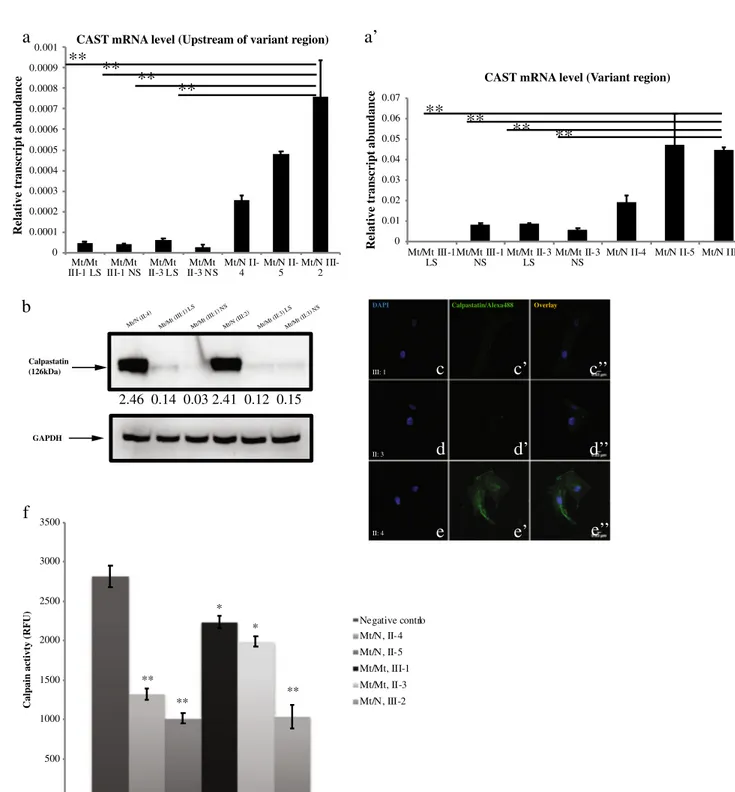
Negative control Mt/N, II-4 Mt/N, II-5 Mt/Mt, III-1 Mt/Mt, II-3 Mt/N, III-2 ** ** ** * * Overlay III: 1 DAPI Calpastatin/Alexa488 II: 3 II: 4 Mt/N (II:4) Mt/M t (III: 1) LS Mt/Mt (III:1) NS Mt/N (III:2) Mt/Mt (II:3) LS Mt/M t (II:3) NS Calpastatin (126kDa) 2.46 0.14 0.03 2.41 0.12 0.15 GAPDH
a’
c
c’
c’’
d
d’
d’’
e
e’
e’’
Fig. 4 mRNA quantification results of CAST gene upstream of the variant region (a) and the variant region (a’), for homozygote (mt/mt) and heterozygote (mt/wt) individuals. The residual expression of CAST mRNA in the affected cases was presented (b). Semi-quantitative protein expressions for different genotypes. Calpastatin was normalized to GAPDH, which was set to 1.00 and the intensity ratio of calpastatin to GAPDH was calculated. LS lesioned skin, NS normal skin. Immunofluorescence stainings of CAST for homozygous proband (III-1)
(c, c’, c”), for homozygous proband’s aunt (II-3) (d, d’, d”) and for hetero-zygote carrier (III-2) (e, e’, e”). Calpastatin activity assay. Calpastatin ac-tivity was indirectly derived from inhibition of calpain acac-tivity in protein lysates from homozygote and heterozygote cases. Negative control contains no lysate and indicates baseline calpain activity. Data shown are mean relative fluorescence units from three independent experiments; bars, SE; **P < .01, **P < .05, affected cases, carriers vs. negative controls (f)
Real-time qPCR analysis
In contrast to the carriers, we could detect only trace expres-sion of CAST mRNA in the affected cases, probably due to mechanisms of nonsense mediated mRNA decay (Fig.4a, a’).
Furthermore, we demonstrated a significant difference be-tween the expression of mRNA upstream of the variant region and the variant region in affected cases (both Normal Skin (NS) and Lesioned Skin (LS)) and Mt/N III-2. Similarly, we found that expression of CAST mRNA was significantly dif-ferent in affected cases compared to Mt/N II-4 (*P < .05) and Mt/N II-5 (**P < .01).
Protein quantification and immunofluorescence
To compare the abundance of calpastatin in fibroblasts of in-dividuals with homozygous and heterozygous genotypes, to-tal protein samples were resolved on SDS-PAGE and immunoblotted using calpastatin and GAPDH-specific anti-bodies. A semi-quantitative protein expression assay revealed a drastically low amount of calpastatin in patients’ normal and lesioned skin cells as compared to cells from the heterozygous unaffected cell line (Fig.4b). Hence, we detected more than a two-fold decrease in calpastatin expression levels in the pa-tients homozygous for c.544G>T (Mt/Mt III:1 and Mt/Mt II:3) compared with heterozygous carriers (Mt/N II:4 and Mt/N III:2). Confocal microscopy results also confirmed re-duced expression pattern of calpastatin in fibroblasts from affected patients compared to carriers (Fig.4c–c”, d–d”, e–e”).
In vitro calpastatin assay findings
In vitro calpastatin activity of fibroblast cell extracts obtained from patients homozygous for c.544G>T (III:1 and II:3) and unaffected heterozygote cases were evaluated indirectly by fluorogenic calpain proteolysis assay using a Suc-Leu-Leu-Val-Tyr-AMC fluorescence probe. Our results demonstrated increased calpain activity and, thus, decreased calpastatin ac-tivity in affected individuals compared with control samples (**P < .01) (Fig.4f).
Discussion
PLACK syndrome has recently been described as a new form of peeling skin syndrome (PSS), characterized by generalized peeling skin, leukonychia, acral punctate keratoses, cheilitis and knuckle pads (Lin et al.2015). To date, four homozygous pathogenic variants (loss of function mutations) in CAST gene (c.424A>T, c.607dup, c.1750delG, c.461dupGCAT) have been reported with PLACK syndrome (Lin et al.2015; Alkhalifah et al.2017). This study presents the fifth family with PLACK syndrome and is associated with a novel
homozygous nonsense mutation (c.544G>T p.Glu182*) in the CAST gene.
CAST encodes calpastatin, which is an endogenous specific inhibitor of calpain, a calcium-dependent cysteine protease (Goll et al.2003). Calpastatin can bind to calcium activated calpains (Potz et al.2016). Calpains are involved in a range of cellular processes, including cell proliferation (Kovács and Su
2014), migration (Franco and Huttenlocher 2005), wound healing (Nassar et al. 2012), apoptosis and survival (Tan et al.2006). It is also involved in the regulation of epidermal cell differentiation (Carragher and Frame2004). A proposed mechanism underlying PLACK syndrome is that the absence of a functional calpastatin prevents the inhibition of calpains leading to elevated keratinocyte apoptosis and skin hyperker-atosis (Lin et al.2015; Wang et al.2015).
Modulation of calpastatin expression in mice caused no phenotypic abnormalities under normal conditions indicating that calpastatin may only function as a negative regulator of calpain only under pathological conditions (Takano et al.
2005). In order to determine the functional consequences of CAST mutations in vitro, Lin et al. performed siRNA-mediated knockdown of CAST in the immortalized keratinocyte cell line, HaCaT and performed a mechanical induced stress assay. This showed that mechanical stress re-vealed breakage of the intercellular connections in CAST knock down cell monolayers in contrast to control cell lines (Lin et al.2015). Calpain overactivity is involved in the path-ophysiology of a large number of disease processes of the brain, eyes, heart, lungs, pancreas, kidneys, vascular system and skeletal muscle. The situation of stress leads to increased calcium ion release causing over-activation of calpain, which may cause organ dysfunction by promoting cellular apoptosis and degradation of cytoskeletal structure (Potz et al. 2016). Therefore, calpain was suggested as a potential therapeutic target for a wide variety of disease processes. In line with this, inhibition of calpain by over-expression of calpastatin inhibits the abnormal breakdown of cytoskeletal proteins (spectrin, MAP2 and neurofilaments) and ameliorates motor axon loss in amyotrophic lateral sclerosis (ALS) hSOD1G93A trans-genic mouse model (Rao et al.2016). Another study supports the hypothesis that calpastatin may play a role in regulating the initial metastatic dissemination of breast cancer (Storr et al.
2011).
It is noteworthy that our novel mutation and the associated clinical features differ in that leukonychia was absent although it was reported in all of the previous cases. Mild cerebral atrophy and muscle involvement was also present in one of the affected family members, whereas it was not reported in the study by Lin et al. (2015). However, Alkhalifah et al. (2017) did report muscle involvement in their study. The ab-sence of leukonychia and the preab-sence of mild cerebral atro-phy in our patient suggest significant phenotypical variability within individuals with CAST mutations.
In this study, we demonstrated that calpastatin expression in fibroblasts from affected family members was reduced at both the mRNA and protein level. In vitro calpastatin activ-ity revealed reduced calpain proteolysis in both affected in-dividuals. The molecular and biochemical studies in con-junction with the described clinical features in this study emphasize the importance of tight regulation of proteolytic pathways by calpastatin in maintaining the structural integ-rity of the skin.
Acknowledgments The authors wish to express their gratitude to the family members for participating in this study and Prof. Dr. Esin ASAN for her valuable comments on semithin sections and electronmicrographs and TUBITAK-IGBAM for exome sequencing and bioinformatic advice. Funding This work was supported by the Uludag University Scientific Research Unit with Project number KUAP(T)-2014/36.
Compliance with ethical standards
Conflict of interest The authors declare that they have no conflict of interest.
Ethical approval All procedures performed in studies involving human participants were in accordance with the ethical standards of the institu-tional research committee and with the 1964 Helsinki declaration and its later amendments or comparable ethical standards.
Informed consent Informed consent was obtained from all individual participants included in the study. Additional informed consent was ob-tained from all individual participants and their guardians for whom iden-tifying information is included in this article.
References
Akgün M, Faruk Gerdan Ö, Görmez Z, Demirci H (2016) FMFilter: a fast model based variant filtering tool. J Biomed Inform 60:319–327 Alkhalifah A, Chiaverini C, Del Giudice P, Supsrisunjai C, Hsu CK, Liu
L, Charlesworth A, McGrath JA, Lacour JP (2017) PLACK syn-drome resulting from a new homozygous insertion mutation in CAST. J Dermatol Sci 88:256–258
Bowden PE (2011) Peeling skin syndrome: genetic defects in late termi-nal differentiation of the epidermis. J Invest Dermatol 131:561–564 Cabral RM, Kurban M, Wajid M, Shimomura Y, Petukhova L, Christiano AM (2012) Whole-exome sequencing in a single proband reveals a mutation in the CHST8 gene in autosomal recessive peeling skin syndrome. Genomics 99:202–208
Carragher NO, Frame MC (2004) Focal adhesion and actin dynamics: a place where kinases and proteases meet to promote invasion. Trends Cell Biol 14:241–249
Cassidy AJ, van Steensel MA, Steijlen PM, van Geel M, van der Velden J, Morley SM, Terrinoni A, Melino G, Candi E, McLean WH (2005) A homozygous missense mutation in TGM5 abolishes epidermal transglutaminase 5 activity and causes Acral peeling skin syndrome. Am J Hum Genet 77:909–917
Cingolani P, Platts A, Wang le L, Coon M, Nguyen T, Wang L, Land SJ, Lu X, Ruden DM (2012) A program for annotating and predicting the effects of single nucleotide polymorphisms, SnpEff: SNPs in the genome of Drosophila melanogaster strain w1118; iso-2; iso-3. Fly (Austin) 6:80–92
DePristo MA, Banks E, Poplin R, Garimella KV, Maguire JR, Hartl C, Philippakis AA, del Angel G, Rivas MA, Hanna M, McKenna A, Fennell TJ, Kernytsky AM, Sivachenko AY, Cibulskis K, Gabriel SB, Altshuler D, Daly MJ (2011) A framework for variation discov-ery and genotyping using next-generation DNA sequencing data. Nat Genet 43:491–498
Goll DE, Thompson VF, Li H, Wei W, Cong J (2003) The calpain system. Physiol Rev 83:731–801
Görmez Z, Bakir-Gungor B, Sagiroglu MS (2014) HomSI: a homozy-gous stretch identifier from next-generation sequencing data. Bioinformatics 30:445–447
Franco SJ, Huttenlocher A (2005) Regulating cell migration: calpains make the cut. J Cell Sci 118:3829–3838
Hashimoto K, Hamzavi I, Tanaka K, Shwayder T (2000) Acral peeling skin syndrome. J Am Acad Dermatol 43:1112–1119
Israeli S, Zamir H, Sarig O, Bergman R, Sprecher E (2011) Inflammatory peeling skin syndrome caused by a mutation in CDSN encoding corneodesmosin. J Invest Dermatol 131:779–781
Judge MR, McLean WHI, Munro CS (2004) Disorders of keratinization. In: Rook’s textbook of dermatology (Burns T, Breathnach S, Cox C, Griffiths C eds), vol 2. Blackwell Scientific, Oxford, pp 54–56 Kharfi M, El Fekih N, Ammar D, Jaafoura H, Schwonbeck S, van
Steensel MA, Fazaa B, Kamoun MR, Fischer J (2009) A missense mutation in TGM5 causes acral peeling skin syndrome in a Tunisian family. J Invest Dermatol 129:2512–2515
Kovács L, Su Y (2014) The critical role of calpain in cell proliferations. J Biomol Res Ther 3:112
Kurban AK, Azar HA (1969) Familial continual skin peeling. Br J Dermatol 81:191–195
Krunic AL, Stone KL, Simpson MA, McGrath JA (2013) Acral peeling skin syndrome resulting from a homozygous nonsense mutation in the CSTA gene encoding cystatin A. Pediatr Dermatol 30:e87–e88 Levy SB, Goldsmith LA (1982) The peeling skin syndrome. J Am Acad
Dermatol 7:606–613
Li H, Durbin R (2009) Fast and accurate short read alignment with Burrows-Wheeler transform. Bioinformatics 25:1754–1760 Li H, Handsaker B, Wysoker A, Fennell T, Ruan J, Homer N, Marth G,
Abecasis G, Durbin R, 1000 Genome Project Data Processing Subgroup (2009) 1000 genome project data processing subgroup. Th e Seq uen ce Alignment/Map format and SAMto ols. Bioinformatics 25:2078–2079
Lin Z, Zhao J, Nitoiu D, Scott CA, Plagnol V, Smith FJ, Wilson NJ, Cole C, Schwartz ME, McLean WH, Wang H, Feng C, Duo L, Zhou EY, Ren Y, Dai L, Chen Y, Zhang J, Xu X, O'Toole EA, Kelsell DP, Yang Y (2015) Loss-of-function mutations in CAST cause peeling skin, leukonychia, acral punctate keratoses, cheilitis, and knuckle pads. Am J Hum Genet 96:440–447
Nassar D, Letavernier E, Baud L, Aractingi S, Khosrotehrani K (2012) Calpain activity is essential in skin wound healing and contributes to scar formation. PLoS One 7:e37084
Oji V, Eckl KM, Aufenvenne K, Nätebus M, Tarinski T, Ackermann K, Seller N, Metze D, Nürnberg G, Fölster-Holst R, Schäfer-Korting M, Hausser I, Traupe H, Hennies HC (2010) Loss of corneodesmosin leads to severe skin barrier defect, pruritus, and atopy: unravelling the peeling skin disease. Am J Hum Genet 87: 274–281
Potz BA, Abid MR, Sellke FW (2016) Role of calpain in pathogenesis of human disease processes. J Nat Sci 2: pii: e218
Quinlan AR, Hall IM (2010) BEDTools: a flexible suite of utilities for comparing genomic features. Bioinformatics 226:841–842 Rao MV, Campbell J, Palaniappan A, Kumar A, Nixon RA (2016)
Calpastatin inhibits motor neuron death and increases survival of hSOD1(G93A) mice. J Neurochem 137:253–265
Richards S, Aziz N, Bale S, Bick D, Das S, Gastier-Foster J, Grody WW, Hegde M, Lyon E, Spector E, Voelkerding K, Rehm HL, ACMG Laboratory Quality Assurance Committee (2015) Standards and
guidelines for the interpretation of sequence variants: a joint consen-sus recommendation of the American College of Medical Genetics and Genomics and the Association for Molecular Pathology. Genet Med 17:405–424
Storr SJ, Mohammed RA, Woolston CM, Green AR, Parr T, Spiteri I, Caldas C, Ball GR, Ellis IO, Martin SG (2011) Calpastatin is asso-ciated with lymphovascular invasion in breast cancer. Breast 20: 413–418
Takano J, Tomioka M, Tsubuki S, Higuchi M, Iwata N, Itohara S, Maki M, Saido TC (2005) Calpain mediates excitotoxic DNA fragmenta-tion via mitochondrial pathways in adult brains: evidence from calpastatin mutant mice. J Biol Chem 280:16175–16184
Tan Y, Wu C, De Veyra T, Greer PA (2006) Ubiquitous calpains promote both apoptosis and survival signals in response to different cell death stimuli. J Biol Chem 281:17689–17698
Wang H, Cao X, Lin Z, Lee M, Jia X, Ren Y, Dai L, Guan L, Zhang J, Lin X, Zhang J, Chen Q, Feng C, Zhou EY, Yin J, Xu G, Yang Y (2015) Exome sequencing reveals mutation in GJA1 as a cause of keratoderma-hypotrichosis-leukonychia totalis syndrome. Hum Mol Genet 24:243–250
The list of public and restricted databases
100 Genomes (A Deep Catalog of Human Genetic Variation): available at http://phase3browser.1000genomes.org/index.html) (last accessed 25 January 2019)
ClinVar: available athttps://www.ncbi.nlm.nih.gov/clinvar(last accessed 25 Januaryr 2019)
CentoMD 5.3: available at (https://www.centomd.com) (last accessed 25 January 2019)
dbSNP (Single Nucleotide Polymorphism database): available athttps:// www.ncbi.nlm.nih.gov/snp) (last accessed 25 January 2019) ExAC (Exome Aggregation Consortium): available athttp://exac.
broadinstitute.org) (last accessed 25 January 2019)
ESP (NHLBI GO Exome Sequencing Project): available athttp://evs.gs. washington.edu/EVS) (last accessed 25 January 2019)
gnomAD (Genome Aggregation Database): available athttp://gnomad. broadinstitute.org/(last accessed 25 January 2019)
HGMD (Human Gene Mutation Database) Professional version 2018.4: available athttps://portal.biobase-international.com/hgmd/pro/start. php(last accessed 25 January 2019)
LOVD v.3.0 (Leiden Open Variantion Database version 3.0): available at http://www.lovd.nl/3.0/home(last accessed 25 January 2019) OMIM (Online Mendelian Inheritance In Man): available athttps://www.
omim.org(last accessed 25 January 2019)
TOPMED (NHLBI Trans-Omics for Precision Medicine): available at http://www.nhlbiwgs.org) (last accessed 25 January 2019) UCSC Genome Browser: available atwww.genome.ucsc.edu) (last
accessed 25 January 2019)
Open Access
This article is distributed under the terms of the Creative Commons Attribution 4.0 International License (http://creativecommons.org/ licenses/by/4.0/), which permits unrestricted use, distribution, and reproduction in any medium, provided you give appropriate credit to the original author(s) and the source, provide a link to the Creative Commons license, and indicate if changes were made.
Publisher’s note Springer Nature remains neutral with regard to jurisdic-tional claims in published maps and institujurisdic-tional affiliations.