Cukurova Medical Journal
Cukurova Med J 2016;41(Suppl 1):82-83ÇUKUROVA ÜNİVERSİTESİ TIP FAKÜLTESİ DERGİSİ DOI: 10.17826/cutf.254644
Yazışma Adresi/Address for Correspondence: Dr. Elif Karadeli, Baskent University, Faculty of Medicine, Department of Radiology, Adana, Turkey E-mail: [email protected]
Geliş tarihi/Received: 05.05.2015 Kabul tarihi/Accepted: 21.06.2015
OLGU SUNUMU/CASE REPORT
Perforation without manifestations of acute abdomen after ERCP
ERCP sonrası akut abdomen bulgusu olmayan perforasyon
Elif Karadeli
1, Sermin Tok
1, Gürcan Erbay
11Baskent University, Faculty of Medicine, Department of Radiology, Adana, Turkey Cukurova Medical Journal 2016;41(Suppl 1):82-83.
Abstract Öz
Perforations are rare after endoscopic retrograde cholangiography (ERCP) but they are serious complications. Perforations may occur due to patient-related factors or procedural technique. The mortality rate is higher than 37.5%. We are presenting a patient with perforation detected on a computed tomography (CT) scan after ERCP for obstructive jaundice that was treated conservatively. Perforation after ERCP can be operated or treated conservatively according to the patient’s condition.
Perforasyonlar, endoskopik retrograd kolanjiyopankreatografi (ERCP) sonrası nadiren gözlenmesine rağmen ciddi komplikasyonlar oluşturabilirler. Perforasyonlar hastayla ilişkili faktörler veya prosedüral teknikler vasıtasıyla oluşabilir. Ölüm oranı %37,5 tan daha yüksektir. Geleneksel olarak tedavi edilmiş obstruktif sarılığı olan hastalar için; ERCP sonrası bilgisayarlı tomografi (CT) taramasında tespit edilen perforasyonlu hastayı sunuyoruz. ERCP sonrası perforasyon hastanın durumuna bağlı olarak, geleneksel yöntemlerle tedavi edilebilceği gibi ameliyatta edilebilir.
Key words: CT, ERCP, perforation. Anahtar kelimeler: CT, ERCP, perforasyon.
INTRODUCTION
The complications of endoscopic retrograde cholangiography (ERCP) are pancreatitis, infections, bleeding, perforation and cardiopulmonary complications and the rate of perforation has been reported as 0.3-2.1%1,2. Perforations may be secondary to previous surgeries or due to technical reasons1. Here we are presenting a patient with perforation detected on a computed tomography (CT) scan after ERCP for obstructive jaundice that was treated conservatively.
CASE
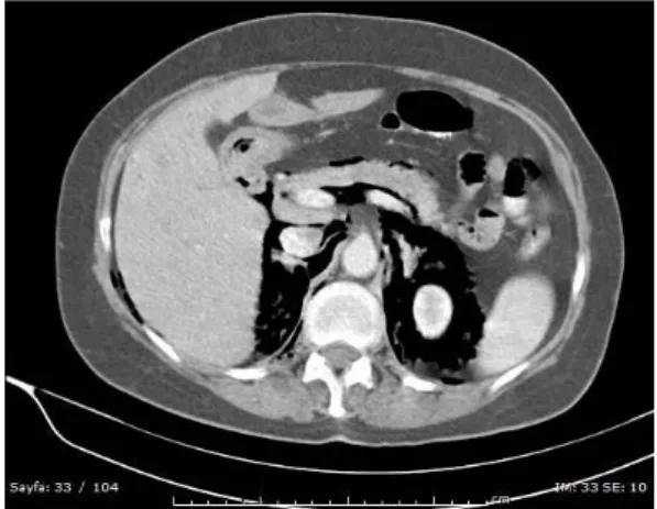
A 56-year-old female patient underwent ERCP for obstructive jaundice. The proximal choledochus could not be passed due to obstruction. The same day, an IV-oral contrasted abdominal CT scan was obtained to investigate the etiology. Diffuse air densities were observed intraabdominally
surrounding the liver, stomach, duodenum, in the retroperitoneal areas bilaterally, surrounding the kidneys and surrounding the colon on the right (Figure 1-2).
Oral contrast agent extravasation was not observed. There was no retroperitoneal or intraabdominal free fluid. The images were evaluated as suggestive of perforation. The patient was treated conservatively as no abdominal pain, oral contrast extravasation or intraabdominal free fluid was observed. During conservative treatment oral intake was stopped, antibiotherapy was administered. No clinical or laboratory pathology was observed during observation. The patient had no complaints and was discharged after one week.
DISCUSSION
Perforation after ERCP can be a fatal complication3. Perforations are classified in many ways and guidewire perforations, periampullary or duodenal
Karadeli et al. Cukurova Medical Journal
perforations are most common3. The diagnosis of perforation can be made upon suspicion during ERCP by showing the contrast agent extravasation or hours or days after the procedure, as in our case.
Figure 1. Retroperitoneal and peripancreatic diffuse free air densities in the IV-oral contrasted axial abdominal CT scan.
Figure 2. Perihepatic and periintestinal diffuse free air densities in coronal CT slices.
Clinically, abdominal pain, vomiting, abdominal distention, fever, leukocytosis may be present and acute phase reactants may be increased. CT scans are important for guidance. Oral contrast must be administered during CT scans4.
If the diagnosis of perforation was made during surgery with major contrast agent extravasation, it should be clipped if possible, if it cannot be clipped then laparoscopic or conventional surgery must be performed5. When cases with abdominal pain are diagnosed with perforation after ERCP with CT scans, treatment can be shaped according to the images. If oral contrast agent extravasation or retroperitoneal fluid is observed surgery must be performed. Conservative treatment can be performed if retroperitoneal air is observed only, as in our case. CT scans can be repeated for clinical monitoring or upon necessity3.
Finally, although perforation after ERCP can be a fatal complication, if the patient has complaints after the procedure is completed, treatment can be surgical or conservative according to the CT findings.
REFERENCES
1. Anderson M A, Fisher L, Jain R. Complications of ERCP. Gastroint Endoscopy. 2012;75:467-73. 2. Li G, Chen Y, Zhou X, Lv N. Early management
experience of perforation after ERCP. Gastroenterol Res Pract. 2012;2012:657418.
3. Wu HM, Dixon E, May GR, Sutherland FR. Management of perforation after endoscopic retrograde cholangiopancreatography (ERCP): a population-based review. HPB (Oxford). 2006;8:393-9.
4. Tonolini M, Pagani A, Bianco R. Cross-sectional imaging of common and unusual complications after endoscopic retrograde cholangiopancreatography. Insights Imaging. 2015;6:323-38.
5. Koc B, Bircan HY, Adas G, Kemik O, Akcakaya A, Yavuz A et al. Complications following endoscopic retrograde cholangiopancreatography: minimal invasive surgical recommendations. PLoS One. 2014;9:e113073..