A. U. Vet. Fak. Derg. 32 (2) : 301-310, 1985
THE IN VITRO CULTIVATION OF MOUSE OVA FROM ONE CELL TO BLASTOCYST
çetin Kılıçoğlu
*
Fare ovumlannın tek hücreliden blastosist aşamasına kadar in vitro kültürü
.Özet: Preimplantasyon aşamasındaki fare embriyoları yaşamları için gerekli ortamı sağlayan vasatlar içersinde gelişme gösterebilirler. Ancak em-briyolar vasatlarda oluşabilecek değişikliklere diğer doku ve hücrelerden daha hassastırlar.
Bu çalışmada PAISG ve HCG enjekte edilmiş dişi farelerin erkek farelerle çiftleştirildikten sonra oviductlarının yıkanması sonu elde edilen tek hücreli döllenmiş ovumlarının blastosist oluncaya kadar in vitro kültüre edilmeleri in-lenmiştir.
Bunun için servikal dislokasyonla öldürülen dişi farelerin oviductları 50-70[1.1. PB!
+
4 mg ımı. BSA+
0.3 mg. ımı. hyaluronidase içeren petri kutularına konmuş, oviductun ampullasının bir iğne yardımıyla yırtılma-masıyla bu vasat içine dökülen tek hücreli döllenmiş yumurtalar bir pipet yar-dımıyla toplanarak parafinle örtülü 50 - 100fL 1. M 16-+-
BSA vasatında,37 Cderecede,
%
5lik COıli ortam içersinde tutulmuşlar ve gelişmeleri periodik olarak izlenmiştir.SUJnınary: The mouse embıyo can develop normally in vitro in a simple, chemically defined medium which fully meets the specijic nutritional require-ments of the embryos during pre-implantation development. However the embryos are far more sensitive to variations in the culture medium than other cells aııd tissues.
Introduction
There has been an inercasing interest in the in vitro eulture of fcrtilized mammalian cmbryos over the past 50,years, with major ardvanees in ecU and tissue culturc teehniqucs satisfyign the rcquire-ment~ of cmbryos at thcir differcnt clcavagc stages .
302 çETİN KıuÇOÖLU
Mark and Long (10) first reported studying the mouse embryo in vitro but they only ob~erved the ova for 12 hours and did not see any cleavage division. Lewis and Wright (9) used drops of plasma and embryo extract to observe and to photograph the early stages of the mouse embryo, .but they also did not see any cleavage division. Hammond (8) was the first investigator to successfully cu1ture mouse embryos through several stages of division in vitro. As a cu1ture medium, he employed a simple, physiological salt solution contaning sodium chloride, potassium chloridc and magnesium chloride with a glucose concentration of 1 mg. per mL. supplemcnted with aboııt 5
%
egg white. Using this medium, he observocd hatching of the blasto-cyst from the zona pellucida.A very impartant factor for the in vitro cultivation of all mamma. lian cells is the composition of the culture media. The most commonly used chemically defined media for culturing mouse embryos are based upon the Krebs~Ringer bicarbonate solution (7,17). Whitten (13,14) cultured 8-cell mouse embryos to blactocysts in a modified Krebs Ringer solution supplemented with lactate, glucose, erystalline boyine serum albumin and antibiotics. Later Brinster (1,2) cu1tured 2-cell mouse eggs to blastocysts and after Whittingham (16) achieved fertilization of mouse eggs in vitro, Whitten and Biggers (15) and Whittingham (16) successfully cultured mouse embryos through the entire pre-implantation period of development.
The aim of this study is to make well-design ed studies which will yicld quantitative information. it sounds more logical to make the largest number and most detailed studies on the embryos of la-boratory animals and the n to attempt to extrapolate this information to the embryos of larger species and of course if a good foundation of knowledge is available about the cmbryos of laboratöry animals it will be possiblc to desing more meaningful experiments in which the eggs of larger animals are used. In thisway the most efficicnt use can be made of embryos from the eostly large domestic animals.
Materials and Methods
Eight weeks old II Fı hybrid females from C5 7B1/6j'i!9 x CBA/Caöö were superovulated with 5 LU. Pregnant Mare's Serum Gonadotrophin (PMSG, Folligon Intervet) by intra peritoneal fol-lowed 48 hours later by 5 LU. Human Chorionie Gonadotrophin
THE IN VJTRO CUL TIVA TION OF MOUSE ... 303
males (CFLP) at the time of HCG and chccked for vaginal plugs the following moming (= Day I). A total of 325 fertlized I-cell eggs were recovered from donor f(~malcs with vaginal plugs autopsied between 9.30- 10.30 Day I. The donors were killcd by cervical dislocation and the oviducts were placed in a petri dish containing 50-7011-1. PBI+4 mgImI. BSA (Bovine Serum Albumin) + 0.3 mgImI. Hyaluronidase (Bovine Testis). Eaclı oviduct was chcckcd for a transparent swelling. The l-cell embryos were located within the ampullary portion of the oviduct, embedded in cumulus and usually stuck together as a singlc large mass of ova and cumulus cells. To remove this cumulus masses from the ampulla the expanded ampulla was tom and the mass ex-pelIcd in the hyaluronidase solution. Liberation of the intact cumulus mass was efIected using a pair of watchmaker forceps and a disposable 25. gau. needIe. The eggs wcre released from the cumulus cells after 2-2 i
12
minutes at 3rC when an average of 30 eggs wcre recovered(ı
I) The denuded eggs were then \Vashed through 6 drops of Mı
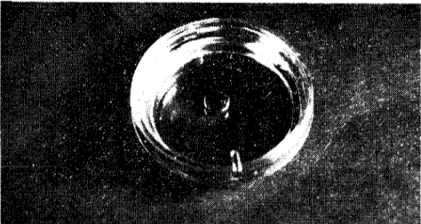
6 -i- 4 mgImI. BSA to complcte cumulus dispersal and remove the PBI + hyaluronidase (II).The washed eggs wcre the n quickly transferred to the final cu l-turc microdrop 50-100[.L1.M 16+BSA equilibrated earlier with 5
%
C02in air under paraffin oil (0.83-0.87 S.G.) in a steriIc petri dish(Fig. ı). The composition of the lIvf 16 culture medium used to culture I-cell eggs to blastocysts is given in Table
ı.
ng. ı.The final cuIıurc microdrop under parafrin oil in a sterilc petri dish.
304 ÇETİN KIUÇOGLU
TabIc ı.The composition of M. 16 culture medium. (for 100 mis.)
NaCl CaCI,2H,O KCl NaH,PO,2H,0 Na pyruvate Glucose PenicİHine Streptomyin MgCI,6H,0 NaHCO, Ka lactate Phenol red 0.472 g. 0.026 g. 0.011 g. 0.006 g. 0.003 g. 0.1 g. 0.006 mL. 100.000 LU./mI. 0.005 mL. 50 mg /ml. 0.01 g. 0.21 g. 0.305 g. Trace To this must be added 4 mg/mL. BSA before use.
The paraffjn oil prevents evaporatian of the culture drop and helps maintain sterility of the culture medium. To maintajn the pH of the medium between 7.2 and 7.4 embryos were cultured throughout at 3rC in an atmasphere of 5
%
COzin air. The petri dishes were incubated in a sealed anaerobie culture jar eontaining the required gas mixture. This system gaye the opportunity to examine and pho-tograph the embryos as development proeeeded.Table 2. The timing and location of embryos during pre-implantation development.
in vivo (II).
Copulation Ovulation No Location
age age cell
---_."-."----. - --_._--0-24 22-29 i Ampulla 24--38 24--57 2 Uppcr oviduct 38-50 44-59 3-4 Midlower oviduct 50-64 50.59 5.8 Lower Oviduct-uterus 60-80 77.80 Morulac Uterus 74.82 77.82 Blastocyst Uterus i Results
90
%
of the total i eeU embryos developed into blastoeysts foUow-jng eulture in vitro. The rate of development was similar that in vivo. The hatehing of blastoeysts from their zona pellueida started Iate on the fourth dayand proeeeded through the fifth day of eulti-vation. The rate of l-eeU eggs eultured in vitro is presented in TabIe 3.THE IN VITRO COUL TIVA TION OF MOUSE ...
Tablc 3. In "itro nılturc of I-ccll mousc cgg'.
305 III 2 21 24 13 29 10 7 15 9 17 l!.l 3 7 13 4 20 i 4 ı-ccll 2- eclis 4- eclis 8- eclis 16-32 eclis Early morulae Latc morulac Blastocyst Hatehed blastoeyst Degenerated First day controls 10.30 40 17.30 27 13 Second day controls 10.30 17.30 Third day controls 10.30 17.30 Fourth day controls . 10.30 17.30
Discussion and Condusion
The results of this study supported the observations (1,2,3,4,16, 17) that a simple, ehemieal!y defined medium sueh as M 16 ean support ful! development of pre-implantation mouse cmbryos in vitro.
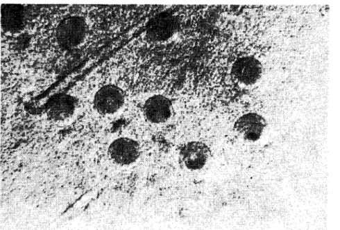
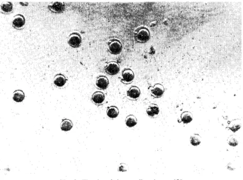
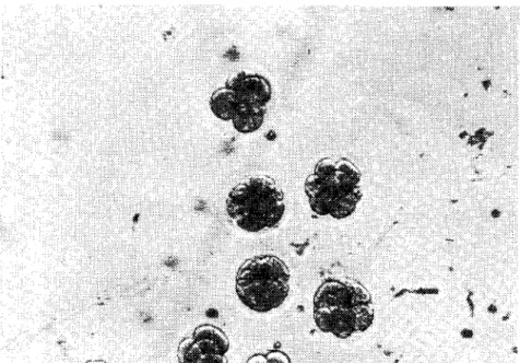
The relationship between time and development stagc of mouse ova in vivo (10,12) as given in Table 2 was very similar to that observed in the present study during the eulture in vitro of fertilized I-eel! mouse embryos (Fig.2-7).
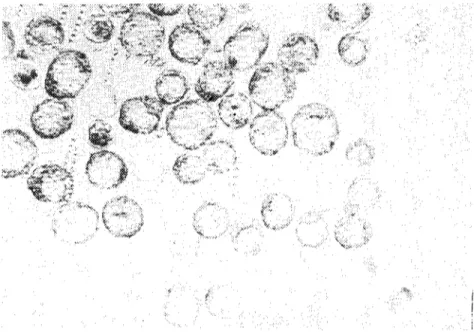
Fig. 2. One-ecU cmbryos and cumulus eclis.
306 çETiN KIUÇOGLU
'.£.
Fig. 3, The denuded one-cell embryos, ISOx
Şekil 3. eumulus hücrelerinden arındırılmış tek hücreli embriyolar.
'<:'/
;:)..
'~'~
'.~
Fig. 4. Two-cell embryos in M 16 culture medium.
THE IN VITRO CULTIVATION OF MOUSE ... 30i
Fig. 5. 4 and 8-ccl! cmbryos 111 M 16 clIlturc medilim. 180x
Şekil 5. M 16 kültüründeki 4 ve 8 hücreIi embriyolar.
Fig. 6. Hatching blastocyst. 3(jOx
308 çETIN KIUÇOGlU
Fig. 7. Hatched blastocysts and the free zona pclucidas. ıaOx
Şekil 7. Serbest zona pclucidalar ve zona pclucidasız blastosistler.
The salts of aıı culture media dosdy resemble the ions in blood (6). Whitten (13) using 8-ceıı mouse embryos found that omission of cakium, magncsium or potasium from the medium prevented growth and that embryonic development was delayed in the absence ofphosp-hate. Brinster (6) has a1so showed that the reduction, İn cakium con-centration resulted in a significant decrease in the number ofb1astocysts developing from 2-ceıı mouse embryos.
The effect of hydrogen ion concentration on the development of mammalian embryos has been studied primarily in the mouse. Whitten (13) found that the development of 8-ceıı mouse embryos occured betwecn pH 6.9 and 7.7. Brinster (2) found that the maximum development of 2-ccıı mouse embryos into blastocysts occured between pH 5.78 and 7.78 in an atmosphere of 5
%
CO2 in air. Howeverfurther studies (3) demonstrated that the optimum pH for develop-ment of the 2-ceıı mouse embryo was dependant on the concentration of pyruvate or 1actate in the meditim:
lt has been shown that 8-ceıı mouse embryos could develop İnto b1astocysts when cultivated in Krebs-Ringer solution containing 1 mg.
THE IN VITRO CULTlVATION OF MOUSE... 309
per mL. glucose and crystalline bovine serum albumin at a concent-ration between 0.03 and 6
%
(13). Brinster (4) has studicd the cffects of various exogenous amino nitrogen sources on the development of 2-cell mouse embryos into blastocysts. He found that a range of concentration between I-lA mg. per mL. of bovine serum albumin allowed maximum development. Howcver, there is no essential requirement for the devclopment of 2-eelI mouse embryos. In 1968 Brinster (5) confirmed that when glutathione is the only fixed nitrogen source in the culture medium 2-celI mouse embryos could develop into blastocysts.As a result, methods for the recovery and manipulation of mouse embryos have been described (I -6,iO,15) but information concerning many.specific aspects of development of the pre-implantation mamma-lian embryos are stilI unknown. The detailed studies which wilI per-formed on convenient economical laboratory animals cspecialIy the mouse, should provide suitablc experimental research models and indicate profitable are as to exploid later using the embryos of farm animals.
References
i. Brinster, R.L. (I 9(3). A medllOdfor in vilro cullivalion of mouse ova from lıvo-eel! lo
blas-loeysl. Expl. Cdl Res., 32: 205
2. Brinster, R.L. (1965al. Sıudies on Ihe developmenl of mouse embryos in vilro. IV Inleraelion
of erıergy sourees.J.Reprad. Fert., 10: 227.
3. Brinster, R.L. (1965 b). Sludies on Ihe developmenl of mo/ıse embryos iıı vi/ro. i Effecl of
osmolarity aııd hydrogeıı ioıı conemiraıion. J. Exp. Zool., 158: 49.
4. Brinster, R.L. (ı 965 c). Sludies on Ihe developmelll of embr)'osiıı/'ilro. III Th~ ejfecl ~f fixed
nilrogm source.J.Exp. Zool., 158: 69.
5. Brinster, R.L. (1968). Effecı of glulaılzione all ıhe deı'elopmenl of Iwo-cel! mouse embryos
in vi/ro.J.Reprod. Ferı., 17: 521.
6. Brinster, R.L. (1969). ıWammalian embryo cullure. lıı: The .~ammaliaıı Oviduel. p 419.
Eds. E. Hafe;:, R. I. Blmıdau. The University of Chicago Prc.'s.
7. Danlel, j.e. (1971). Meıhods iıı Mammalian Embryology. W. H. Freeman. San Francisco.
8. Hammond, j. (1949). Recovery aııd cullure qflubal mouse ova. Nalure, 163: 28.
9. Lewis, W.H. and Wright, E.S. (1935). On ılze deve!opmeııı ofllu mouse. Conlr. Embryol.
Carnegie Insı., 25: 113.
LO. Mark, F.L. and Long, j.A. (1912). Sludies on ear!.)'slages of deı'elopmenliıırals aııd mice.
310 ÇETİN KIUÇOGLU
1ı.Rafferty, K.A. (1970). Meıhods iıı Experimellıal Embryology of ıhe 'ıdause. The Johns
Hop-kins Press. Baltimorc-Lonclon.
12. Ünal, E.F. (1983). Farelerde EmbriYo TralLlferi. Doktora Tezi.
13. Whitten, W. K. (ı9:i6). Cullure of lubal mouse ova. "iaturc, Loncl., ı77: 96.
14. Whitten, W. K. (1957). Cullure oflubal ova. :'\aturc, Loncl., 179: 1081.
15. Whitten, W.K. and Biggers, J.D. (1968). Cmnpleıe deve/o/mıeııl in vilro of ıhe
pre-implan-lalion slages of mouse in a simple chemically defined medium. J. Reprocl. Fert., 17: 399.
lG. Whittingham, D.G. (19G8). Ferıilizalioıı of mouse eggs in vilro. Nalıırc, Luncl., 220: 592.
