Ankara Üniv Vet Fak Derg
47, 73-S0. 2000
TREATMENT
OF ULNAR FRACTURE
WITH DISLOCA TION OF
CA PUT
RADII
(MONTEGGIA LESION) IN 2 DO GS AND 3 CATS*
Mehmet SACLAM!
Barış KÜRÜMJ
Hasaıı BiLGiLP Arkuıı CANDAŞ4
iki köpek veüçkedide kaput radii dislokasyOllu ile ulııa kırığuıııı (Monteggia lezyonu) sağaltımı
Özet: Bu çalışmada klinik ve radyolojik muayeneler sonucunda MonteKgicl lezyonu oldulfu belirlenen 2 köpek ve 3 kedi konu edildi.
Olguların 2köpek ve J kedi 'de tip I, i kedi 'de tıı) i/, i kedi 'de ise tip III Mon-tegKül lezyonu saptandı.
Genel anestezi altmda ulna 'daki kırıkların redüksiyonu ve fikz.asY0l1u i
köpek 'te Steinman pin, 3 kedi 'de Kirschner tellerinin intramedüller UYKulanması iLe, i kijpek 'te ise, dinamik kompresyon plagı kullamlması ile salfLandı. Kaput radii 'nin redüksiyonu 3oLgu 'da uLna ile birlikte semiserklaj, J oLKuda vida iLe, i
oLKU da ise ligamentum annulare radü'ye sentetik bir iplik iLe diki,ç UYKulanması iLe saıf Landı.
Postoperatif 3 hafta süreyLe olgulara destekli bandaj UYKulandı. OIKulcırın postoperatil60-90. Künler arasında ön ekstremitelerini çok rahat kullandıkları iz-lendi. DIKularda uLna 'da yetersiz kallus, kaput radii 'nin relükzasyonu, pe-riartiküler ossifikasyon, osteoartritis, cubiti ekleminin hareket aralılfııun da-ralması, radius ulna arasmda sinostozis oluşumu Kibi komplikasyonlarla karşlla,çILmadl.
Sonuç oLarak, kedi ve köpeklerde rastlanılan 5olguya ait bilKilerin sunuldulfu bu çalı,ı'mada, Monteggiu lezyonu hakkmda meslekta,çLarımıw detayLı biLKi su-nuLarak, bu aLandaki sımI'Lı sayıdaki literatüre katkı saıflanmaya çalı,I'ı!ml"tır.
Anahtar kelimeler: Caput radü lukzasyonu, kedi, köpek, MontegKüı Le"Zyonu, su/{altıln, ulnar kırık.
Summary: Two dOKS and three cats with Monteggia lesions were diaKnosed due to clinicaL and radiological examinations which were subjected in this study.
, This stııdy presented in 25ıh World Smail Animal Congress, 25-291h April 2000, Aınsterdaın-Netherlands.
i. Assıst. Prof.. DYM., PhD., Depe of Orthopaedics & Traumatology Faculty of Yeterinary Medicıne, Ankara University. Dışkapı-Ankara 061LO, TURKEY.
2. DYM. PhD., Depe of Orthopaedics & Traumatology Faculty of Yeterinary Medicine, Ankara University, Dışkapı-Ankara 061LO. TURKEY.
3. DYM, Res.Ass., Dept. of Orthopaedics & Traumatology Faculty of Yeterinary Medicine, Ankara University. Dışkapı-Ankara 061LO.TURKEY.
4. ProLDL DYM. Depe of Orthopaedics&Traumatology Faeulty of Yeterinary Medicine, Ankara University, Dışkapı-Ankara 061LO, TURKEY.
74 M. SAGLAM. H. BILGILI. B. KÜRÜM. A CANDAŞ
Type I, type ii and type III MontegE;ia lesions were diagnosed in two dogs and three cats.
Reduction and ./ıxation o{ ulnar ./i"aetures were perl'ormed under general
anesthesia usinK Steinman pin in one dog, intramedu!lar Kirschner wires in three
cats and DCP in one dOE;.The reduction o{ the radicı! head was maintained by
.\vnthetie substitution o{ the annular ligament in one case by trans/ixatio/1 ol' ulna ~ith a sereı'v i/1 O/1ecase and with semi cerclaKe in three cases. Coaptatio/1 bcll7-daKes were used post operatively for three week.\'. It is observed that ('(Ises beKwı to use their leKs pmperly between 60 and 90 days post operatively.
Complications such as incomplete ca!lus formation. recurrent subluxatiO/1 ol'
radial head, periarticular ossi/ication. osteoarthritis. poor function o{ art. eubiti
and synostosis between radius and ulna were not developed.
As a conc/usion. this study presents detailed knowledge on MonteKgicl lesions, in which there are limited studies on this subject.
Key words: Caput radii luxation, cat, dog. Monteggia /esion. treatment. u/nar./i"acture.
Introduction
The luxation of caput radii and fracture of ulna and also named as "Monteggia fracture-dislocation" is not a common lesion (1,3,5,6,
13).
Monteggia lesion was fırst described by Giovanni Battisla Monteggia in 1814 and rcpre-scnts about 0.7% of e1bow fractures and dis-locations and 7% of fractures of the radius and ulna in human (9,14,15).
Four types of Montcggia Iesions have been described in human by Bado (1962), according to the dislocation direction of caput radii and the !evel of ulnar fracture according to the elas-sifıcation made (11,14).
Type i: Crania1 dislocation of caput radii with caudal angulation and ulnar diaphyseal fracture. lt is the most common type seen in cats and dogs.
Type (J: Caudal dislocaLİon of caput radii and ulnar diaphyseal fracturc.
Typc
ın:
Craniolateral and lateral dis-location of caput radii and ulnar diaphyseal fracture.Type IV: Cranial dislocation of caput radii with proximal 1/3 fracture of radius and ulnar diaphyseal fracture.
Close reduction and external fixation can be uscd in cases with slight dislocation, but this technique is not successful enough for treat-ment of lesions (2,14). üpen reduction and im-mobilization is reported lo be the best operaıive technique in Monteggia Icsions (2,4,6,8,14). ReducLİon of the caput radii and immobilization of the ulnar fracture must be made by using in-tramedullary pins, bone screws and plates. Re-duction of the radial head can be mainlained by repairing of lig. anmılare radii or ulnar lrans-fıxation of the ligament using screws, pins or cerelage wİres (4,5,7,12,14).
Monteggia lesion is not a common prob-lem in veterinary surgery and there are limiled studies conccming the subjecı (1,2,6,14). In a study concerning 28 cases; type I, ıype II and type III Monıeggia Icsions were described ın 24, 1 and 3 cases respectively (14).
In the present elinical study, it was aimed to determine the treatments and prognosis of Monteggia lesions diagnosed in 2 dogs and 3 cats.
Materials and Methods
The study performed on 2 dogs and 3 cats with different breed, age and sex which were brought lo ürıhopaedics and Traumatology
TREATMENT OF ULNAR FRACTURE WITH OISLOCATION OF CAPUT RAOll (MONTEGGl/\ LESION) 75 IN 2 OOGSAI\O 3 CATS
Clinics of Veterinary Faculty of Ankara Uni-versity.
Cases were brought with the etiology be-cause of trafik accidents in 2 cases (Case i and 4) and falIing down windows in 3 eases (Case 2, 3 and 5). Deformation and pain in art. cubiti and crepitation in ulna was notieed in their clin-ical examination.
According to the radiographie examina-tions, in 2 dogs (Case 1 and 4) and one eat (Case 3) Type I, in one cat (Case 2) Type II and İn one cat (Case 5) Type III Monteggia lesions were diagnosed (Fig. 1,2,5,6,7,10).
Af ter the cases were premedicated by xy-lazine hydrochlorid (Rompun, Bayer, Türkiye, 23,32 mg/ml, O,i mg/kg IM), anesthesia was performed by ketamine hydroehlorure (Ketalar, Eczacibasi, Türkiye, 50 mg/ml, LOmg/kg IM). All cases were prepared in lateral recumbeney and operation site was prepared.
Thc skin incision is started from the lateral humeral epicondyle foııowed the joint and cen-tered between ulna and radius to the mid points of these lwo bones. After the ski n incision sub-cutaneous fat tissue and fascia was elevated and muscles were exposed. Af ter the incision of ap-onellrosis, musculus extensor digitalis eom-munis and musculus extensor digitalis lateral is were reached from the septum intermuscularis.
Fracture line on ulna was exposed with the elevation of these two muscles. The reduction and fixation of the ulnar fracture has been ac-complished by using Steinmann pin in 1 dog (Case i) (Fig. 3,4) and Kirschner wires in 3 cas-es. (Case 2, 3 and 5) intrameduııary with ret-rograd method. Fixation of ulnar fraeture was accomplished by dynamie compression plate (DCP) and serew system in one dog (Case 4) (Fig. 8).
Proximal part of the incision was pro-longed including the art. eubiti. Musculus ex-tensor digitalis communis and musculus ex-tensor digitalis Iateralis were elcvated to the sides and reaehed to the joint with a myotomie under the humeral condyle (7,10) within these direetions. Attention was paid to protect the
ra-dial nerve. The reduction of cranially, eaudally or lateraııy disloeated radius was aeeomplished manually. In 3 eases (Case I, 3 and 5) semi-cerclages, in one case (Case 4) a serew was used in thation of caput radii and in one ease (Case 2) repair of join annular ligament was performed with a 3/0 propilen suture material (Ethicon,UK, polyglieolic asid) with sutures myotomied m. supinator had it's original an-atomical eondition.
Post operatively the fixation of the joint in it's physiological angle was performed by bandages whieh is supported with plastic co-aptation material, in dogs and handages sup-portcd with cartoon in cats.
Betwecn postoperative 71!ı and iOılı days,
skin sutures, and after 3 week s banda ges were removcd.
Results
Because of the post operativcly applied padded bandages it is ohserved that hınction loose of front limbs deereased by removing of these bandages.
After observing enough caııus formation and healing of ulna in radiographs taken in 45-6011ı days, in one cat (Case 2) intramedullar pin
was been left in ulna because of the 6 months age of the ease.
In the other 3 cases (Casc I, 3 and 5) in which intramedullar pins wcre used and in i
easc (Case 4) in which DCP was used, iixation matcrials were removed with a second opera-tion on 611ı months (Fig. 9).
The ecrclage wire which was used in rc-pairment of annular ligament was Idt in cases (Case ( 1, 3 and 5) and no complications about this was observed.
In clinical examinations which were done in postoperative 60-901!ı days, it was observed
that cases were using their front limbs easily and comfortably. Detailed knowledgc about cases are presented in tabı e i.
76
,
~,\"
'.\
i \ \ \,
j
M. SAGLAM. H BILGILI, B KÜRÜM. A C;\NDAŞ
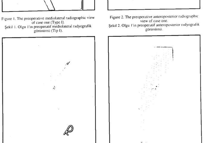
Fıgure I. The preoperative mediolateral radiographic view of case one (Type I).
Şekil ı.Olgu i'in preoperatif mediolateraI radyografik görünümü (Tip I).
//
\
'J
.
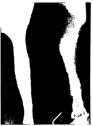
Fıgııre 3. The p()sıoper~Hivc mediolateraI radiographic vicw of case one.
Şekil 3. Olgu i'in posıoperaıif mediolateral radyografik görünümü
Figure 2. The preoperative anteroposıerior r~\dlOgraphıc vicw of ease one.
Şekil 2. Olgu i'in preoperatif anteroposıerior radyografik görünümü
Figure 4. The postoperative anteroposıerior radiogr~\phıc view of case one.
Şekil 4. Olgu ı'in posteperatif anıeroposıerior radyografik görüniiıııü
TREATMENT OF ULNAR FRACTURE WITH DISLOCATION OF CAPUT RADII (MONTEGGIA LESION) 77 1\1 ~ DOGS AND 3 CATS
Fıgure 5. The preoperative anteroposterior andmediolateral radiographie views of case 2 (Type II). Şekil 5. Olgu 2'nin preoperatif anteroposterior ve
ınediolateral radyografik görüniimleri (Tip II)
Fıgure 7. The preoperatif anteroposterior ve mediolateral radiographic views of case 4 (Type i).
Şekıl 7. Olgu 4'ün preoperatif anteroposterior ve ınediolateral radyografik görünümleri (Tip i).
Figure 6. The preoperative ınediolateral radiographic view of case 3 (Type i).
Şekil 6. Olgu 3'ün preoperatif mediolateral radyografik görünümü (Tip 1).
Figure 8. The postoperative anteroposterior and mediolateral radiographic views of case 4. Şekil 8. Olgu 4'iin postoperatil' anteroposterior ve
M. SAGLAM. H. BILGILI. B. KÜRÜ\tl. A. CANDAŞ
Fıgure 9. The anteroposterıor and ınediolateral radiographıc vıcws of C,1se 4 when plate and snews reınoved on 6th months.
Şekil 9 Olgu 4'lin po.,toperatif 6. ayda plak ve vidalarının uzakla~tırılmasından sonraki anteroposterior vc mediolateral radyografik göriinlimleri.
figure ıll. The preoperalİ ve anteroposterior and mediolaıcral r<ıdiographic vicws of casc 5 (Typc III)
Şekil ıll. Olgu S'in preopcratif anteroposterior ve mcdiolatcral r<ıdyografik görlinliınleri (Tip III)
Discussion
The dispersion of Monteggia 1esions in to-tal yeterinary cases, are limited according to the reports. The most common Monteggia lesion is Type i. In 3 cases Type I, in one case Type II and in one case Type III lesions were obseryed in this study. Second type of Monteggia Iesion was recorded İn one dog but not in any cat (4,11,12,14).
Definitiye diagnosis can be made only by radiographic examination. it has been reported that closed reduction and extcmal fixation may be considered in cases without displacement but the chanee of this method is so wcak in eas-es with displacemcnts (2,14).
Definitiye diagnosis about the type of the lesions was madc by doublc sided radiographs. Operatiye treatment mu st be undertaken for the eorrection of the displacements.
In operalİye treatment while some S tır-geons are recommending the fixation of the ul-nar fracture with a intrameduHar pin firstly and than the immobilization of radius with cercIage
TREATMENT OF lJLNAR FRACTURE WITH D1SLOCATION OF CAPUT RADII (MONTEGGIA LES/O:\') 7') IN:2 DOGS AND 3 CATS
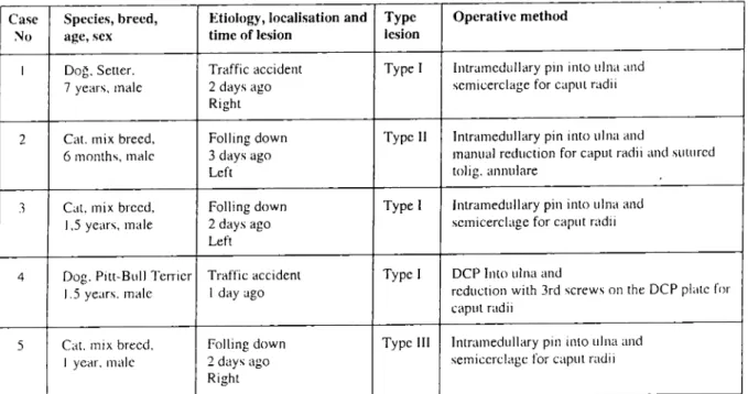
Table I. Data of cases. Tablo I. Olgulara ait veriler.
Case Species, brecd, Etiology, loealisation and Type Operative method
:'0/0 age, sex time of lesi on lesion
i Doğ. Setter. Traffic aceident Type i Intramedullary pııı into ulna and 7 years. male 2 days ago semicerclage for capuı radii
Righı
2 Cal. mix breed. Folling down Type ii Intramedullary pin into ulna and
6 months. male 3 days ago manua! reduction for caput radii and suıured
Left tolig. annulare
3 Cat. mix brecd, Folling down Type i Intramedullary pin into ulna and 1,5 years, male 2 days ago semicerclage for caput radii
Left
4 Dog. Pitt-Bull Terrier Tratlic accident Type i DCP Into ulna and
1.5 years. male ıdayago reduction wİth 3rd screws on the DCP plaıe for
i
raput radii
5 Cal. mix brecd. Folling dow n Type iii Intramedullary pin into ulna and
i year. male 2 days ago semicerclage for eaput radii Right
wires turn around ulna and radius (6), some oth-er surgeons recommends the use of a long screw to the proximal fracture of the olecranon (2) or a plate to the ulna and different methods and materials are also been recommcnded (4,5,7,12,14). In chronic cases, some rec-ommend the excision of caput radii (2,8).
In our study case, the fixation of ulna was performed by a Steinman pin 1 case, by a Kirschner wire in 3 cases and by DCP in 1 case and no functional problem occlIfred about the 3/0 propilen which was used in repairment of lig. ,mmdare and fixation of the dislocated ca-put radii to the u1na with a semicerclage. Ac-cording ta the literature knowledge; complete caııus formatian on ulna, redislocation of caput radii, traumatic periarticu1er ossification, os-teoarthritis, restricted range of motion clbow, asteomyelitis and synosthosis between the ra-dius and ulna are the most common eomplica-tions (2,8).
In cases between postoperative 60-90th day s a complete funetional healing was ob-served and complications, like nonunion, ma-lunion, joint lesion, redislocatian, ankylosis were observed.
As a result of this study in whieh knowl-edge about 5 cases, wc worked was presented, detailed knowledge abaut Monteggia lesion was presented. it is aimed to he1p to the few lit-eratllfe abaut this subject.
Rcferenccs
ı. Archibald J (I 973) ChirurKil' Cmıilll'. Yigot Freres. Paris.
2. Bojrab.J (ı978) Tl'chni,lut's Acıul'lIas de ChirurKie des Pl'lils Aııimuu. Editions Yigot, Pans.
3. Boyd HB, Boals .JC (1969) Thl' MollleKKia Il'swıı.
Clin Orthop. 66. 94-100.
4. Candaş A, Sağlam M, Özba B(ı989)
M()IlII'KKia11'-sion and ir's surKi('(l1 Irl'alml'lll iıı a dOK. AÜ Yel Fak
Derg, 36, 358-366.
5. Denny HR (ı985) A Cuidl' lo CU/ıiııl' Orıhopedıc SUl"
KI',}'. Blackwell Sri. Publ.. Oxford, 170-i7ı.
6. Leonard EP (ı974) Chirurgit' Orıhopl'dllfue du C/llen
1'1du Chaı. Yigoı heres Ed .. Paris.
7. Lipowitz AJ, Caywood DD (1993) Smail Aııimalıı.
lusırull'd: Surgical Apprnachl's aııd Procl'durs.
Mosby. Year Book Ine .. St. Louis. Missouri. 138-141. 8. Olmstead ML (I 995) Smail Aııimal Orıhopaedics.
Mosby-Year Book Ine .. Si. Louis. Missouri. :210.:211. 9. Penrose.JH (195ı)Thl' MO/lIl'ggia jmerurl' wiıh pos.
lerior dislo('(lliOlI oj' ılıl' mdilLi hmd. 1 Bone loiııı
Surg, 33B, 65-73.
iO.Piermattei DL, Greely RG (ı971) Aıias dl's Voit's
D'ucces DallS lu Chirurgit' Osseusl' du Chil'lı e/ du
so
i i Reckling FW (J 982) UnslU!JIeji'aclUre-dislocalions o/
ıiıe/oreann J Bone Joınt SlIrg, 64A, 857-853.
12. Robins G (1993) Tiı/' Elhow joinı. 207-208, In: Ho-lIJton, JE.F, Collinson, R.W. (Ed). Manual of Smail Antmal Arthrology. British Smail Animal veterinary Assoc. PlIbl.. UK.
13. Sağlam M, Bilgili H (I 997) Type LI (~/
MOIlIe}{I;iale-sion und opl'rmivl' Irealment in u cal. AÜ Vet Fak
Derg, 44, 1-4.
14. Schwarz PD, Schradcr SC (1984) Ulnarfrucıure und
dıslocu/lon o/ ıhe proximal rudial epiphysis
(Mon-1/'!iKial/'si(1) in ıhe do,; and cal: a review 0/28 casI's.
J Am Vet Med Assoc, IliS, 190-194.
M. SAGLAM, H. BILGILI. B. KÜRÜM, A CAI\'DAŞ 15. Speed .TS, Boyd HB (I 940) Trmımenı o/Facıures ii
ulna wl1h dislocaıiıııı o/hmd o/rııdius. J Am Vet Med
Assoc, 115,1699-1705.
Corrcspondcnce to i Yazı~ma adresi:
Dr. Hasan Bilgili. J)VM. PhD.
Depı. ofOrlhopaedics & Traumalolot:Y
Faculı)' (i/Veıerinıır)' Medicine
Ankara Universit\' Dı>~kapı-AnkııraOM /o TURKEY
PhOlıe. + 90 312 3170315 exl 403or 32<)
Fax +903123104472

![Synthesis, crystal structure, and characterization of two heterometallic transition metal citrate complexes [M = Co(II) and Cd(II)]](data:image/gif;base64,R0lGODlhAQABAIAAAP///wAAACH5BAEAAAAALAAAAAABAAEAAAICRAEAOw==)