Abstract
Diabetes Mellitus is the most common of the serious metabolic diseases. It is characterized by metabolic abnormalities; by long-term complication. Correlation between erythrocyte membrane Na+K+ATPase
and plasma homocysteine levels in patient. This study was conducted in 29 patients with Type II Diabetes Mellitus and 15 healthy controls. Erythrocyte membrane Na+K+ATPase activity and malondialdehyde were
measured spectrophotometrically. Plasma homocysteine levels were measured with High Performance Liquid Chromatography method. When compared to the control group, mean plasma homocysteine levels were found to be increased (p<0.001) and mean Na+K+ATPase values of
erythrocyte membranes activities were found to be decreased (p<0.05). There was a negative correlation between erythrocyte membrane Na+K+ATPase and plasma
homocysteine levels in patient group (p< 0,01; r= - 0,49), but any correlation was not detected in the control group. Malondialdehyde was measurement in the tip 2 diabetic patients decreased more than control group. We found an increased plasma homocysteine levels and decreased erythrocyte membranes Na+K+ATPase activities in
Diabetes Mellitus group. We think that the decrease in activities of Na+K+ATPase may be due to the increased
plasma homocysteine levels.
It was concluded that to be more impact in diabetes which is a complicated disease, cell membrane lipids of oxidation with starting negative the table as a result of high homocysteine levels and reduced antioxidant capacity.
Keywords: Diabetes Mellitus, Na+K+ATPase,
Homocysteine, Malondialdehyde 1. Introduction
Diabetes Mellitus is a heterogeneous disease in insulin secretion or effect full or partial deficiency results emerging and showing itself mainly with chronic hyperglycemia (KADAYIFCI et al. 1998).
Accepted Date: 05.11.2015 Corresponding author: Naim Uzun, PhD
Ağrı İbrahim Çeçen University, Faculty of Pharmacy, Department of Pharmaceutical Professional Sciences, Ağrı, Turkey
e-mail: [email protected]
Diabetes mellitus continues to be one of the last century's most important diseases, although it has been nearly a century since the use of insulin. Acute and chronic complications of diabetes lead to disability and death caused by diabetes mellitus (TÜRKMEN et al. 1990).
Na+K+ATPase is an integral membrane protein
(LEHNINGER et al. 1993). Na+K+ATPase activity is a
determinant of the intracellular K+ levels. K+ values are
very important in maintaining the intracellular negative electric potential. Na+K+ATPase are a classic example of
cell membrane pump system. This pump is localized in the plasma membrane of all cells but it shows the highest activity in nerve and muscle tissue. Na+K+ATPase takes
three sodium ions out of the cell and two potassium ions into the cell, and one ATP is hydrolyzed during each transport. Na+K+ATPase keep the Na+ K+ gradient of of
the normal level in the plasma membrane. This process called as "Electrogenic" because the ion transport of system is a net transport of electric charge. Electric charge is negative cell membrane inner surface and outer surface is positive. Thus the inner and outer membrane surface is maintained the balance of the electrical potential. Ion transport with ATP hydrolysis is a pair of compact. Na+K+ATPase is a major metabolic energy consumers.
The brain consumes to Na+K+ pump at least half of the
energy (MEISENBERG et al. 1998). Na+K+ATPase
consumes approximately 70% of the total energy requirement that can be electrically stimulated cells (MATHEWS and HOLDE 1990).
Homocysteine is from intermediate metabolites of cysteine amino acid biosynthesis. It is synthesized from methionine which must be taken with food. Cystathionine was synthesized by the condensation of homocysteine with serine amino acid (KEHA and KUFREVIOGLU 2000). Homocysteine was first found in 1932 by Vigneaud. Homocysteine in 1962 was detected in the urine of three children. Cystathionine synthetase deficiency was detected in 1964. Refsum et al, in 1985, determined the amount of homocysteine. Fasting plasma homocysteine levels is 5-15 mol/L in the normal population. Above this value is defined as
Correlation between Erythrocyte Membrane Na
+K
+ATPase and Plasma Homocysteine
Levels in Type 2 Diabetes Patient
NAİM UZUN1*, AHMET KIZILTUNÇ2
1Ağrı İbrahim Çeçen University, Faculty of Pharmacy, Department of Pharmaceutical Professional Sciences, Ağrı, Turkey 2Atatürk University, Faculty of Medicine, Department of Biochemistry, Erzurum, Turkey
hyperhomocysteinemia. It raises homocysteine levels as a result of coming together one or more of the essential factors which can be changed such as smoking, too little physical activity, excess coffee consumption and inadequate amounts of nutritional vitamins taken. The diagnosis of hyperhomocysteinemia may be an important factor in decision making to patients for more healthy life. In addition hyperhomocysteinemia may be treated with a simple vitamin addition. Cancer, renal failure, autoimmune diseases such as diabetes mellitus affect the metabolism of homocysteine (GOUAILLE 1999).
It has been reported that free oxygen radicals will be cause irreversible cell damage (BURRELL and BLAKE 1990) and cell damage will continue chaining (WHITEHEAD et al. 1995). It was determined the relationship between diabetes and malondialdehyde in previous studies (AYALA et al. 2014).
According to us, increased homocysteine concentration causes increase in superoxide anion radical and hydrogen peroxide. These radicals can cause the onset of cell membrane lipid peroxidation. The structure and integrity of the membrane deteriorates as a result of started lipid peroxidation. We believe that the structure of damaged membrane can affect the activity of the Na+K+ATPase.
2. Material and Methods
In this study, Ataturk University Research Hospital receiving treatment with Type II diabetes mellitus diagnosis was performed on 29 patients. As a control group of 15 healthy volunteers were included in the study. The control group was chosen from among persons no systemic and non-pathological disorder. The demographic data of both groups are given in Table 1.
10 ml blood samples were taken 12 hours after fasting forearm vein with plastic syringes and transferred to heparinized glass tubes. Blood samples were centrifuged at 2000 g for 10 minutes. Thus blood cells and plasma were separated. Erythrocyte membranes were prepared according to Wood and Beutler (WOOD and BEUTLER 1967). End of the process, the erythrocyte membrane ghost stored until the time to be analyzed for the determination of protein and inorganic phosphate (Pi)
in 1 ml Tris-EDTA Buffer (pH 7.4) at -80 °C. Na+K+ATPase activity was performed according to Muriel
and Mourella (MURIEL and MOURELLA 1990). The supernatant from ghost suspension was taken from for determination of Pi. Pi determination was made according
to Munoz (MUNOZ et al. 1983). The reaction medium was prepared for standard graphics with determined amounts. Standard chart was prepared according to absorbance received at 390 nm.
Protein assay was performed with the method of Bradford (MINCH 1989). Standard curves were constructed according to the absorbance at 595 nm taken. Calculation of protein in the samples was conducted utilizing the standard curve.
Plasma homocysteine assay was performed with High Performance Liquid Chromatography (HPLC) method. (Instruction manual for HPLC analysis of homocysteine in plasma/serum (fast elution). Chromsystems. Instruments & Chemicals). Malondialdehyde was measured by spectrophotometry (YOSHIOKA et al. 1979).
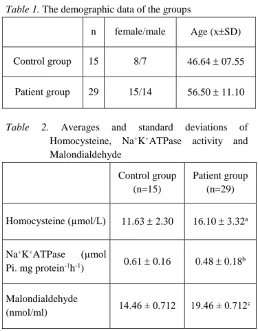
Statistical analyzes were performed with IBM-type computers by using SPSS 10.5 package program. Significance test of a difference between the two averages were made with Mann-Whitney U test. The significance limits of 0.05 were taken. Degree of importance of p was determined. Correlation analysis was performed with Sunderman Test, r and p values were determined; the results were given in Table 2. The correlation between homocysteine concentrations and Na+K+ATPase activity
was given in figure 1.
There is no conflict of interest with any other person or institution of research. Declaration of Helsinki principles were adhered to in the research.
3. Results
Table 1. The demographic data of the groups
n female/male Age (xSD)
Control group 15 8/7 46.64 07.55
Patient group 29 15/14 56.50 11.10
Table 2. Averages and standard deviations of Homocysteine, Na+K+ATPase activity and
Malondialdehyde Control group (n=15) Patient group (n=29) Homocysteine (µmol/L) 11.63 2.30 16.10 3.32a Na+K+ATPase (µmol Pi. mg protein-1h-1) 0.61 0.16 0.48 0.18b Malondialdehyde (nmol/ml) 14.46 ± 0.712 19.46 ± 0.712 c a: p< 0.001, b: p<0.05, c: p<0.01
Figure 1. The graph of correlation between homocysteine concentration and Na+K+ATPase activity
(r=-0.49, p<0.01) 4. Discussion
The mobility of integral proteins, lateral diffusion, and the scale association and dissociation equilibrium of membrane proteins regulates by membrane fluidity and viscosity (CORNELIUS and SKOU 1984).It knows that cell membrane enzyme activities were affected by from changes in the vicinity of the enzyme (MOORE et al. 1989).
Active membrane proteins are functional to be active when in liquid phase lipid layer (CANTLEY and HAEST 1987). Likewise, Na+K+ATPase only have been
demonstrated to be active when in liquid phase lipid layer (GRISHAM and BARNETT 1973).
It has been reported that acidic phospholipids are making the Na+K+ATPase active but activation is attached
to the structure of the hydrocarbon chain and phospholipid polar group (WALKER and WHEELER 1975). Na+K+ATPase activity is bound to the phospholipid it has
been shown in many studies (DEUTICE and HAEST 1987). Also it has been reported to increase enzyme activity with increasing concentrations of membrane linoleic acid (JOHANNSSON and SMITH 1981).
The between Polyunsaturated fatty acids and saturated fatty acids imbalance causes a change in Na+K+ATPase activity. Therefore changes in activity of
Na+K+ATPase can be an indicator of membrane fluidity
(GUTIERREZ et al. 1993).
It have been reported that the decrease in Na+K+ATPase activity in patients with diabetes may be
derived from the increase in the concentration of Lysophosphatidylcholine (lysoPC) (RABINI et al. 1994).
Decrease in the enzymatic activity of Na+K+ATPase plays a major role in the pathophysiology
of chronic complications of diabetes (WINEGRAD 1987).
Carrier-mediated transport is reduced membrane cholesterol/phospholipid ratio in the increased. Many in the studies have been reported that increase in this ratio causes significantly decrease in the activity of Na+K+ATPase (MOURELLA and FRANCO 1991).
Our findings (Table 2) support that Na+K+ATPase activity is decreases by increasing type 2
diabetes and radical oxygen species of emerging with advancing age.
Protein or sulfhydryl groups (-SH) may be exposed to oxidation, (KONG et al. 1991) exposure to oxidation of the -SH group disturbed the activity of Na+K+ATPase, (KAKO et al. 1998) because there are 36
sulfhydryl groups in the structure of Na+K+ATPase.
Free radicals are impairing between the functions and structure of the cell membrane in the form of a chain reaction (OSHI 1980). It is obvious that Na+K+ATPase
activity in the damaged cell membrane will reduce, and this situation supports our findings.
A study carried out, Na+K+ATPase activity had
found significantly lower in the diabetic, it has been suggested that on the decrease in activity was caused by cholesterol and lysophosphatidylcholine. It has been identified that high cholesterol concentrations were significantly lower activity and lysophosphatidylcholine at high concentrations were significantly higher activity in vitro conditions. Due to low concentrations of lecithin cholesterol acyl transferase is that low Na+K+ATPase
activity in diabetic patients and this situation is indicated in patients with diabetes may explain the chronic complications of atherosclerosis (KIZILTUNC et al. 1997).
Na+K+ATPase is one from enzymes membrane
the destructive effects of free radicals act. We think this would be effective in two ways. First, membrane indirectly damaged with occur peroxidation of membrane lipids and thus Na+K+ATPase activity will decrease.
Secondly, free radicals can cause the reduction of activity interact with sulfhydryl groups in the structure of Na+K+ATPase.
Kitao and his friends have reported that peroxidation damaged to the erythrocyte membrane lipids and there was a role in the Na+K+ATPase inhibition play
of this damage (KITAO and HATTORI 1983).
In the recent study demonstrated significantly higher in diabetic than non-diabetic homocysteine. Plasma homocysteine levels affect both the duration and metabolic control of type II diabetes (PASSARO et al. 2000).
Homocysteine initiates oxidative stress, or homocysteine contributes to the onset of oxidative stress weakening the antioxidant defense system. According to
redox thiol (sulfhydryl) the structure theory, as a pro-oxidant of reduced homocysteine acts and as an antioxidant of the reduced system assumes function. It is more reactive reduced homocysteine than oxidized homocysteine or protein bound homocysteine. The balance between oxidized homocysteine with reduced free homocysteine and total plasma homocysteine levels provides stay at specific border with a strict system where there is adequate folate. If the value increases intracellular levels of homocysteine, diseased conditions associated with significantly higher values may possibly occur. The majority of the work is now focused on the total thiol concentration in plasma. Homocysteine is rapidly oxidized (reduced homocysteine) in the increased plasma levels of it (STAMM and REYNOLDS 1999).
Na+K+ATPase decreases significantly in the
presence of diabetes. It concluded it will have a negative impact on cell membrane and Na+K+ATPase activity of
high plasma homocysteine level. In the our studies, in diabetic patients, plasma homocysteine concentration significantly higher than healthy control group and erythrocyte membrane Na+K+ATPase activity was found
low (Table 2). It was showed a significant negative correlation between homocysteine and Na+K+ATPase in
diabetics (Figure 1).
High homocysteine levels and decreased antioxidant capacity and lipid peroxidation of cell membrane will be affected negatively Na+K+ATPase
activity, and it will increase further the impact of diabetes that is a complex disease. To clarify the relationship between homocysteine and cell membrane integrity, performing works in larger populations is required. REFERENCES
KADAYIFCI A., KARAASLAN Y. and KÖROĞLU E. (1998), Pratisyen Hekimin El Kitabi Hekimler Yayın Birliği. Ankara. 1–541.
TÜRKMEN F., AKKUŞ İ., BÜYÜKBAŞ S. and ÇIĞLI A. (1990), Diyabetes Mellitusda Biyokimyasal Değişiklikler ve Komplikasyonlar. Turkiye Klinikleri J Med Sci. 10, 1–10.
LEHNINGER A.L., NELSON D.L. and COX M.M. (1993), Principler of Biochemistry. Worth puplisher. New York. 268–293.
MEISENBERG G., WILLIAM H. and SIMMON S. (1998), Principles of medical biochemistry. Mosby. Toronto 1–668.
MATHEWS K.C. and HOLDE K.E. (1990), Biochem. Redwood City. The Benjamin Cumminings Publishing. 229- 230.
KEHA E. and KÜFREVIOĞLU Ö.İ. (2000), Biyokimya. Aktif Yayınevi. Erzurum. 200–384.
GOUAILLE C.B. (1999), Determination of homocysteine why, when and how. Sweden. Bryne Offset. 1-54. BURRELL C.J. and BLAKE D.R. (1990), Reactive
oxigen metabolites and the human myocardium. J Heart. 60, 4–8.
WHITEHEAD T.P., ROBINSON D., ALLAVAY S., SYMS J. and ANN H. (1995), Effect of red wine ingestion on the antioxidant capacity of serum. Clin Chem. 41, 32–35.
AYALA A., MARIO F. MUÑOZ, and SANDRO ARGÜELLES. (2014), Lipid Peroxidation: Production, Metabolism, and Signaling Mechanisms of Malondialdehyde and 4-Hydroxy-2-Nonenal. Oxidative Medicine and Cellular Longevity. ID 360438, 31 pages.
WOOD L. and BEUTLER E. (1967), Temperature dependence of Na+, K+ activated erytrocyte adenosine triphosphase. J Lab Vlin Med. 70, 287–294.
MURIEL P. and MOURELLA M. (1990), The role of membrane composotion in ATPase activities of cirrhotic rat liver effect of silymarin. J Appl Toxicol. 10, 281–284.
MUNOZ M.A., BALON M. and FERNANDEZ C. (1983), Direct determination of inorganic phosphorus in serum with a single reagent. Clin Chem. 29/2, 372–374.
MINCH M.J. (1989), Experiments in biochemistry: Projects and procedures. Prentice Hall College Div. New Jersey 1–716.
YOSHIOKA T., KAWADA K., SHIMADA T. and MORI M. (1979), Lipid peroxidation in maternal and cord blood and protective mechanism against activated-oxygen toxicity in the blood. Am J Obstet Gynecol; 135, 372-376.
CORNELIUS F. and SKOU J.C. (1984), Reconstitution of Na/K-ATPase into phospholipid vesicles with full recovery of its specific activity. Biochem Biophys Acta. 772, 357–373.
MOORE R.B., BRUMMITT M.L. and MANKAD V.N. (1989), Hydroperoxides selectively inhibit human erytrocyte membrane enzymes. Arch Biochem Biophys. 273, 527–534.
CANTLEY B. and HAEST W.M. (1987), Lipid modulation of transport proteins in vertebrate cell membranes. Ann Rev Physiol. 49, 211–235. GRISHAM C.M. and BARNETT R.E. (1973), The role of
lipid phase transition in the regulation of the Na/K-ATPase. Biochem. 12/14, 2635–2637. WALKER J.A. and WHEELER K.P. (1975), Polar
phospholipid dependent Na/K-ATPase. Biochem Biopys Acta. 394, 135–144.
DEUTICE B. and HAEST W.M. (1987), Lipid modulation of transport proteins in vertebrate cell membranes. Ann Rev Physiol. 49, 221–235. JOHANNSSON A., SMITH G.A. and METCALPHE J.C.
(1981), The effect of bilayer thickness on the activity of Na+, K+, ATPase. Biochem. Biophys. Acta. 641, 91–96.
GUTIERREZ V.R., STREFEL P., VILLAR J., GARCÍA-DONAS M.A., ACOSTA D. and CARNEADO J. (1993), Cell membrane fatty acid composition in type 1 diabetic patients: relationship with sodium transport abnormalities and metabolic control. Diabetologia. 36, 850–856 .
RABINI R.A., GALASSI R., FUMELLI P., DOUSSET N., SOLERA M.L., VALDIGUIE P., CURATOLA G., FERRETTI G., TAUS M. and MAZZANTI L. (1994), Reduced Na+-K+-ATPase activity and plasma lysophosphatidylcholine concentrations in diabetic patients. Diabetes. 43, 915–919.
WINEGRAD A.I. (1987), Does a common mechanism induce the diverse complications of diabetes? Diabetes. 36, 396–406.
MOURELLA M. and FRANCO T. (1991), Erytrocyte defects precede the onset of CCI4 ınduced liver cirrhosis protection by silymarin. Life Sci. 48(2), 1083–1090.
KONG K., LESNEFSKY J.E., YE J. and HORWITZ L.D. (1991), Prevention of lipid peroxidation does not prevent oxidant-induced myocardial contractile dysfunction. Am Physiol Soc. H: 2371–2377. KAKO K., KATO M., MATSUOKA T. and MUSTAFA
A. (1988), Depression of membrane- bound Na/K- ATP ase activity induced by free radicals and by ischemia of kidney. Am Physiol Soc. 330– 337.
OSHI F.A. (1980), Vitamin E-A radical’s defense. J Med. 303, 454–455.
KIZILTUNÇ A., AKÇAY F., POLAT F., KUŞKAY S. and ŞAHIN Y.N. (1997), Reduced lecithin: Cholesterol acyltransferase and Na+, K+, ATPase activity in diabetic patients. Clin. Biochem. 30/2, 177–182.
KITAO T. and HATTORI K. (1983), Inhibition of erytrocyte ATPase activity by aclacinomycin and reverse effects of ascorbate on ATPase activity. 39, 1362–1364.
PASSARO A., D'ELIA K., PARESCHI P.L., CALZONI F., CARANTONI M., FELLIN R. and SOLINI A. (2000), Factors influencing plasma homocysteine levels in type 2 diabetes. Diabetes
Care. 23(3), 420–421.
STAMM E.B. and REYNOLDS R.D. (1999), Plasma total homocysteine may not be the most appropriate Index for cardiovascular disease risk. J Nutrition. 129, 1927–1930.