Accepted: 2014.12.10 Published: 2015.03.24
2276
3
2
26
Montelukast Inhibits Pentylenetetrazol-Induced
Seizures in Rats
ABCEG 1
Betul Cevik
ABEF 2
Volkan Solmaz
CDF 1
Durdane Aksoy
ABDFG 3
Oytun Erbas
Corresponding Author: Volkan Solmaz, e-mail: [email protected] Source of support: Departmental sources
Background: Montelukast is an antiinflammatory drug with an antioxidant property. In this study, we aimed to reveal wheth-er montelukast has a preventive effect against seizures and post-seizure oxidative stress in pentylenetetrazol (PTZ)-induced seizures in rats.
Material/Methods: Of the 48 male Sprague-Dawley rats used in the study, 24 were assigned to EEG recordings (group A) and 24 were assigned to behavioral studies (group B). In group A, the electrodes were implanted on dura over the left frontal cortex for EEG recording. After 10 days, in group A, i.p. saline, 25, 50, or 100 mg/kg montelukast+35 mg/kg PTZ was administered to the rats. EEG was recorded and spike percentage was evaluated. In group B, i.p. saline, 25, 50, or 100 mg/kg montelukast+70 mg/kg PTZ was administered to the rats. Racine’s Convulsion Scale (RCS) and onset times of first myoclonic jerk (FMJ) was used to evaluate the seizures. Malondialdehyde (MDA) and superoxide dismutase (SOD) levels were determined in the brain tissue of animals.
Results: Animals treated with 50 or 100 mg/kg montelukast had significantly lower RCS and significantly increased FMJ onset time compared to the saline-treated animals. Moreover, groups given 25, 50, or 100 mg/kg montelukast had significantly lower MDA and higher SOD levels compared to the saline-treated group. The differences were more pronounced in the 100 mg/kg montelukast-pretreated group (p<0.001).
Conclusions: Montelukast showed anticonvulsant action and led to amelioration of oxidative stress markers in PTZ-induced seizures in rats.
MeSH Keywords: Malondialdehyde • Oxidative Stress • Pentylenetetrazole • Seizures • Superoxide Dismutase
Abbreviations: CysLTs – cysteinyl leukotrienes; CysLTR1A – cysteinyl leukotriene receptor 1 antagonist; LT – leukotri-ene; EEG – electroencephalography; FMJ – first myoclonic jerk; GABA – gama-aminobutyric acid; MDA – malondialdehyde; NBT – nitrobluetetrazolium; PTZ – pentylenetetrazole; RCS – Racine’s Convulsion Scale; ROS – reactive oxygen species; SOD – superoxide dismutase; TBARS – thiobarbituric acid reactive substances
Full-text PDF: http://www.medscimonit.com/abstract/index/idArt/892932 Authors’ Contribution: Study Design A Data Collection B Statistical Analysis C Data Interpretation D Manuscript Preparation E Literature Search F Funds Collection G
1 Department of Neurology, Faculty of Medicine, Gaziosmanpasa University, Tokat, Turkey
2 Department of Neurology, Turhal State Hospital, Tokat, Turkey
3 Department of Physiology, Faculty of Medicine, Istanbul Bilim University, Istanbul, Turkey
Background
Epilepsy is one of the most common neurodegenerative dis-eases and is characterized by recurrent spontaneous seizures arising from abnormal electrical activity in the brain. Despite the increasing number and variety of antiepileptic drugs, nearly 30% of epilepsy patients who receive the appropriate medical treatment have seizures that persist [1,2]. Increasing data from experimental and clinical reports suggest that oxidative stress is involved in the pathophysiology of epilepsy and that anti-oxidants or radical scavengers ameliorate the seizure activity and postseizure neuronal damage in various epileptic models [2]. These data indicate that there is a great need to develop novel antiepileptic drugs with broad spectrum of activity such as neuroprotective and antioxidant actions [2].
Antileukotriene drugs (leukotriene receptor antagonists and synthesis inhibitors) are antiinflammatory drugs with antiox-idant and neuroprotective properties [3–5]. Montelukast is a potent, highly selective, and orally active leukotriene (LT) D4/E4 receptor antagonist (cysteinyl leukotriene receptor 1 antago-nist (CysLTR1A)), used to treat bronchial asthma and allergic rhinitis with high safety/tolerability profiles [6]. The cysteinyl leukotrienes (CysLTs) – LTC4, LTD4, and LTE4 – are potent in-flammatory lipid mediators derived from the 5-lipoxygenase pathway of arachidonic acid metabolism. The CysLTs and their receptors (CysLTR1 and CysLTR2 receptors) are closely associ-ated with neuronal injury after various neurological diseases such as cerebral ischemia, subarachnoid hemorrhage, brain tumors, and epilepsy in animal models [7]. A number of re-ports have demonstrated a markedly enhanced transcription and activation of CysLTs synthesis during seizures both in ex-perimental animals suffering from spontaneous epilepsy or chemically induced epilepsy as well as in clinical subjects [8,9]. The aim of the present study was to investigate the anticon-vulsant potential of different dosages of montelukast and its action on markers of oxidative stress on the brain in PTZ-induced seizure in Sprague-Dawley rats.
Material and Methods
Animals and laboratory
Of the 48 male Sprague-Dawley rats weighing 200–250 g, 24 were assigned to EEG recordings (group A) and 24 were assigned to the behavioral studies (group B). The rats were kept on a 12 hour–12 hour light–dark cycle (light from 07:00 to 19:00) in quiet rooms, with 22–24°C ambient temperature. They were provided standard laboratory food and tap water ad libitum.
The experimental procedures used in the present study were approved by the Gaziosmanpasa University Animal Ethics Committee. All experiments were carried out according to the rules listed in the Guide for the Care and Use of Laboratory Animals, as confirmed by National Institutes of Health (U.S.).
Experimental procedures
Forty-eight rats were randomly divided into 2 groups: Animals in Group A were used for EEG recordings and those in the Group B for behavioral assessment. A single dose of 10–30 mg/kg in-traperitoneal or subcutaneous injection of PTZ leads to syn-chronous bilateral spike-and-slow waves in electroencephalog-raphy (EEG) at 7–9 Hz. When the dose increases over 40 mg/ kg, clinically obvious behavioral seizures at various stages are seen within the 20 min after the injection [10,11]. In Group A, animals were deeply anesthetized by intraperitoneal (i.p.) in-jection of ketamine (80 mg/kg) (Alfamine®, Ege Vet, Alfasan International B.V. Holland) and xylazine (4 mg/kg) (Alfazyne®, Ege Vet, Alfasan International B.V. Holland). Then, a small hole was opened with a drill stereotaxically. The electrodes (Polyamide-coated stainless steel wires 0.1 mm in diameter with an electrical resistance of <1Ω/10 mm) were implanted on dura over the left frontal cortex (2.0 mm lateral to the mid-line, 1.5 mm anterior to the bregma) and the reference elec-trode was implanted over the cerebellum (1.5 mm posterior to the lambda, on midline) for EEG recording [12]. Then, the electrodes were fixed by using a dental acrylic (a mixture of numerous alloys used for dental restoration). All animals were treated by single-injection of crystallized penicillin (intramuscu-lar) to prevent postsurgical infections. Ten days after the elec-trodes were implanted, the 24 rats in Group A were divided ran-domly into 4 groups (n=6 in each): Group A1, A2, A3, and A4. Group A1 was treated with i.p. saline, while Groups A2, A3, and A4 were treated with 25, 50, and 100 mg/kg i.p. monte-lukast (Singulair, Merck Sharp & Dohme BV Waarderweg 39, Haarlem – The Netherlands), respectively. The drugs were ad-ministered 30 min prior to the PTZ injection (35 mg/kg, i.p.) (Sigma-Aldrich). All these 4 groups received 35 mg/kg PTZ i.p. and EEG was recorded. EEG recordings were taken in conscious rats in a special container 5 min after PTZ administration. The EEG recording was made during a 60-min period (Figure 1) [13]. The EEG signals were amplified 10 000 times and filtered with a range of 1–60 Hz. EEG records were taken by using the Biopac MP 150 amplifier system and 2 clinical neurophysiol-ogists (BC and DA) scored the EEG data for spike percentage. We defined “spike percentage” as a reproducible way of quan-tifying epileptiform activity that quantifies the percentage of 1-s bins with at least 1 spike-wave in them, also termed as pike-wave percentage [14]. Following the euthanization, the location of the electrode was confirmed histologically.
The other 24 rats from Group B were then divided into 4 groups (n=6 in each): Group B1, B2, B3, and B4. Group B1 treated with i.p. saline, while Group B2, B3, and B4 were treated with 25, 50, and 100 mg/kg i.p. montelukast, respectively. The drugs were administered 30 min prior to the PTZ injection (70 mg/kg, i.p.). Racine’s Convulsion Scale (RCS) and the onset times of ‘first myoclonic jerk’ (FMJ) was used to evaluate the seizures (only in animals treated with PTZ 70 mg/kg) as follows: 0= no con-vulsion; 1=twitching of vibrissae and pinnae; 2=motor arrest with more pronounced twitching; 3=motor arrest with gener-alized myoclonic jerks; 4=tonic-clonic seizure while the ani-mal remains on its feed; 5=tonic-clonic seizure with loss of the righting reflex; 6=lethal seizure [15]. Rats were observed for onset times of FMJ as previously described. The onset times were recorded as seconds. Almost all animals showing tonic generalized extension died. The observation period for PTZ-induced seizures were limited to 30 min in duration [16], af-ter which, the animals were euthanized.
Measurement of brain lipid peroxidation (malondialdehyde) Lipid peroxidation was determined in brain samples by mea-suring malondialdehyde (MDA) levels as thiobarbituric acid re-active substances (TBARS) [17]. Briefly, trichloroacetic acid and TBARS reagent were added to the brain samples, then mixed and incubated at 100°C for 60 min. After cooling on ice, the
samples were centrifuged at 3000 rpm for 20 min and the ab-sorbance of the supernatant was read at 535 nm. MDA levels were calculated from the standard calibration curve using tet-raethoxypropene and expressed as nmol/g protein.
Determination of brain superoxide dismutase activity Total superoxide dismutase (SOD) activity was determined ac-cording to the method of [18]. The principle of the method is the inhibition of nitroblue tetrazolium (NBT) reduction by the xanthine-xanthine oxidase system as a superoxide generator. One unit of SOD was defined as the enzyme amount causing 50% inhibition in the NBT reduction rate. SOD activity was ex-pressed as U/mg protein.
Statistical analysis
Data were analyzed by using SPSS version 15.0 for Windows. The spike percentage, RCS, and the FMJ onset time were eval-uated by one-way analysis of variance (ANOVA). Post hoc Bonferroni test was used to identify the differences between the experimental groups. MDA and SOD levels were analyzed by using the Mann-Whitney U-test. Results are expressed as mean ± standard error of mean (SEM). The value of p<0.05 was accepted as statistically significant.
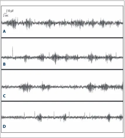
Figure 1. EEG recording (A): PTZ (35 mg/kg) and saline, (B): PTZ (35 mg/kg) and 25 mg/ kg montelukast group; (C): PTZ (35 mg/kg) and 50 mg/kg montelukast group; (D): PTZ (35 mg/kg) and 100 mg/kg montelukast group. 10 µV 2 sec
A
B
C
D
Results
Comparison of the groups in terms of the spike percentage
The spike percentage was significantly different between the saline- and montelukast-pretreated groups (F=12.65, p<0.001). In the Post hoc Bonferroni test, spike percentage was signifi-cantly lower in groups A3 and A4 (50 and 100 mg/kg monte-lukast-pretreated groups, respectively) compared to Group A1 (the saline-pretreated group), which became more pronounced as the montelukast dose increases (p<0.05 and p<0.001, re-spectively) (Figure 1) (Table1).
Assessment of the groups by using Racine’s Convulsion Scale
ANOVA test revealed significant differences in RCS between the groups (F=19.34, p<0.000). In the Post hoc Bonferroni test, RCS was significantly lower in the groups B3 and B4 (50 and 100 mg/kg montelukast-pretreated groups, respectively) compared to the Group B1 (saline-pretreated group), which became more pronounced as the montelukast dose increases (p<0.05 and p<0.000, respectively) (Figure 2).
Comparison of the groups in terms of the first myoclonic jerk onset time
The first myoclonic jerk onset time was significantly different on ANOVA test between the groups (F=15.12, p<0.001). In the Post hoc Bonferroni test, FMJ onset time was significantly lon-ger in the groups pretreated with 50 and 100 mg/kg monte-lukast compared to the saline-pretreated group, which also became more pronounced as the montelukast dose increases (p<0.05 and p<0.01, respectively) (Table 2).
Comparison of the brain MDA level and SOD activity between the groups
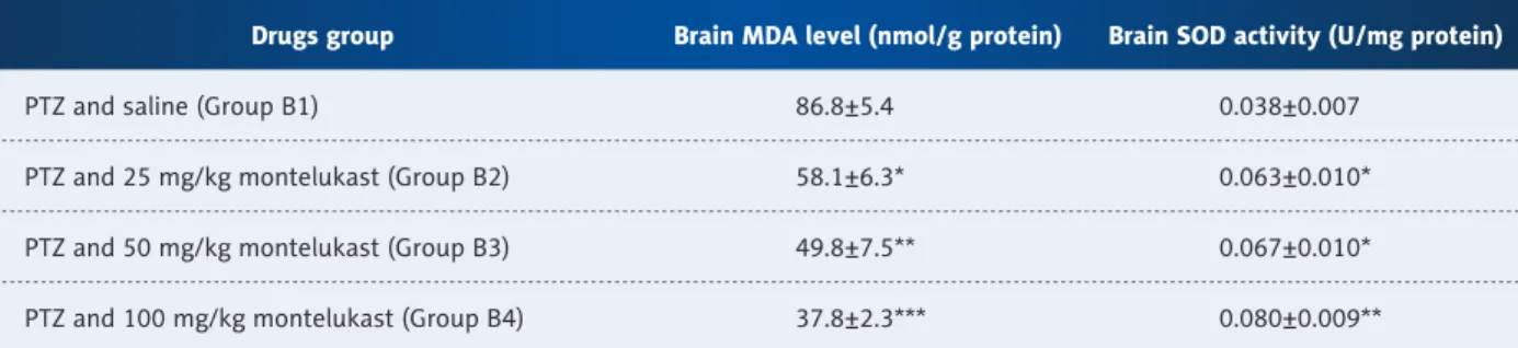
There were significant differences in brain MDA level and SOD activity between the groups (F=13.73, p<0.01, and F=12.59, p<0.05, respectively). The brain MDA level was significantly lower in the groups pretreated with 25, 50, and 100 mg/kg montelukast compared to the saline-pretreated group (p<0.05, p<0.01, and p<0.000, respectively), while brain SOD activity was significantly higher in the groups pretreated with 25, 50, and 100 mg/kg montelukast compared to the saline-pretreat-ed group (p<0.05, p<0.05, and p<0.01, respectively), becom-ing more pronounced with increasbecom-ing doses of montelukast for both parameters (Table 3).
Discussion
In this study, montelukast showed anticonvulsant action and ameliorated the oxidative stress markers in PTZ-induced
Drugs/group Spike percentage
PTZ (35 mg/kg) and saline (Group A1) 74.1%±6.3 PTZ (35 mg/kg) and 25 mg/kg
montelukast (Group A2) 69.3%±6.5 PTZ (35 mg/kg) and 50 mg/kg
montelukast (Group A3) 54.2%±7.8* PTZ (35 mg/kg) and 100 mg/kg
montelukast (Group A4) 33.8%±4.5**
Table 1. Comparison of the spike percentages in the EEG traces of the groups with montelukast.
Data were expressed as mean ± SEM. Statistical analyses were performed by one-way ANOVA test. * p<0.05, ** p<0.001 (different from the saline-treated PTZ group).
Figure 2. Racine’s convulsion scale scores of the groups treated with saline, 25, 50, 100 mg/kg montelukast+70 mg/kg PTZ. * p<0.05, ** p<0.000 (different from the saline-treated PTZ group). 7 6 5 4 3 2 1 0 PTZ (70 mg/kg)
and saline PTZ (70 mg/kg)and 25 mg/kg montelukast PTZ (70 mg/kg) and 50 mg/kg montelukast
*
**
PTZ (70 mg/kg) and 100 mg/kg montelukastRacine convulsion scor
e
Drugs group FMJ onset time (sec)
PTZ (70 mg/kg) and saline (Group B1) 74.6±6.3 PTZ (70 mg/kg) and 25 mg/kg montelukast (Group B2) 78.8±8.4 PTZ (70 mg/kg) and 50 mg/kg montelukast (Group B3) 91.7±5.9* PTZ (70 mg/kg) and 100 mg/kg montelukast (Group B4) 162.8±22.3#
Table 2. Effect of montelukast on the FMJ onset time in PTZ-induced rats.
Data were expressed as mean ± SEM. Statistical analyses were performed by one-way ANOVA test. * p<0.05, # p<0.01 (different from the saline-treated PTZ group).
seizures in rats. For the rats treated with 35 mg of PTZ, ptreatment with 50 mg/kg and 100 mg/kg of montelukast re-duced the spike percentages compared with the saline group (Table 1). In the rats treated with 70 mg of PTZ, a significant difference in the RCS and the FMJ onset time was observed with pretreatment with 50 or 100 mg/kg montelukast (Figure 2, Table 2). To the best of our knowledge, no previous study has evaluated the anticonvulsive and antioxidative potential of acute administration montelukast in PTZ-induced seizure in rats. There are very limited studies on the anticonvulsant effect of Montelukast. In one of these studies, Rehni and Singh showed that montelukast sodium, as well as 1, 2, 3, 4, tetra-hydroisoquinoline, a leukotriene D4 synthetic pathway inhib-itor, markedly and dose-dependently suppressed the develop-ment of kindled (with chronic administration of PTZ) seizures and spontaneous recurrent seizures induced by pilocarpine status epilepticus in mice [19], in contrast to the anticonvul-sant effects induced by single-dose montelukast shown in our study. Takahashi et al. reported that pranlukast add-on ther-apy reduces seizure frequencies in patients with intractable partial epilepsy and that this effect possibly results from the pleiotropic effects, including normalization of matrix metal-loproteinase-9 in sera, reduced leakage of pro-inflammatory cytokines into central nervous system, and inhibition of ex-travasation of leucocytes from brain capillaries [20]. Because montelukast is structurally different from pranlukast and it is the most prescribed CysLT1 receptor antagonist in Europe and the USA, we used montelukast in our study, whereas pranlu-kast is only marketed in Japan and other Asian countries [5]. We believe that in the present study, the anticonvulsant fea-ture of montelukast is due to the decrease of lipid peroxida-tion and oxidative stress.
Oxidative stress resulting from enhanced production of re-active oxygen species (ROS) is known both as the cause and the consequence of epileptic seizures. Oxidative stress may disturb the balance of electrical activity in the brain, leading
to seizures. The increased free radicals can induce seizure ac-tivity by direct inactivation of glutamine synthase and gluta-mate decarboxylase, thereby permitting an abnormal build-up of excitatory (glutamate/glutamic acid) and inhibitory (GABA) neurotransmitters [21]. On the other hand, recurrent or pro-longed seizures can increase ROS and superoxide generation in the brain [22]. These increased ROS not only cause long-lasting seizure formation, but if not arrested, can also lead to neuronal degeneration/death through damage to cellular pro-teins, lipids, and DNA, and can decrease the enzymatic (gluta-thione) and non-enzymatic (SOD, glutathione peroxidase, and catalase) antioxidants in the epileptic focus [22]. The high rate of oxidative metabolism, coupled with the low antioxidant de-fenses, low repair mechanism, and the richness in polyunsat-urated fatty acids, makes the brain highly vulnerable to the free radical damage [2]. SOD rapidly removes superoxide an-ions and protects cells from its direct toxic effect. Lipid per-oxidation causes membrane structure alterations that affect membrane fluidity and permeability and membrane protein activity; this might be the proconvulsant effect of lipid per-oxidation. As an end-product of lipid peroxidation, MDA is a well-known parameter for determining free radical formation in tissues [23]. In this study, pretreatment with montelukast (25, 50, and 100 mg/kg, i.p.) dose-dependently decreased the MDA levels and increased SOD activity compared to saline-pretreated group in the brain homogenate of PTZ-induced rats (Table 3), indicating the attenuation of lipid peroxidation and increase in antioxidant defense mechanisms. This theory is supported by the low MDA and high SOD levels in the mon-telukast groups found in our study.
Several experimental reports have also demonstrated the neu-roprotective effects for montelukast and other members of the same class of drugs in various neurodegenerative conditions such as acute and chronic ischemic brain injury [5], traumat-ic brain injury [24], Huntington disease-like symptoms [25], ischemia-reperfusion-induced vasculitic neuropathic pain [26], spinal cord injury [3], and experimental autoimmune enceph-alomyelitis [4].
Drugs group Brain MDA level (nmol/g protein) Brain SOD activity (U/mg protein)
PTZ and saline (Group B1) 86.8±5.4 0.038±0.007 PTZ and 25 mg/kg montelukast (Group B2) 58.1±6.3* 0.063±0.010* PTZ and 50 mg/kg montelukast (Group B3) 49.8±7.5** 0.067±0.010* PTZ and 100 mg/kg montelukast (Group B4) 37.8±2.3*** 0.080±0.009**
Table 3. Effect of montelukast on changes in MDA level and SOD activity in brain tissues of rats.
Data were expressed as mean ± SEM. Statistical analyses were performed by one-way ANOVA test. * p<0.05, ** p<0.01, *** p<0.000 (different from saline-treated PTZ group).
Conclusions
In conclusion, the results of the present study demonstrate the neuroprotective potential of montelukast via antioxida-tive (attenuated lipid peroxidation and increased SOD activ-ity) and a dose-dependent anticonvulsive effect on the PTZ-induced seizures in rats. The results of the present study may add to the evidence that LTD4 plays a role in the pathogenesis
of epilepsy and that LTD4 antagonist may be an effective al-ternative treatment for epilepsy. Further studies are needed to determine these effects of montelukast.
Conflict of interests
The authors declare that there is no conflict of interests re-garding the publication of this paper.
References:
1. Reddy DS, Kuruba R: Experimental models of status epilepticus and neu-ronal injury for evaluation of therapeutic interventions. Int J Mol Sci, 2013; 14: 18284–318
2. Shin EJ, Jeong JH, Chung YH et al: Role of oxidative stress in epileptic sei-zures, Neurochem Int, 2011; 59: 122–37
3. Erşahin M, Çevik Ö, Akakın D et al: Montelukast inhibits caspase-3 activity and ameliorates oxidative damage in the spinal cord and urinary bladder of rats with spinal cord injury. Prostaglandins Other Lipid Mediat, 2012; 99: 131–39
4. Wang L, Du C, Lv J et al: Antiasthmatic drugs targeting the cysteinyl leu-kotriene receptor 1 alleviate central nervous system inflammatory cell in-filtration and pathogenesis of experimental autoimmune encephalomyeli-tis. J Immunol, 2011; 187: 2336–45
5. Zhao R, Shi WZ, Zhang YM et al: Montelukast, a cysteinyl leukotriene recep-tor-1 antagonist, attenuates chronic brain injury after focal cerebral isch-aemia in mice and rats. J Pharm Pharmacol, 2011; 63: 550–57 6. Al Saadi MM, Meo SA, Mustafa A et al: Effects of Montelukast on free
radi-cal production in whole blood and isolated human polymorphonuclear neu-trophils (PMNs) in asthmatic children. Saudi Pharm J, 2011; 19: 215–20 7. Simmet T, Tippler B: On the relation between cerebral
cysteinyl-leukotri-ene formation and epileptic seizures. Brain Res, 1991; 540: 283–86 8. Bazan NG, Birkle DL, Tang W, Reddy TS: The accumulation of free
arachidon-ic acid, diacylglycerols, prostaglandins, and lipoxygenase reaction products in the brain during experimental epilepsy. Adv Neurol, 1986; 44: 879–902 9. Bishnoi M, Patil CS, Kumar A, Kulkarni SK: Co-administration of acetyl-11-keto-beta-boswellic acid, a specific 5-lipoxygenase inhibitor, potenti-ates the protective effect of COX-2 inhibitors in kainic acid-induced neu-rotoxicity in mice. Pharmacology, 2007; 79: 34–41
10. Pitkanen A, Schwartzkroin PA, Moshe SL: Models of seizures and epilepsy. Elsevier Academic Press, New York, 2006
11. Phillis JW, Horrocks LA, Farooqui AA: Cyclooxygenases, lipoxygenases, and epoxygenases in CNS: their role and involvement in neurological disorders. Brain Res Rev, 2006; 52: 201–43
12. Paxinos G, Watson C: The Rat Brain in Stereotaxic Coordinates. 4th ed.
Academic Press, Spiral Bound, New York, 1998
13. Souza MA, Mota BC, Gerbatin RR et al: Antioxidant activity elicited by low dose of caffeine attenuates pentylenetetrazol-induced seizures and oxida-tive damage in rats. Neurochem Int, 2013; 62: 821–30
14. Aeby A, Poznanski N, Verheulpen D et al: Levetiracetam efficacy in epilep-tic syndromes with continuous spikes and wavess during slow sleep: ex-perience in 12 cases, Epilepsia, 2005; 46: 1937–42
15. Erbas O, Yılmaz M, Korkmaz HA et al: Oxytocin inhibits pentylentetrazol-induced seizures in the rat. Peptides, 2013; 40: 141–44
16. Kaputlu I, Uzbay T: L-NAME inhibits pentylenetetrazole and strychnine-in-duced seizures in mice. Brain Res, 1997; 753: 98–101
17. Demougeot C, Marie C, Beley A: Importance of iron location in iron-induced hydroxyl radical production by brain slices. Life Sci, 2000; 67: 399–410 18. Sun Y, Oberley LW, Li Y: A simple method for clinical assay of superoxide
dismutase. Clin Chem, 1998; 34: 497–500
19. Rehni AK, Singh TG: Modulation of leukotriene D4 attenuates the develop-ment of seizures in mice. Prostaglandins Leukot Essent Fatty Acids, 2011; 85: 97–106
20. Takahashi Y, Imai K, Ikeda H et al: Open study of pranlukast add-on ther-apy in intractable partial epilepsy. Brain Dev, 2013; 35: 236–44 21. Obay BD, Taşdemir E, Tümer C et al: Dose dependent effects of ghrelin on
pentylenetetrazole-induced oxidative stress in a rat seizure model. Peptides, 2008; 29: 448–55
22. Grosso S, Longini M, Rodriguez A et al: Oxidative stress in children affect-ed by epileptic encephalopathies. J Neurol Sci, 2011; 300: 103–6 23. Rumià J, Marmol F, Sanchez J et al: Oxidative stress markers in the
neo-cortex of drug-resistant epilepsy patients submitted to epilepsy surgery. Epilepsy Res, 2013; 107: 75–81
24. Biber N, Toklu HZ, Solakoglu S et al: Cysteinyl-leukotriene receptor antag-onist montelukast decreases blood-brain barrier permeability but does not prevent oedema formation in traumatic brain injury. Brain Inj, 2009; 23: 577–84
25. Kalonia H, Kumar P, Kumar A, Nehru B: Protective effect of montelukast against quinolinic acid/malonic acid induced neurotoxicity: possible behav-ioral, biochemical, mitochondrial and tumor necrosis factor-α level altera-tions in rats. Neuroscience, 2010; 171: 284–99
26. Muthuraman A, Ramesh M, Sood S: Ameliorative potential of montelukast on ischemia-reperfusion injury induced vasculitic neuropathic pain in rat. Life Sci, 2012; 90: 755–62