Balloon Dilatation of Iatrogenic Ureteral Strictures after
Upper Urinary Tract Reconstruction
Üst Üriner Trakt
Rekonstrüksiyonu Sonrası Oluşan İyatrojenik Üreteral
Darlıkların Balon Dilatasyonu
Enes Duman1, Erkan Yıldırım1, İshak Akıllıoğlu2, Selçuk Yücesan2
1
Baskent University Medical Faculty, Konya Resurge Center, Department of Radiology, 2Department of Pediatric Surgery, KONYA
Cukurova Medical Journal 2015;40(2):311-316.
ABSTRACT
Purpose: Open pyeloplasty is the gold standard treatment of uretero-pelvic junction (UPJ) stenosis. Although failure
after open pyeloplasty or uretero-neosistosmy is not common, percutaneous or endoscopic interventions must be necessary in some cases. We present treatment of 6 pediatric patients by balloon dilatation after failed surgery.
Material and Methods: Between 2008 and 2013 6 children were treated. Five patients had undergone open pyeloplasty
for UP stenosis and, one has uretero-vesical anastomosis stricture after ureteroneosistostomy. Percutaneous antegrade balloon dilatation and double J stent placement were performed in three of six patients. In the other three patients retrograde endoscopic balloon dilatation and double J stent placement were performed.
Results: Primary technical success was %50 and %100 for antegrade and retrograde approach respectively. Antegrade
approach has failed in 3 patients (%50) and, retrograde approach and treatment were successful in all of these patients. Secondary and tertiary balloon dilatations were done through antegrade approach in 3 patients. Mean duration of double J stent was 114 days. As of today, five patients are followed smoothly. One patient who has UP stenosis underwent secondary open surgery.
Conclusion: Balloon dilatation can be used safely in both antegrade and retrograde approaches because of lower
profile of balloon catheters, so it could be considered as a first line treatment option after failed surgical treatment of upper urinary tract stenosis in children.
Key words: UPJ stenosis, Balloon dilatation, Percutaneous, Pyeloplasty
ÖZET
Amaç: Üreteropelvik bileşke darlıklarının tedavisinde açık pyeloplasti altın standarttır. Üretero-neosistostomi ya da açık
pyeloplasti sonrası başarısızlık nadir olmasına rağmen bazı vakalarda perkütan ya da endoskopik müdahaleler gerekli olabilir. Biz, 6 pediatrik hastanın başarısız acık cerrahi sonrası balon dilatasyonu ile tedavisini sunuyoruz.
Materyal ve Metod: 2008 – 2013 yılları arasında 6 çocuk hasta tedavi edildi. Beş hastada üreteropelvik darlık
nedeniyle açık pyeloplasti sonrası, bir hastada ise üreteroneosistostomi sonrası gelişen üretero-vezikal anastomoz striktürü tespit edildi. Altı hastanın üç tanesine perkütan antegrad balon dilatasyonu uygulandı ve “Double J” stent yerleştirildi. Diğer üç hastaya retrograd endoskopik balon dilatasyon yapıldı ve “Double J” stent yerleştirildi.
Bulgular: Primer teknik başarı antegrad ve retrograd yaklaşım için sırası ile % 50 ve % 100 idi. Antegrad yaklaşım 3
hastada (%50) başarısız oldu , retrograd yaklaşım ve tedavi tüm hastalarda başarılı oldu. Sekonder ve tersiyer balon dilatasyonlar üç hastada antegrad yaklaşımla yapıldı. “Double J” stentin ortalama kalma süresi 114 gün idi. Bugüne dek beş hastanın izlemi sorunsuz olarak devam etti. UP darlığı olan bir hasta sekonder açık cerrahiye gitti.
Sonuç: Balon dilatasyon, balon kateterlerin yumuşak yapıları nedeni ile hem antegrad hem de retrograd yaklaşımlarda
güvenle kullanılabilir. Bu nedenle de, çocuklardaki cerrahi olarak başarısız olmuş üst üriner trakt stenozlarının tedavisinde ilk seçenek olarak düşünülebilir.
Anahtar kelimeler: Üreteropelvik bileşke darlıkları, balon dilatasyon, perkütan , pyeloplasti
INTRODUCTION
The urinary tract obstruction usually occurs at uretero-pelvic junction (UPJ), and it usually detected at antenatal ultrasound screening. UPJ obstruction is mostly being congenital or caused by postoperative strictures after surgery1. Open pyeloplasty is the gold standard treatment; however, endoluminal procedures and laparoscopic pyeloplasty are increasingly being used with varying degrees of success2-4. Although failure after open pyeloplasty or uretero-neosistosmy is not common, percutaneous or endoscopic secondary interventions must be necessary in some cases. The success rates of secondary open salvage procedures after failed primary intervention are reported to exceed 90%. But, peripelvic scarring and adhesion causes longer operation times and increases the patient morbidity5. Relatively noninvasive treatment options such as endopyelotomy or balloon dilatation have increasingly been used for the first line treatment of these patients, and secondary open procedures preserved for cases those noninvasive procedures failed.
Balloon dilatation, first described in 1982, can be performed retrograde with cystoscopic assistance, antegrade through a nephrostomy, or percutaneously6. We assessed short term results of our experience of balloon dilatation using both antegrade and retrograde approach for treatment of 6 pediatric patients after failed surgery.
MATERIALS and METHODS
Medical carts of children were retrospectively evaluated. Between 2008 and 2014 6 children who have recurrent upper urinary tract stenosis after primary open surgery (4 boys and 2 girls) were treated with balloon dilatation. Because of secondary open surgery has higher morbidity
dilatation which is relatively less invasive. The second important reason why we preferred the balloon dilatation was that fibrosis at the anostomosis side is the primary cause of the stricture. The ages of patients were between 8 month and 11 years old. Five patients had undergone open pyeloplasty for UPJ stenosis and, one has uretero-vesical anastomosis stricture after ureteroneosistostomy. An informed consent was obtained before the procedures.
Percutaneous antegrade balloon dilatation and double J stent placement were performed in three of six patients (Fig. 1). In the other three patients, antegrade passing from the tight stenosis with guide wire was failed and, retrograde endoscopic balloon dilatation and double J stent placement were performed. Percutaneous antegrade procedures were performed under conscious sedation and local anesthesia. After preparation of patients in sterile fashion US guided middle or upper calyx puncture with 20 G needle were done and, 5 Fr vascular sheaths were placed. Then, 5Fr vascular catheter and 0.035 inch hydrophilic guide wire (Terumo Europe N.V, Leuven, Belgium) were used to pass the stenosis. After successful passing to the ureter a 0.018 inch guide wire (V-18 control wire, Boston scientific, Heredia, Costa Rica) were inserted to the bladder. After reliable access gained, noncompliant balloon were inflated about 5 minutes. Cutting balloon (Peripheral cutting balloon, Boston scientific, Galway, Ireland) was used in two patients because of failure of conventional noncompliant balloons. After dilatation of stricture with balloon an appropriate size double J stent (4 or 6 Fr) were placed, and 6 or 8 Fr nephrostomy tube left in place to control of urinary passage. Endoscopic retrograde procedures were performed under general anesthesia. After visualization of the
hydrophilic guide wire (Terumo Europe N.V, Leuven, Belgium) was inserted to the ureter. Then, a 6 Fr 45 cm long vascular sheath were placed into the ureter under fluoroscopic guidance. Then, the stricture was passed with 5 Fr catheter and hydrophilic guide wire. Balloon dilatation was done over a 0.018 inch guide wire (V-18 control wire, Boston scientific, Heredia, Costa Rica). Cutting
balloon (Peripheral cutting balloon, Boston scientific, Galway, Ireland) was used in one patient. After optimal dilatation of stricture with balloon an appropriate size double J stent (4 or 6 Fr) were placed. Patients were followed up with US and scintigraphy. The procedure details and scintigraphic results of patients summarized in table 1.
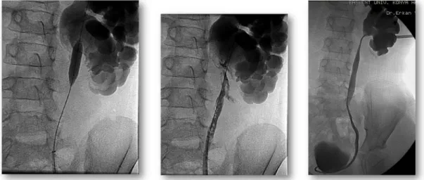
Figure 1 a-c. Percutaneous antegrade balloon dilatation in a 5 year old male patient a) crossing of tight stenosis using
hydrophilic guide wire, b) cutting balloon dilatation of stenosis over a 0,018 guide wire, c) control pyelography after dilatation, note the clear passage of contrast material through UPJ.
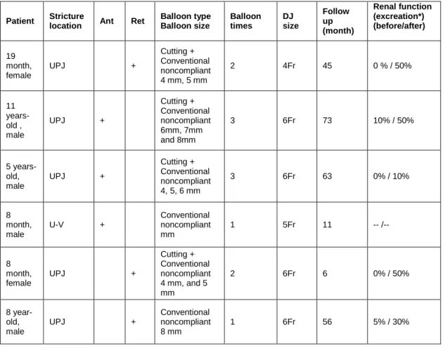
Table 1. Procedure details and follow-up results of six patients
*Response to the diuretic
RESULTS
Six patients underwent balloon dilatation and double J stent placement during 5 year. Mean age of patients was 4 years old. All patients have recurrent stenosis occurred after surgery. Primary technical success was %50 and %100 for antegrade and retrograde approach respectively. Antegrade approach was failed in 3 patients (%50) and, retrograde approach and treatment were successful in all of these patients. Secondary and tertiary balloon dilatations were done through antegrade approach in 3 patients (3 times in two and, 2 times in one patient) because of inadequate
clinical and radiological response. The mean follow up time was 42 month. In one patient, contrast material extravasations occurred after retrograde balloon dilatation (Fig. 2). No other complications including hematuria, pyelonephritis or double J migration occurred. Mean hospital stay was 4 day. Mean duration of double J stent was 114 days. All double J stents were removed sistoscopically. As of today, five patients are followed smoothly. One patient who has UP stenosis underwent secondary open surgery.
Patient Stricture
location Ant Ret
Balloon type Balloon size Balloon times DJ size Follow up (month) Renal function (excreation*) (before/after) 19 month, female UPJ + Cutting + Conventional noncompliant 4 mm, 5 mm 2 4Fr 45 0 % / 50% 11 years-old , male UPJ + Cutting + Conventional noncompliant 6mm, 7mm and 8mm 3 6Fr 73 10% / 50% 5 years-old, male UPJ + Cutting + Conventional noncompliant 4, 5, 6 mm 3 6Fr 63 0% / 10% 8 month, male U-V + Conventional noncompliant mm 1 5Fr 11 -- /-- 8 month, female UPJ + Cutting + Conventional noncompliant 4 mm, and 5 mm 2 6Fr 6 0% / 50% 8 year-old, male UPJ + Conventional noncompliant 8 mm 1 6Fr 56 5% / 30%
Figure 2 a-c. Retrograde cutting balloon dilatation in 7 years old boy a) cutting balloon dilatation of stenosis, b) control
retrograde pyelography shows extravasations of contrast material at the stenosis side, c) control antegrade pyelography, 1 month after the procedure, shows the resolution of extravasations and clear passage of contrast material to the ureter.
DISCUSSION
The purpose our study is to report of our experience in the treatment of recurrent UPJ and UV stenosis after surgery in children. Pyeloplasty is the gold standard treatment method for UPJ stenosis, with a success rate of % 907. Veenboer PW et al have reported the possible causes of recurrence are epithelial overgrowth or flap-valve mechanism8. The treatment options of recurrence are include; surgical pyeloplasty (open or laparoscopic), endoscopic pyelotomy with holmium laser, cold knife and electrocautery, and balloon dilatation. Endoscopic pyelotomy is less invasive than the surgical treatment with low complication rates. Endoscopic pyelotomy can be performed antegrade or retrograde access. However, as Veenboer PW et al mentioned in their report retrograde endopyelotomy can be difficult because of small diameter of ureter and limited view in small children. So they suggest that antegrade approach should be choosing in these patients. However, in our study, percutaneous antegrade approach was failed in 3 patients because of tight stenosis. The
possible causes of failure are over dilatation of collecting system and non visualization of UP junction due to total occlusion. Over dilatation causes poor back-up force to the guide wire and catheter when trying to pass the occlusion. Technical success was 100% in retrograde approach. The insertion of a long sheath and small diameter of ureter (about 3- 4 mm) provides a strong back-up force, catheter and guide wire goes directly to the UP junction so passing from a short segment tight stenosis or totally occlusion is much easier than antegrade approach. There are some
reports in the literature supporting our
consideration. MacKenzie et al. and Alberto Parente et al. reported series about successful treatment of congenital UP stenosis with retrograde balloon dilatation9,10. The advantages of the procedure include less invasiveness, short procedure time, lower complication rates and, short hospital stay. Only in one patient developed extravasations of contrast material after cutting balloon dilatation. No other major and minor complications such as bleeding or sepsis occurred.
The one important point is necessity of repeated balloon dilatations. We performed repeated balloon dilatation (3 times) to two patients.
Massive hydronephrosis and poor renal function seem to reduce success rates after endopyelotomies11. All patients in our study have massive hydronephrosis, but only in one of six patients the treatment failed and patient underwent secondary open pyeloplasty operation. In other five patient massive hidronephrosis regressed to grade 1 hydronephrosis and in four patients who has recurrent UPJ stenosis, renal functions improved at least 25% on scintigraphy. We didn’t performed scintigraphy to one patient who has UV stenosis after ureteroneosistostomy.
The major limitation of study is the small number of patients.
CONCLUSION
Endopyelotomy and balloon dilatation are the mostly used alternative treatment options in patients who has recurrent stenosis after surgical treatment of upper urinary tract stenosis. Balloon dilatation can be used safely in both antegrade and retrograde approaches because of lower profile of balloon catheters, so it could be considered as a first line treatment option after failed surgical treatment of upper urinary tract stenosis in children.
REFERENCES
1. Flaschner SC, King LR. Ureteropelvic junction. In: Kelalis PP, King LR, Belman AB editors. Clinical Pediatric Urology.Philadelphia, PA, Saunders, 1992.
2. Tan HL, Najmaldin A, Webb DR. Endopyelotomy for pelviureteric junction obstruction in children. Eur Urol. 1993;24:84–8.
3. Tan HI. Laparoscopic Anderson-Hynes dismembered pyeloplasty in children. J Urol. 1999;162:1045–8. 4. Slama MRB, Salomon L, Hoznek A, et al.
Extraperitoneal laparoscopic repair of ureteropelvic junction obstruction. Initial experience in 15 cases. Urology. 2000;56:45–8.
5. Park JS, Kim WS, Hong B, Park T, Park HK. Long-term outcome of secondary endopyelotomy after failed primary intervention for ureteropelvic junction obstruction. International Journal of Urology. 2008;15:490–4
6. Kadir S, White RI Jr, Engel R. Balloon dilatation of ureteropelvic junction obstruction. Radiology. 1982;143:263–64.
7. Tan BJ, Smith AD. Ureteropelvic junction obstruction repair: when, how, what? Curr Opin Urol. 2004;14:55–9.
8. Veenboer PW, Chrzan R, Dik P, Klijn AJ, de Jong TP. Secondary endoscopic pyelotomy in children with failed pyeloplasty. Urology. 2011;77:1450–4. 9. Mackenzie RK, Youngson GG, Hussey JK, Mahomed
AA. Is there a role for balloon dilatation of pelvi-ureteric obstruction in children? J Pediatr Surg. 2002;37:893–6.
10. Parente A, Angulo JM, Romero RM, Rivas S, Burgos L, Tardáguila A. Management of ureteropelvic junction obstruction with high-pressure balloon dilatation: long-term outcome in 50 children under 18 months of age. Urology 2013;82:1138–43.
11. Lam JS, Cooper KL, Greene TD, et al. Impact of hydronephrosis and renal function on treatment outcome: antegrade versus retrograde endopyelotomy. Urology. 2003;61:1107–12
Yazışma Adresi / Address for Correspondence:
Dr. Enes Duman
Başkent University Faculty of Medicine Department of Radiology
KONYA
E-mail: [email protected]