CUAJ • September-October 2015 • Volume 9, Issues 9-10 © 2015 Canadian Urological Association
Bilateral renal leiomyoma with 5 year follow-up: Case report
Original research
E734
Cite as: Can Urol Assoc J 2015;9(9-10):E734-6. http://dx.doi.org/10.5489/cuaj.3011 Published online October 13, 2015.
Abstract
Renal leiomyomas are exceptionally rare benign tumours of the kid-ney. Although the renal leiomyomas usually do not metastasize, the differential diagnosis between renal leiomyomas and malign lesions (leiomyosarcoma or renal cell carcinoma) cannot be done by radio-logical examinations, but is possible by historadio-logical examination. Surgery is the preferred treatment. After surgery, the prognosis is excellent without recurrence. Although uterine leiomyomas can be multicentric, renal leiomyomas have been single lesions. We report an incidentally detected case of bilateral renal leiomyoma in a 50-year-old woman with a 5-year follow-up. We also review the literature and discuss clinical, radiological and histological features of renal leiomyomas.
Introduction
Leiomyomas are benign neoplasms of the smooth muscles,
usually occurring in the uterus.1 Renal leiomyomas are
exceptionally rare neoplasms that occur at a much lower frequency (0.001%) and show a marked female predomi-nance.1 They rarely present with flank pain and hematuria.2
Renal leiomyomas are usually detected as single-kidney-masses.3 There are no reports in literature regarding
long-term survival or subsequent recurrence with sarcomatous differantiation.
We present a 5-year case of bilateral renal leiomyoma detected via contrast-enhanced computed tomography (CECT), magnetic-resonance-imaging (MRI), and histopatho-logic images.
Case report
A 50-year-old female, with migraine, presented with a non-specific complaint of left flank pain. The hematological,
bio-chemical parameters, and urine analyses were within normal limits. Ultrasonography revealed hypoechoic 30 × 25-mm left mid-pole and 11 × 10-mm right lower-pole renal-masses. CECT of the abdomen revealed a 30 × 35-mm well-circum-scribed mid-polar exophytic left renal-mass enhancing less than renal parenchyma on corticomedullary phase with a suspicious renal pelvis invasion and a 10 × 10-mm right lower pole exophytic mass showing a similar enhancing pattern (Fig. 1).
Gadolinium-enhanced MRI revealed a 30 × 35-mm well-circumscribed mild exophytic mid-pole renal-mass with renal pelvis invasion; no pathological lymph nodes were detected. On T1-weighted images, both lesions were isointense to muscle tissue and hypointense to renal paren-chyma. Enhanced T1-weighted images revealed a periph-eral and centrally reticular enhanced left renal mass. Both lesions had no intracellular fat on inphase/outphase images. Malignancy was suggestive due to intense diffusion restric-tion on diffusion-weighted images (Fig. 1).
The left renal mass had renal pelvis invasion on MRI; therefore left radical nephrectomy was performed. Active surveillance was planned for the right renal-mass.
A microscopic examination of the left renal mass revealed a leiomyoma. Necrosis, nuclear pleomorphism, and mitot-ic activity were not detected. Immunohistochemmitot-ically, the tumour stained strongly with smooth-muscle-actin (SMA) (Fig. 2). HMB45, S100 and desmin were negatively stained. MRI and CECT findings were re-evaluated according to the histopathological findings. Both lesions were homogeneous and had nodular areas and isointense to adjacent muscles on T1 and T2-weighted images. These lesions were isointense and hypointense to renal cortex on T1 and T2-weighted images, respectively. Gadolinium-enhancements of these lesions were peripheral and in a septate pattern through the arterial to late venous phase. MRI features of both lesions, such as hyperintensity on high-b-values and apparent-diffusion-coefficient (ADC) maps, were similar. Evaluation of the lesions according to hypercellular leiomyoma MRI
Mehmet Resit Goren, MD;
*Gurcan Erbay, MD;
¥Cevahir Ozer, MD;
*Vinil Goren, MD;
§Nebil Bal, MD
±*Department of Urology, Baskent University Adana Medical and Research Center, Adana, Turkey; ¥Department of Radiology, Baskent University Adana Medical and Research Center, Adana, Turkey; ±Department of Pathology, Baskent University Adana Medical and Research Center, Adana, Turkey; §Department of Radiology, Minister of Health Adana Numune Training and Research Hospital, Adana, Turkey
CUAJ • September-October 2015 • Volume 9, Issues 9-10 E735 characteristics revealed similar findings.4 The needle biopsy
of the right renal mass revealed leiomyoma with similar histopathological findings.
The patient was followed up for the right renal-mass with ultrasonography every 3 months and with MRI annually. The MRI findings of the right renal mass did not alter from the initial MRI at the 5-year follow-up (Fig. 1).
Discussion
Leiomyomas are benign mesenchymal tumours, which are
more common in the uterus.5 Although leiomyomas can
occur in any organ of the genitourinary system, they com-monly affect the kidney.5 Leiomyomas originate from smooth
muscle cells of the renal capsule, pelvis, calices, and blood vessels.6
Renal leiomyomas account for 1.5% of benign lesions and 0.29% of all treated renal tumours, with autopsy evi-dence in 4.2% to 5.2% of cases.7 However, current reports
of renal leiomyomas consists mostly of case reports and case series.7
Renal and uterine leiomyomas have similar ultraso-nographic features. Renal leiomyomas are incidentally
detected with ultrasonography.3 Uterine leiomyomas can
be hypoechoic to normal myometrium.8 However, it can
be iso-oechoic or even hyperechoic. Calcification can be seen as echogenic foci with shadowing, and cystic areas of necrosis or degeneration may be seen.8
CECT is commonly used for the differential diagnosis of a renal mass. CECT cannot precisely diagnose leiomyomas, even uterine leiomyomas.3 CECT features of uterine
leiomyo-mas are soft-tissue density, exhibit coarse peripheral or cen-tral calcification. The enhancement pattern is variable and they also may distort the usually smooth uterine contour.3
MRI is the most accurate imaging modality for leiomyo-mas.9 Uterine leiomyomas can be seen as
hypointense/isoin-tence to normal myometrium on T1 images and they might have characteristic high signal intensity on T1-weighted
images.9 Variable enhancement can be seen with
gado-Bilateral renal leiomyoma
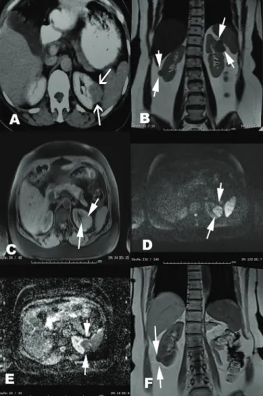
Fig. 1. Radiological images of the renal leiomyomas. A: Axial contrast
enhancing computed tomography shows a 30 × 35-mm well-circumscribed mid-polar exophytic left renal-mass enhancing less than renal parenchyma on corticomedullary phase. B: Coronal T1-weighted pre-contrast magnetic resonance imaging (MRI) shows bilateral exophytic renal-masses isointense to muscle tissue. C: Axial fat-saturated post-contrast T1-weighted image shows an enhancing left renal-mass with renal pelvis involvement. D: Axial diffusion weighted MRI shows hyperintense left renal-mass. E: Apparent-diffusion-coefficient map images show restriction of the left renal-mass mimics malignancy. F: 5-year follow-up MRI imaging of the right renal-mass shows no differentiation of size and signal characteristics.
Fig. 2. Histopathological sections of left renal leiomyoma. A Neoplastic
differentiation consists of clustered spindle cells adjacent to renal capsule. Hematoxylin and eosin (H&E) ×40. B: Tumour composed of spindle-shaped cells with cigar-shaped nuclei. H&E ×400. C: A thick wall vessel seen within the tumour. H&E ×100; D: Smooth-muscle-actin (SMA) positive staining cells in the tumour; SMA ×200.
linium administration.9 Renal cell carcinoma (RCC) of the
kidney has similar MRI features.10 The following
radiologi-cal features may suggest renal leiomyomas: homogeneous density or signal, peripheral mass with well-defined mar-gins, less heterogenous or homogeneous enhancement than adjacent renal parenchyma, continuous and homogeneous enhancement.11
In our case, lesions were hypointense with low-b-values, hyperintense with high-b-values on diffusion-weighted-images, and low signal-intensity on all ADC maps compared with the normal renal parencyhma. Both lesions were simi-lar to uterine leiomyoma. However, RCC has simisimi-lar MRI features and it was not possible to rule out malignancy.12
Macroscopically, renal leiomyoma is a solid,
well-cir-cumscribed, encapsulated mass with a whorled surface.13
Focal areas of hemorrhage and calcification can be seen. Histologically, the tumour shows intersecting fascicles of spindle cells with cigar-shaped nuclei and eosinophilic cyto-plasm without significant nuclear pleomorphism or tumour necrosis.13
The histological differential diagnosis of renal leiomyoma includes other benign and malignant renal spindle cell neo-plasms.14,15 Immunohistochemical markers, such as desmin
and actin, can be used to differentiate renal leiomyoma from
mesenchymal tumours.1 Renal leiomyomas can be
differen-tiated from angiomyolipoma with the lack of a fat compo-nent and lack of expression of melanocytic markers, such as
HMB-45 and Melan-A.1,13 Nuclear pleomorphism, atypical
mitotic figures, cytological atypia, and tumour necrosis are seen in leiomyosarcoma, but not in leiomyoma.1,13
In renal masses, including renal leiomyoma, surgery is still the gold standard.16 Renal biopsy is a way to inform
treatment decision in small renal masses (SRM) (≤4 cm).17,18
Percutaneous renal tumour biopsy (PRTB) could be an option for bilateral SRMs, such as in our case. PRTB is safe and at least 80% of first PRTBs are diagnostic.18 The frequent benign
pathology found with excised SRMs and the lack of specific-ity in imaging have led to an increasing acceptance of the role for pre-treatment PRTB.18 However, multiple tumours
may have different histological feature and therefore repeat biopsies may be required to identify tumour histology.
Conclusion
Renal leiomyomas are rare, benign non-metastasizing tumours. They have excellent prognosis after the surgery. This bilateral renal leiomyoma case reflects clinical and radiological features. In our case, although the MRI features did not change during follow-up, the differential diagnosis is still possible by histopathological examination.
Competing interests: The authors all declare no competing financial or personal interests.
This paper has been peer-reviewed.
References
1. Patil PA, McKenney JK, Trpkov K, et al. Renal leiomyoma: A contemporary multi-institution study of an infrequent and frequently misclassified neoplasm. Am J Surg Pathol 2015;39:349-56. http://dx.doi. org/10.1097/PAS.0000000000000354
2. Romero FR, Kohanim S, Lima G, et al. Leiomyomas of the kidney: Emphasis on conservative diagnosis and treatment. Urology 2005;66:1319. http://dx.doi.org/10.1016/j.urology.2005.06.078 3. Onur MR, Akin MM, Onur AR. Renal leiomyoma: Ultrasonography and computed tomography features with
histopathologic correlation. Eurasian J Med 2013;45:214-7. http://dx.doi.org/10.5152/eajm.2013.42 4. Koc Z, Erbay G, Ulusan S, et al. Optimization of b value in diffusion-weighted MRI for characterization of benign and malignant gynecological lesions. J Magn Reson Imaging 2012;35:650-9. http://dx.doi. org/10.1002/jmri.22871
5. Nagar AM, Raut AA, Narlawar RS, et al. Giant renal capsular leiomyoma: Study of two cases. Br J Radiol 2004;77:957-8. http://dx.doi.org/10.1259/bjr/13222406
6. Alvarez Maestro M, Martinez-Pineiro L, Dominguez Franjo P, et al. [Leiomyoma of the renal capsule: A case report and literature review]. Actas Urol Esp 2010;34:116-8. http://dx.doi.org/10.1016/ S2173-5786(10)70020-2
7. Kuroda N, Inoue Y, Taguchi T, et al. Renal leiomyoma: An immunohistochemical, ultrastructural and comparative genomic hybridization study. Histol Histopathol 2007;22:883-8.
8. Wilde S, Scott-Barrett S. Radiological appearances of uterine fibroids. Indian J Radiol Imaging 2009;19:222-31. http://dx.doi.org/10.4103/0971-3026.54887
9. Murase E, Siegelman ES, Outwater EK, et al. Uterine leiomyomas: Histopathologic features, MR imag-ing findimag-ings, differential diagnosis, and treatment. Radiographics 1999;19:1179-97. http://dx.doi. org/10.1148/radiographics.19.5.g99se131179
10. Oliva MR, Glickman JN, Zou KH, et al. Renal cell carcinoma: t1 and t2 signal intensity characteristics of papillary and clear cell types correlated with pathology. AJR Am J Roentgenol 2009;192:1524-30. http://dx.doi.org/10.2214/AJR.08.1727
11. Cong Z, Gong J, Wang Y. Radiological findings of renal leiomyomas: A report of five surgically confirmed cases. Abdom Imaging 2011;36:604-8. http://dx.doi.org/10.1007/s00261-010-9653-1 12. Gurel S, Narra V, Elsayes KM, et al. Subtypes of renal cell carcinoma: MRI and pathological features.
Diagn Interv Radiol 2013;19:304-11. http://dx.doi.org/10.5152/dir.2013.147
13. Fu L, Humphrey PA, Adeniran AJ. Renal leiomyoma. J Urol 2015;193:997-8. http://dx.doi. org/10.1016/j.juro.2014.12.083
14. Tamboli P, Ro JY, Amin MB, et al. Benign tumors and tumor-like lesions of the adult kidney. Part II: Benign mesenchymal and mixed neoplasms, and tumor-like lesions. Adv Anat Pathol 2000;7:47-66. http:// dx.doi.org/10.1097/00125480-200007010-00007
15. Prasad SR, Surabhi VR, Menias CO, et al. Benign renal neoplasms in adults: Cross-sectional imaging findings. AJR Am J Roentgenol 2008;190:158-64. http://dx.doi.org/10.2214/AJR.07.2724 16. Brunocilla E, Pultrone CV, Schiavina R, et al. Renal leiomyoma: Case report and literature review. Can
Urol Assoc J 2012;6:E87-90. http://dx.doi.org/10.5489/cuaj.11159
17. Ljungberg B, Bensalah K, Canfield S, et al. EAU guidelines on renal cell carcinoma: 2014 update. Eur Urol 2015;67:913-24. http://dx.doi.org/10.1016/j.eururo.2015.01.005
18. Jewett MAS, Rendon R, Lacombe L, et al. Canadian guidelines for the management of the small renal mass (SRM). Can Urol Assoc J 2015;9:160. http://dx.doi.org/10.5489/cuaj.2969
Correspondence: Dr. Mehmet Resit Goren, Department of Urology, Baskent University Adana Medical and Research Center, Adana, Turkey; [email protected]
CUAJ • September-October 2015 • Volume 9, Issues 9-10
E736