Breast Involvement in Acute Lymphoblastic Leukemia
Akut Lenfoblastik Lösemide Meme Tutulumu
Aysin Pourbagher
Baskent University Faculty of Medicine, Department of Radiology, ADANA
Cukurova Medical Journal 2015;40 (Ek Sayı 1):64-70.
ABSTRACT
Extra-medullary infiltration of breast by acute lymphoblastic leukemia is very rare. Mammography, ultrasound and magnetic resonance imaging findings of leukemic breast involvement have been previously reported in the literature. However; ultrasound elastography findings of leukemic breast involvement have not been reported yet. Herein, we report ultrasound elastography and magnetic resonance imaging findings of a case with bilateral breast infiltration by T-cell acute lymphoblastic leukemia.
Key words: Ultrasound elastography, Magnetic Resonance Imaging, Acute lymphoblastic leukemia
ÖZET
Memenin akut lenfoblastik lösemi tarafından ekstramedüller tutulumu nadirdir. Memenin lösemi tarafından tutulumunda mamografi, ultrasonografi ve manyetik rezonans görüntüleme bulguları literatürde daha önceden bildirilmiştir. Literatürde henüz memenin lösemi tarafından tutulumunda ultrason elastografi bulgularını değerlendiren yayın bulunmamaktadır. Bu olgu sunumunda bilateral meme tutulumu olan T-hücreli akut lenfoblastik lösemili olgunun ultrason elastografi ve manyetik rezonans görüntüleme bulguları sunulmuştur.
Anahtar kelimeler: Ultrason Elastografi, Manyetik Rezonans Görüntüleme, Akut Lenfoblastik Lösemi
INTRODUCTION
Breast involvement with leukemia is rare. Acute myeloid leukemia is the most common type involving the breasts1. Extra-medullary leukemic infiltration of breast by T-cell acute lymphoblastic leukemia (ALL) is very rare2. It may be the first sign of relapsing leukemia or may occur as an isolated breast tumor prior to systemic leukemia3. The main therapy protocols are chemotherapy and/or radiotherapy4. So, it should be differentiated from other breast masses. ammography, ultrasound (US) and magnetic resonance (MR) imaging findings of leukemic breast involvement have previously been reported 2, 5, 6. The imaging
features of breast leukemia with mammography, US and MR imaging are not specific. At mammography, common imaging findings include lobulated or irregular shaped mass without microcalcifications, diffuse infiltration or architectural distortion. Common US findings are single or multiple hypoechoic masses with indistinct or microlobulated margins7. US may also show diffuse hypoechoic areas including island like low echo areas8. Common MR imaging findings include markedly enhancing masses with washout pattern8. This is the first report evaluating US elastography findings of leukemic breast involvement.
CASE
20-year-old female who had a history of bone marrow transplantation due to T-cell ALL 2 years ago, presented with palpable lumps in both breasts 8 months ago.
On physical examination, she had palpable hard masses in both breasts. We performed breast US, US elastography, dynamic contrast enhanced MR imaging and diffusion weighted MR imaging at our department.
B-mode US and US elastography images were acquired with a scanner Acuson S2000, Siemens, Erlangen, Germany.
MR images were acquired with a 1, 5 Tesla scanner (MR Symphony, Avanto, Erlangen, Germany). Single shot spin echo planar imaging sequence at b values of 0, 200,500, and 800 s/mm
(2)
were obtained for diffusion-weighted MR images and the apparent diffusion coefficient (ADC) map of the lesion was obtained.
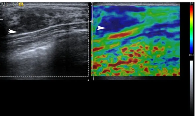
On B-mode US she had bilateral, similar appearing, oval to rounded shaped, homogenous, hypoechoic, multiple solid masses with indistinct borders (fig. 1). The masses showed slightly posterior acoustic enhancement. All of the lesions had uniform hypoechoic features and didn’t contain any calcifications. The largest lesion was in 2cm diameter. On US elastography images, when we set the color coded scale ranging from red (softest components) to blue (hardest components), the entire hypoechoic lesion was blue with a green surrounding area.
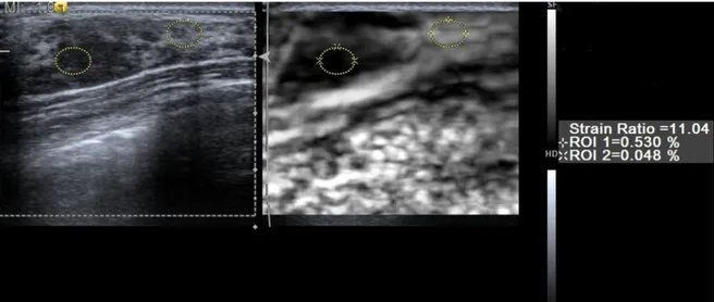
The lesions had a mean strain ratio (SR) of ranging from 6.85 to 11.04 when compared with the normal appearing parenchyma (fig. 2). According to the Tsukuba score she had point 49.
The differential diagnosis included leukemic
fat-saturated T2weighted images compared with adjacent breast tissue. All of the masses showed restricted diffusion on diffusion weighted MR images (figs. 3a, 3b). The mean ADC value was 0.42± 0.19 x10⁻³ mm2/sec. After contrast administration the lesions showed diffuse enhancement pattern (fig. 4).
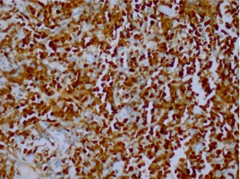
The BI-RADS score of the lesions were 4 both with US and MR imaging findings. We performed US-guided core-needle biopsy. Histopathological examination of breast core biopsies revealed diffuse infiltration composed of small cells with dark nuclei and scant cytoplasm (fig. 5a). In immunohistochemical study; neoplastic cells are positive with CD3, CD2 and tdt (fig. 5b). CD20 was negative. The final diagnosis was leukemic infiltration of breast.
She received radiotherapy for breast infiltration and also she was under chemotherapy protocol. The lesions recurred 6 months after response to radiotherapy. She passed away after sepsis with brain metastases last month.
DISCUSSION
Involvement of breast by ALL is a very rare condition2. Breast leukemia may occur as the first sign or relapse of systemic leukemia or may occur simultaneously with systemic leukemia. Intramammary leukemic relapse may occur after therapy or stem cell transplantation3. Breast leukemia may also occur as an isolated breast tumor prior to systemic leukemia3. On US, it typically presents as hypoechoic microlobulated or indistinct margins. On MR imaging leukemic breast lesions commonly markedly enhance. Differential diagnosis of palpable breast lesions of a patient with leukemia includes leukemic involvement of the
2 pattern of enhancement after contrast administration and restricted diffusion have been previously reported 3,10. All of the lesions in our case showed restricted diffusion on diffusion weighted MR images due to probably high cellularity of the lesion. On diffusion weighted MR images threshold ranging from 0.90 to 1.76×10⁻³ mm2/sec were recommended. Mean ADC values of our case is highly suspicious for malignancy11.
On color coded elastography images, our case shows type 4 pattern and according to the Tsukuba scoring system almost completely blue lesions are highly suspicious for malignancy9. It
helps us exclude multiple benign lesions of breast like fibroadenomatosis.
A threshold SR value of 3.8 has been shown to differentiate benign breast lesions from malignant lesions with a sensitivity of 93.5% and specificity of 75.9% 12. High SR was associated with malignancy. Our case shows also high SR ranging from 6.85 to 11.04.
Treatment of leukemic breast involvement is similar to other localizations of leukemic infiltration and chemotherapy and/or radiotherapy are the main treatment options4. Thus, it should be
Figure 1. Bmode US image shows solid hypoechoic lesion with indistinct margings on the right. US elastography image
demonstrated completely blue mass with surrounding tissue in green on the left. It has point 4 due to Tsukuba scoring system (arrow).
Figure 2. The mean strain ratio compared with the normal appearing parenchyma was 11.04.
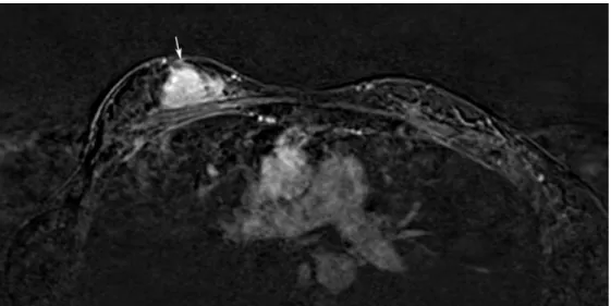
Figure 4. Dynamic contrast enhanced T1 weighted MR images show solid enhancing lesion in the right breast (arrow).
Figure 5a. he x400: The photomicrograph reveals diffuse infiltration effacing normal breast tissue. Infiltration is
composed of small cells with dark nuclei and scant cytoplasm ( HE X400)
Figure 5b. tdt x400: Immunhistochemical diffuse tdt positivity was shown
.
differentiated from other benign breast tumors like fibroadenomas.
Breast involvement by hematologic malignancies should be considered in the patients with leukemia. In conclusion, the US elastography findings of ALL involving breast includes multiple, hypoechoic lesions coding as a stiff lesion on elastography images and the lesions show restricted diffusion on diffusion weighted MR images.
REFERENCES
1. Toombs BD, Kalisher L. Metastatic disease to the breast: clinical, pathologic, and radiographic features. AJR Am J Roentgenol. 1977;129:673-6.
2. Memis A, Killi R, Orguc S, Ustun EE. Bilateral breast involvement in acute lymphoblastic leukemia: color
4. Cunningham I. A clinical review of breast involvement in acute leukemia. Leuk Lymphoma. 2006;47:2517– 26.
5. Basara I, Orguc S. Giant breast involvement in acute lymphoblastic leukemia: MRI findings. J Breast Cancer. 2012;15:258-60.
6. Yang WT, Muttarak M, Ho LW. Nonmammary malignancies of the breast: ultrasound, CT, and MRI. Semin Ultrasound CT MR. 2000;21:375-94.
7. Glazebrook KN, Zingula S, Jones KN, Fazzio RT. Breast imaging findings in haematological malignancies. Insights Imaging. 2014;5:715-22.
8. Kim SJ. Magnetic resonance imaging features of breast leukemia. Magn Reson Med Sci. 2013;12:309-13.
11. Partridge SC, McDonald ES. Diffusion weighted magnetic resonance imaging of the breast: protocol optimization, interpretation, and clinical applications. Magn Reson Imaging Clin N Am. 2013;21:601-24.
12. Ebner L, Bonel HM, Huber A, Ross S, Christe A. Diagnostic Performance and Additional Value of
Elastosonography in Focal Breast Lesions: Statistical Correlation between Size-Dependant Strain Index Measurements, Multimodality-BI-RADS Score, and Histopathology in a Clinical Routine Setting. ISRN Radiol. 2014;2014:396368.
Yazışma Adresi / Address for Correspondence:
Dr. Aysin Pourbagher
Baskent University Faculty of Medicine Department of Radiologyı
ADANA
E-mail: [email protected]
Geliş tarihi/Received on : 05.05.2015 Kabul tarihi/Accepted on: 19.06.2015