IZMIR
KAT
IP CELE
BI
UNIVER
SIT
Y
2018
EMİNE AF
RA
DEMİRCİ
M.Sc. THESIS JANUARY 2018EFFECTS OF BETA-TRICALCIUM PHOSPHATE CONCENTRATION ON OSTEOGENIC DIFFERENTIATION OF HUMAN MESENCHYMAL STEM
CELLS IN CHITOSAN BASED COMPOSITE SCAFFOLDS
Thesis Advisor: Asst. Prof. Dr. Ozan KARAMAN IZMIR KATIP CELEBI UNIVERSITY
GRADUATE SCHOOL OF NATURAL AND APPLIED SCIENCES
Emine Afra DEMİRCİ
Department of Biomedical Technologies Biomedical Technologies Programme
iii
IZMIR KATIP CELEBI UNIVERSITY
GRADUATE SCHOOL OF NATURAL AND APPLIED SCIENCES
JANUARY 2018
EFFECTS OF BETA-TRICALCIUM PHOSPHATE CONCENTRATION ON OSTEOGENIC DIFFERENTIATION OF HUMAN MESENCHYMAL STEM
CELLS IN CHITOSAN BASED COMPOSITE SCAFFOLDS
Thesis Advisor: Asst. Prof. Dr. Ozan KARAMAN Department of Biomedical Technologies
Biomedical Technologies Programme M.Sc. THESIS
Emine Afra DEMİRCİ Y150101010
İZMİR KATİP ÇELEBİ ÜNİVERSİTESİ FEN BİLİMLERİ ENSTİTÜSÜ
KİTOSAN TABANLI KOMPOZİT İSKELELERDE BETA-TRİKALSİYUM FOSFAT KONSANTRASYONUNUN İNSAN MEZENKİMAL KÖK
HÜCRELERİNİN OSTEOJENİK FARKLILAŞMASI ÜZERİNDEKİ ETKİLERİ
YÜKSEK LİSANS TEZİ Emine Afra DEMİRCİ
Y150101010
Biyomedikal Teknolojileri Anabilim Dalı Biyomedikal Teknolojileri Programı
OCAK 2018
v
Emine Afra DEMİRCİ, a M.Sc. student of IKCU Graduate School Of Natural And Applied Sciences, successfully defended the thesis entitled “Effects Of Beta-Tricalcium Phosphate Concentration On Osteogenic Differentiation Of Human Mesenchymal Stem Cells In Chitosan Based Composite Scaffolds”, which she prepared after fulfilling the requirements specified in the associated legislations, before the jury whose signatures are below.
Thesis Advisor: Assist. Prof. Dr.Ozan KARAMAN İzmir Kâtip Çelebi University
Jury Members : Prof. Dr. Bahattin Tanyolaç Ege University
Assist. Prof. Dr. Saliha AKSUN İzmir Kâtip Çelebi University
Date of Submission: 25.12.2017 Date of Defense : 05.01.2018
vii
ix
ACKNOWLEDGEMENTS
I would like to express my sincere gratitude to my supervisor, Assist. Prof. Dr Ozan Karaman, for his encouragement, constructive guidance and words of motivation throughout the duration of my research study and moreover for the inspiration he provided to ensure the completion of this work.
I present my thanks to Bonegraft Biological Materials Company for their support and B-TCP suppy for my thesis. Also I would like to thank to Asst. Prof. Dr. İsmail Hakkı Akgün for freeze drying process.
I would like to endless thanks for understanding and their continuous moral support during my thesis to my lovely cat and family. I am grateful to Res. Assis. Metehan Atagür for his help for performing characterization and criticism. I also express my sincere thanks to Ziyşan Buse Yaralı for her support and patience in PCR studies.
xi Table of Contents
Page
ACKNOWLEDGEMENTS ... ix
Table of Contents ... xi
List of Tables ... xiii
List of Figures ... xv Abbreviations ... xvii SUMMARY ... xix ÖZET ... xxi 1. INTRODUCTION ... 1 1.1 Tissue Engineering ... 1
1.2 Bone Tissue Engineering ... 2
1.2.1 Bone Structure and Properties ... 2
1.2.2 Cellular structure of the bone ... 4
1.2.3 Healing Process of the Bone ... 5
1.3 Tissue Scaffolds in Bone Tissue Engineering ... 5
1.3.1 General Properties of Tissue Scaffolds ... 5
1.3.2.1 Metallic Biomaterials ... 6
1.3.2.2 Bioceramics ... 7
1.3.2.3 Polymers ... 8
1.4 Cells Used in Tissue Engineering ... 9
2. MATERIAL AND METHOD ... 12
2.1 Preparation of scaffolds ... 12
2.1.1 Preparation of Chitosan scaffolds ... 12
2.1.2 Preparation of Chitosan/β-TCP Composite Scaffolds ... 12
2.1.3 Characterization of Scaffolds ... 12
2.1.3.1 Morphological Study ... 12
2.1.3.2 Mechanical Analysis ... 13
2.2 In-vitro Cell Studies ... 13
2.2.1 Toxicity Studies ... 13
2.2.1.2 Cytotoxicity Assay ... 13
2.2.1.3 Genotoxicity Assay ... 14
2.2.2 Differentiation Studies ... 14
2.2.2.1 Culture of the Bone Marrow Mesenchymal Stem Cells ... 14
2.2.2.2 Cell Seeding and Culture ... 14
2.2.2.3 Cell Morphology and Cell Attachment ... 15
2.2.2.4 DNA Quantification Assay ... 15
2.2.2.5 Alkaline Phosphatase Assay (ALP) ... 15
2.2.2.6 Expression of Osteogenic Specific Genes ... 16
3. RESULTS AND DISCUSSION ... 17
3.1 Preparation of Scaffolds ... 17
3.1.1 Morphology and Pore Size ... 17
3.1.2 Mechanical Strength ... 19
3.2 In-vitro Cell Study ... 19
3.2.1 Toxicity Studies ... 19
3.2.1.1 Cytotoxicity Assay ... 19
3.2.1.2 Micronucleus Assay ... 20
3.2.2 Differentiation Studies ... 21
3.2.2.1 Cell Morphology and Cell Attachment ... 21
3.2.2.2 DNA Quantification Assay ... 24
3.2.2.3 Alkaline Phosphatase Assay (ALP) ... 25
3.2.2.4 Expression of Osteogenic Specific Genes ... 26
4. CONCLUSION ... 30
xiii List of Tables
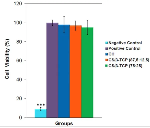
Page Table 1: PCR Primers used for expression of osteogenic specific genes... 16
xv List of Figures
Page Figure 1: Bone tissue engineering involves the use of cells and biomaterials to
treat critical sized bone defects. ... 2
Figure 2: Schematic overview of bone ... 3
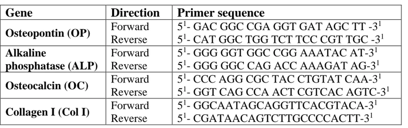
Figure 3: SEM images of the prepared scaffolds. a) Pure CS , b) CS/β-TCP (87,5:12,5) c) CS/β-TCP (75:25). ... 18
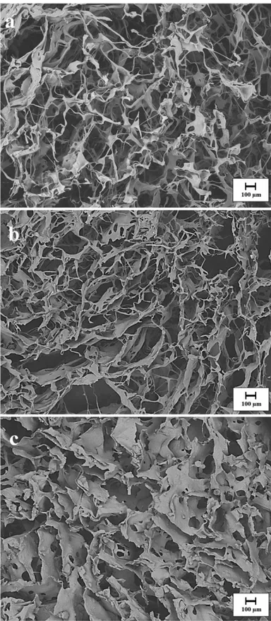
Figure 4: Compressive strength of CS and CS/β-TCP composite scaffolds. ... 19
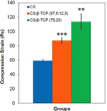
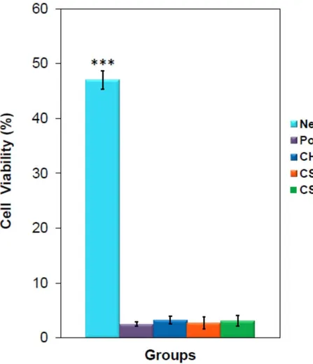
Figure 5: Percentage cell viability after 24-h exposure. ... 20
Figure 6: Percentage of micronuclei of the cells among the groups. ... 21
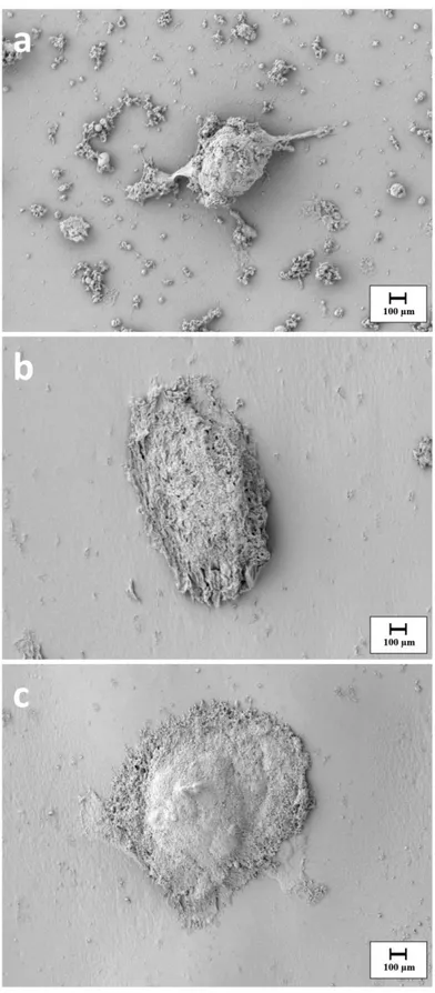
Figure 7: SEM images of BMSCs seeded on CS (a) as control, CS/ β-TCP (87,5:12,5) (b) and CS/ β-TCP (75:25) (c) composite scaffolds. ... 22
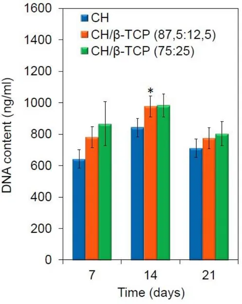
Figure 8: Cell proliferation represented in terms of DNA quantification observed during 21 day of culture. ... 23
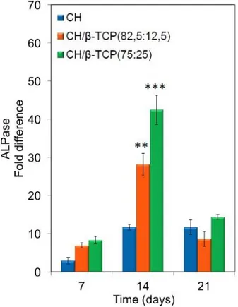
Figure 9: Alkaline Phosphatase activity of CS and CS/β-TCP composite scaffolds after 7, 14 and 21 days of cell seeding.. ... 24
Figure 10: Relative Expression of ALP mRNA of MSCs cultured on CS and CS/ β-TCP scaffolds for 21 days of incubation.. ... 25
Figure 11: Relative Expression of Col 1 mRNA of MSCs cultured on CS and CS/ β-TCP scaffolds for 21 days of incubation.. ... 26
Figure 12: Relative Expression of OP mRNA of MSCs cultured on CS and CS/ β-TCP scaffolds for 21 days of incubation.. ... 27
Figure 13: Relative Expression of OC mRNA of MSCs cultured on CS and CS/ β-TCP scaffolds for 21 days of incubation.. ... 28
xvii Abbreviations
β-TCP β-tricalcium phosphate HA Hydroxyapatite
ALP Alkaline Phosphatase
ECM Extracellular Matrix
MSC Mesenchymal Stem Cell
ESC Embryonic Stem Cells
ASC Adult Stem Cells
CH Chitosan
DMEM Dulbecco’s Modified Eagle’s Medium
FBS Fetal Bovine Serum
HDPE High Density Polyethylene
MTT 3-(4, 5-dimethylthiazol-2-yl)-2, 5-diphenyltetrazolium bromide
a-MEM Minimum Essential Medium Eagle Alpha Modification
SDS Sodium Dodecyl Sulfate
OC Osteocalcin
OP Osteopontin
Col-1 Collegen 1
xix
EFFECTS OF BETA-TRICALCIUM PHOSPHATE CONCENTRATION ON OSTEOGENIC DIFFERENTIATION OF HUMAN MESENCHYMAL STEM CELLS IN CHITOSAN BASED COMPOSITE SCAFFOLDS
SUMMARY
Today, tissue transplant applications are widely used for repair of damaged hard tissue. Despite the gold standard of autografts, interest in synthetic bone grafts produced by tissue engineering techniques is increasing day by day, due to limiting factors such as damage to the tissue site and limited graft availability in allografts, and the risk of developing an immune system response in allografts.Many bioceramic materials, including β-tricalcium phosphate (β-TCP), hydroxyapatite (HA) and calcium sulfate, are widely used in bone tissue engineering. β-TCP has been the most preferred bioceramics in recent years due to its high osteo-compatibility, fast degradation rate and high mechanical strength.
A major disadvantage of existing implant materials is their sintered solid and hard form, which makes it difficult for the surgeon to adapt the surgical graft material to the desired shape during surgery. This causes bone loss and trauma to healthy peripheral tissues and prolonged surgical time. Polymer-ceramic based composite scaffolds are produced to overcome this problem. It is aimed to increase cell adhesion by mimicking the extra cellular matrix with its polymeric character and to imitate bone structure with ceramic character and show osteoconductive and osteoinductive properties.
Within the scope of this study, chitosan based scaffolds with different β-TCP ratios were prepared by freeze drying method. For characterization of the scaffolds produced, morphological characteristics were investigated by Scanning Electron Microscope (SEM) and pore diameters were calculated. At the same time, compression test was performed to determine the mechanical properties.
The biocompatibility of the produced scaffolds was supported by in vitro cytotoxicity and genotoxicity tests. Bone marrow-derived mesenchymal stem cells were used and cultured on scaffolds for the investigation of the effect of β-TCP content on stem cell differentiation. DNA quantification was performed to examine cell proliferation, and SEM analysis was performed to examine cell morphology. In order to examine osteogenic differentiation, the expression of osteogenic specific genes together with ALP analysis on days 7, 14 and 21 was examined.
xxi
KİTOSAN TABANLI KOMPOZİT İSKELELERDE BETA-TRİKALSİYUM FOSFAT KONSANTRASYONUNUN İNSAN MEZENKİMAL KÖK HÜCRELERİNİN OSTEOJENİK FARKLILAŞMASI ÜZERİNDEKİ
ETKİLERİ
ÖZET
Günümüzde hasarlı sert dokunun onarımı için doku nakli uygulamaları yaygın bir şekilde kullanılmaktadır. Otogreftler altın standartlara sahip olmasına rağmen doku alınan bölgede meydana gelen hasar ve sınırlı greft bulunabilirliği, allogreftlerde ise immün sistem yanıtı oluşma riski gibi sınırlayıcı faktörlerden dolayı doku mühendisliği teknikleriyle üretilen sentetik kemik greftlerine olan ilgi her geçen gün artmaktadır. β-triskalsiyum fosfat (β-TCP), hidroksiapatit (HA) ve kalsiyum sülfat dahil olmak üzere birçok biyoseramik materyal yaygın olarak kemik ikame maddeleri olarak kullanılmaktadır. Yüksek osteokompatibilite, hızlı bozunma hızı ve yüksek mekanik dayanıma sahip olması nedeniyle β-TCP son yıllarda en çok tercih edilen biyoseramik olmuştur.
Mevcut implant malzemelerinin önemli bir dezavantajı, sinterlenmiş katı ve sert formda olmalarıdır buda cerrahın uygulama sırasında cerrahi alanda greft malzemesinin istenilen şekli vermesini güçleştirmektedir. Bu durum kemik kaybı, sağlıklı çevre dokularda travma ve cerrahi sürenin uzaması gibi olumsuzluklara neden olmaktadır. Bu problemin üstesinden gelmek için polimer-seramik tabanlı kompozit iskeleler üretilerek, hem polimerik özelliği ile ekstraselüler matriksin taklit edilmesiyle hücre tutunumunu artırması hem de seramik özelliği ile kemik yapının taklit edilmesi ve osteokondüktif ve osteoindüktif özellik göstermesi hedeflenmiştir. Bu çalışma kapsamında farklı β-TCP oranına sahip kitosan tabanlı doku iskeleri dondurarak kurutma yöntemiyle hazırlanmıştır. Üretilen iskelelerin karakterizasyonu için Taramalı Elektron Mikroskobu (SEM) ile morfolojik özellikleri incelenmiş ve por çapları hesaplanmıştır Aynı zamanda mekanik özelliklerinin belirlenmesi için basma testi gerçekleştirilmiştir.
Üretilen iskelelerin biyouyumluluğu yapılan in-vitro sitotoksisite ve genotoksisite testleri ile desteklenmiştir. Kök hücre faklılaşması üzerindeki etkisinin incelenmesi için Kemik iliği Kökenli Mezenkimal Kök Hücreleri kullanılmış ve üretilen iskeler üzerine ekilmiştir. Hücre çoğalmasını incelemek için DNA kantifikasyonu, morfolojisini incelemek için SEM analizi yapılmıştır. Osteojenik Farklılaşmanın incelenmesi için 7,14 ve 21. Günlerde ALP analizi ile birlikte osteojenik spesifik genlerin ekspresyonu incelenmiştir.
1 1. INTRODUCTION
1.1 Tissue Engineering
Primary goal of regenerative medicine is to repair morphologicly and functionally of deformed tissues or organs in order to regain the normal tissue function. Tissue engineering is the combined application of biology, chemistry and engineering principles for repair or reconstruction of living tissues using cell or biosynthetic molecules alone or in combination [1].
Organ or tissue transplantation is still a widely used method, although significant improvements have been made in the medical techniques used in the treatment of tissue damage in the body. Surgical interventions for removal of diseased tissues from damaged areas are mostly successful. However, the amount of tissue that can be taken is limited, also in cases where a second operation is needed, it both afflicts the patient and carries the risk of infection. Alternative tissue sources that can be used including tissues or organs from other humans or from some animals. However, the immunological effects (the responses of the immune system) that can occur after implantation and the difficulty of donor organ harvesting are also causing problems in the use of these sources. For all these reasons, the approach of tissue engineering in the last years has come to the forefront [2].
In tissue engineering, repair of damaged tissue is provided by bone scaffolds. These tissue scaffolds act as an artificial extracellular matrix (ECM) that allows cells to multiply, differentiate, and thus maintain their function. Cells obtained from the appropriate source and replicated in cell culture to the desired number are seed on tissue scaffolds and then the resulting structure is implanted into the site of tissue damage [3].
1.2 Bone Tissue Engineering
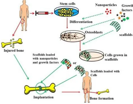
The high regenerative capacity of the bone ensures that a significant part of the fractures heal, especially in young people, without major intervention. However, in large bone injuries and severe fractures, surgical intervention is required [4]. Current treatments for bone injuries include the use of autologous bone grafts, allogenic grafts and alternative metal or ceramic materials. However, in recent years, the "bone tissue engineering" approach has come to the forefront, as autografts increase donor site morbidity and allografts have a risk of bearing disease [5]. Tissue engineered approach requires growth factors that support cell growth and mineralized bone tissue formation with tissue scaffolding, osteoblasts, or osteoblast differentiable cells to achieve bone regeneration. These three components can all be used, as well as bone tissue engineering studies carried out using only cells and tissue scaffolding [6].
Figure 1: Bone tissue engineering involves the use of cells and biomaterials to treat critical
sized bone defects [7].
1.2.1 Bone Structure and Properties
Bone tissue plays a role in many different functions within the body, with its complex organization. It is primarily responsible for the protection of vital organs and the movement of muscle tissue [8]. As a material of the bone, its properties give
3
physiological effects. It also acts as a reservoir for important minerals such as calcium, phosphate, and regulates ion concentrations in the extracellular matrix (ECM). It is a rich source of pluripotent mesenchymal stem cells (MSC) that can differentiate into bone marrow, bone, cartilage, tendon, muscle, dermis and adipose tissue. Besides, bone marrow is the place where hematopoietic cells, which are formed by red blood cells and white blood cells, are involved in nutrient transport and immunological response. [9].
An adult bone tissue consists mainly of 80% cortical (hard), 20% trabecular (spongiform) bone. Cortical bone is a very solid and dense structure that surrounds the bone marrow in long bones and some other bone types, and only 10% is porous, allowing fewer cells and blood vessels to settle. The structural unit of the cortical bone is called "osteon". Osteons are formed by the combination of layers called "lamella". Blood vessels are located in the center of each osteotomy, which is defined as "Havers channel". The nutrient exchange in the bone is carried out through the blood vessels and the structures called "canaliculi" or micro scale channels. The osteons are longitudinally located within the bone and the cortical bone is anisotropic. Axially the cortical bone exhibits a tension of 79-151 MPa and compressive strength of 131-224 MPa, and the elastic modulus for both ranges from 17-20 GPa. [10].
The trabecular bone, which has a spongy and porous structure, is located in the ribs of the bones, the spine and the tip of long bones. Trabecular bone, composed of pores with internal connections, extends in the direction of loading stress. The porous structure, with blood vessels and bone marrow, has a relatively low mechanical strength compared with cortical bone. The strength and elastic modulus of the trabecular bone are between 5-10 MPa and 50-100 MPa, respectively, for tension and compression, depending on the density of the bone.
Nano and micro structure of the bone provides its unique mechanical properties. The bone consists of approximately 70% inorganic minerals and 30% organic molecules. While the calcium phosphate crystals, especially hydroxyapatite (HA), form the inorganic part, the organic phase is largely in the form of collagen type I. HA crystals with a thickness of 2-3 nm precipitate on fibers. Thus, while the HA crystals increase the compressive strength of the composite structure, collagen fibers distribute energies and impart tension properties [9, 10].
1.2.2 Cellular structure of the bone
In the organic-inorganic composite structure of the bone, there are three types of cells: osteoblasts, osteocytes and osteoclasts. Osteoblasts originate from the MSCs and play a role in the secretion of collagenous proteins involved in the structure of the organic matrix of the bone. When mature osteoblasts are surrounded by osteoid, they stop synthesis of the matrix and transformed into osteocytes. Osteocytes play an important role in the regulation of signals against mechanical stimuli. Osteoclasts originate from hematopoietic cells and cause the dissolution of mineral salts and organic matrix by the enzymes they secrete. There is a balance between these cells and the direction of this balance can be changed by the presence of chemical, biological and mechanical factors.
The formation of the bone is divided into 5 phases; stationary phase, activation, resorption, reconstruction, structuring and remodeling. The process begins with a stationary phase, and at this stage the cells on the surface of the bone are inactive and 80% of the cells on the free surface of the bones are present at this stage. In the presence of a biochemical or physical signal, mononuclear monocytes and
5
stimulating the differentiation of osteoclasts. After the osteoclasts break up the organic and inorganic structure and form a cavity, the resorption process is carried out. During the reconstitution process, osteoclasts leave the site and mononuclear macrophage-like cells secrete cement-like structures on the surface. At the last stage, osteoblasts fill the void with the bone matrix and allow new osteons to form.
Cell-cell interactions are very important for the continuation of the functions of the bones. Osteocytes located in the bone matrix sense the mechanical stimuli and produce the necessary signals through biochemical molecules. These cellular signals have a critical prescription for bone continuity [12].
1.2.3 Healing Process of the Bone
Bone injuries cause repetition of events that occur in embryonic bone formation. These biological events are classified into 3 stages; inflammation, repair and restructuring. First, an acute inflammatory response occurs in the area where blood vessels are damaged and a hematoma occurs. Neutrophils and macrophages that come to the region digest cell debris that occur after necrosis and release growth factors and cytokines. These signals stimulate the migration of MSCs in bone marrow and periosteum to damaged area. MSCs differentiate into osteoblasts and the blastema is repaired. After the inflammatory phase, the repair process is reversed and osteoblasts form osteocytes. In the final stage, collagen fibers are reconstructed, shaped and organized as a strong lamellar bone in the mechanical direction. Tissue healing cannot be completed if there are not enough active cells or the damage size is greater than critical size defect. In this case, tissue engineering techniques are required [13-15].
1.3 Tissue Scaffolds in Bone Tissue Engineering
1.3.1 General Properties of Tissue Scaffolds
Tissue scaffolds are 3-dimensional structures designed to provide a temporary artificial extracellular matrix for cells. Materials used in tissue scaffold production should be biocompatible so that they do not cause unwanted tissue reactions when placed in the body.
The scaffolds used in bone tissue engineering applications are expected to be osteoinductive and osteoconductive. Osteoinductive materials are materials that induce the differentiation of stem cells into the osteoblastic cell line at the site of grafting. Osteoconductive materials are materials that promote the formation of the three-dimensional structure of the bone, supporting the production of osteogenic cells. In addition, the mechanical strengths of the tissue scaffolds used in the load-bearing tissues like bones must be high [16]. Another feature that tissue scaffolding should possess is proper porosity. In bone tissue engineering, tissue scaffolds are required to support the flow or diffusion of nutrients, have a porosity of 90% to allow cell migration, and have a pore diameter of at least 100 μm to allow for cell penetration and proper vascularization of the growing tissue [17, 18]. Since scaffolding is not needed when cells reach the capacity to form their own extracellular matrices, the tissue scaffold must be produced from a biodegradable material. However, it is necessary to protect the physical properties of tissue scaffolds used for bone regeneration studies for at least 6 months [19].
1.3.2 Biomaterials Used in Tissue Scaffold Production
Nowadays, various materials are used in the field of bone tissue engineering. Biomaterials divided by 4 group; metals, ceramics, polymers and composites. The similarity of the material that will be used in the production of tissue scaffolds is very important. For example, tissue scaffolds that will form hard tissue such as bone are composed of ceramics, polymers, or composites while tissue scaffolds that form soft tissues (such as skin, tendons, ligaments, and eyes) are composed of natural or synthetic polymers [20].
1.3.2.1 Metallic Biomaterials
Despite their disadvantages such as low biocompatibility, exposure to corrosion, very hard relative to the tissues, high concentrations and metal ion release which can cause allergic tissue reactions, due to crystal structures and strong metallic bonds they have a great share in the biomaterials of metal and metal alloys with superior mechanical properties. They can be used as artificial prosthesis and bone replacement material in orthopedic applications, artificial heart parts in face-jaw surgery, dental implant or cardiovascular surgery, catheter, valve and heart valve.
7
Metallic biomaterials are also preferred in the production of biomedical devices used for diagnostic and therapeutic purposes [21].
The biocompatibility of metal prostheses is related to corrosion in the in-vivo environment.[22] Corrosion occurs when metals undergo an undesirable chemical reaction with their surroundings and form oxygen, hydroxide and other compounds. The human body contains various ions such as fluid, water, dissolved oxygen, protein, chloride and hydroxide [23]. For this reason, the human body is a highly corrosive medium for metals used as biomaterials. The material can weaken as a result of corrosion and corrosion products can penetrate into the tissue and damage the cells. Therefore, metal prostheses to be used in-vivo should be tested in serum, saliva or different synthetic buffer solutions [24, 25].
1.3.2.2 Bioceramics
Bioceramics can be prepared as polycrystalline ceramics (alumina and hydroxyapatite), bioactive glass, bioactive glass ceramics or bioactive composites. These materials, which form an important group of inorganic materials, are used in a wide variety of applications in the health sector. Ceramic composites can be used alone or in combination with other materials that are osteogenic, osteoinductive, or osteoconductive in nature to enhance bone healing. The use of bioceramic materials reduced the wear rate of the components and reduced the amount of ion emission to negligible levels [26, 27].
The bioceramics developed in recent years are divided into 2 groups as bioinert and bioactive. First generation ceramic materials, alumina and zirconia ceramics are often considered bioinert. One of the pioneering applications of bioceramics is the use of high-dynamics, high-purity alumina as a conventional metallic femoral head in hip prostheses. It was seen they have excellent abrasion resistance, excellent corrosion resistance, good biocompatibility and high strength ratios. However, they have major disadvantages such as they are fragile, difficult to process, and non-flexible [28].
Second generation ceramic materials are bioactive ceramics, the main features of which are to provide a surface suitable for bone adhesion and growth. They are mainly used as bone defect fillers. The similarity between the bone mineral phase and the structural and surface properties allows the bone to bind without fibrous connective tissue. Bioactive ceramics are biocompatible and osteoconductive
materials. The most commonly used ceramic materials in this group are; Hydroxyapatite (HA) (Ca10(PO4)6(OH)2), β-TCP(β-TCP, Ca3(PO4)2) [29, 30]. Depending on the synthesis process, different physical and chemical properties of these materials are observed. Although HA has high tissue compatibility and ability to bind to the bone tissue, biological degradation remains relatively slow compared to the rate of new bone formation. HA has good clinical outcomes in many applications, but some complications have also been reported [31]. In some studies, it has been reported that HA has no absorption in the body and remains as a foreign matter for a long time [32]. On the other hand, β-TCP, which is another biocompatible ceramic, has a higher degradation rate than HA. Due to its high osteocompatibility and mechanical strength, β-TCP has become the most preferred ceramic material in recent years [33].
1.3.2.3 Polymers
Polymers which are the most studied materials in tissue engineering are divided into two groups, natural and synthetic. Natural polymers include polysaccharides (starch, alginate, chitin / chitosan and hyaluronic acid derivatives) or proteins (collagen, fibrin jellies and silk). Synthetic polymers are produced under controlled conditions. Generally, the mechanical and physical properties of the polymers produced, such as tensile strength, elastic modulus and speed of disruption, are reproducible [34]. Synthetic polymers do not contain risks such as toxic effects and infection transmission. Natural polymers are highly organized structures with similarity to the extracellular matrix. Natural polymers are biocompatible, biodegradable and biocompatible, so they are more attractive than synthetic polymers [35].
A natural polymer, chitosan, is a cationic and linear polysaccharide obtained by partial deacetylation of chitin found in the shells of insects and shellfish. Chitosan promotes cell adhesion and multiplication through its chemical properties [5]. An important feature of chitosan tissue scaffolds for bone tissue engineering is that it allows the formation of scaffolds with internally-related pores that support both in vitro and in vivo bone tissue formation [19]
Chitosan is also compatible with many anionic and nonionic polymers such as cationic structured dyes, starches, surface active agents and organic compounds such as quaternary ammonium salts. With multivalent anions, it cross-links to form gel
9
as mercury, cadmium, lead, zinc, nickel, chromium, copper, iron, manganese, silver, gold and platinum. With these features, it is an ideal biomaterial feature for building scaffolding [36-38].
The mechanical properties of the tissue scaffold are important for the formation of matrix mineralization in hard tissues such as bone. Because a single material cannot have all the desired properties, it is necessary to develop composite materials. Micro and nano-sized bioceramics contained in natural biopolymers are considered a promising option to develop scaffolds with desirable properties for bone tissue regeneration [39, 40]. Therefore, the use of ceramic materials together with chitosan is one of the subjects to be studied [3].
1.4 Cells Used in Tissue Engineering
In bone tissue engineering, various cell sources such as osteoblasts and stem cells can be used. Osteoblasts, which are the most commonly preferred cells and isolated from the patient's own, have golden standards and do not produce any immunological response at all. However, this method brings with it many disadvantages. First, the number of cells obtained and the potential for replication are limited. Also, transplantation of osteoblasts, where bone-related diseases are the case, is not appropriate since protein expressions are not sufficient. Transplantation of osteoblasts belonging to a different species, which is a different species, is not preferred because of the risk of developing an immunological response, the risk of transport of infection agents, social and ethical problems [41].
The stem cells, which constitute another source of cells, are undifferentiated, have a high proliferation capacity and participate in tissue regeneration. Differentiation potentials of stem cells vary according to species. After the formation and first division of the zygote, the cells that come to the forehead are called "totipotent" and these cells can differentiate into all tissue cells, placenta and supporting tissue cells that will form the embryo. After a few days the cells begin to become specialized and form blastocysts. The cell mass in the blastocyst is defined as embryonic stem cells (ESC) and is pluripotent. Embryonic stem cells can differentiate into every cell originating from 3 germ layers. Finally, the multipotent cells found in fully differentiated tissues are called adult stem cells (ASC). These cells have a limited ability to differentiate, according to their embryonic origin [42].
Adult stem cells take part in tissue renewal, ensuring tissue homeostasis and replacing natural cell losses resulting from injury or apoptosis. In fulfilling these functions, the balance between the mechanisms of self-renewal and differentiation of stem cells has a critical role [43]. Within the niches (physiological microenvironment) in which the cells are located, this balance is controlled by external factors that vary depending on the environment in which the cell is located and the internal factors within the cell [44].
Studies have shown that adult stem cells can be isolated from the bone marrow, periosteum, muscle, fat, brain tissue. Adult stem cells can be divided into tissue-specific and non-tissue-tissue-specific cells. Hematopoietic stem cells, neural stem cells, stem cells found in the liver, "satellite cells" in the muscular tissue, and keratinocyte stem cells in the skin and hair follicle are tissue-specific stem cell groups. The stem cells that bone tissue engineering is particularly interested in are the non-tissue specific cells called "Mesenchymal Stem Cells" (MSC) [45].
MSCs are found in connective tissues within the body and can differentiate into different cells, primarily smooth muscle cells, adipocytes or chondrocytes. The most commonly used MSC source in studies is bone marrow. Caplan has shown that under certain conditions these cells can differentiate into bone, cartilage, fat, muscle, skin, tendon and other mesenchymal cells [9, 46]. Bruder et al. have been shown to MSCs be able to replicate in vitro conditions as well as their differentiating abilities [47]. Pittinger et al. have determined that these cells do not have spontaneous differentiation properties in the progressive passages [48].
Much of the work done for the clinical use of MSCs is aimed at eliminating bone damages. İntention is to create live implants through the biomaterials. Generally clinical trials on this area have been carried out on 'osteogenesis imperfecta' or fragile-bone diseases, and autologous MSC transplantation has been performed to achieve very promising results [49, 50].
Studies in clinical practice have shown that MSCs do not cause allogenic reactions in humans or animals. It is not very certain that these cells are thought to lack tissue-compatibility complexes (MHC-II). However, in order for all these studies to be clinically feasible, it is necessary to understand how to increase data obtained by
11
larger studies and whether MSCs will form malignant structures in the future, depending on their high proliferation capacities [51].
2. MATERIAL AND METHOD 2.1 Preparation of scaffolds
2.1.1 Preparation of Chitosan scaffolds
Chitosan scaffolds were produced following the previously applied procedure [52]. For preparation of 3% chitosan solution 0.1 M aqueous acetic acid (Sigma–Aldrich) was used. Chitosan (Sigma–Aldrich) (Medium Molecular Weight) and acid solution was mixed until polymer dissolved completely. The prepared polymer solution was poured into cylinder vessels that 1 cm in diameter and 10 cm in height and kept at -80°C for 24 hr. The cylinder vessels dried in a freeze dryer for 48 hour at -30°C. After drying, the scaffolds were removed from the cylinder vessels and cut to a height of 2mm.
2.1.2 Preparation of Chitosan/β-TCP Composite Scaffolds
β-TCP powder (100-200 µm) was kindly donated from Bonegraft Biological Materials Company. Β-TCP particles added chitosan solution and ball-mill (Retsch-PM 100) (300 rpm-10 min) was used to ensure homogeneity. Mixing was performed in three different groups using TCP concentration at 12% and 25%. After this, exactly same procedure of the preparation of Chitosan scaffold was followed. The obtained composite scaffolds were cut into specific dimensions.
2.1.3 Characterization of Scaffolds 2.1.3.1 Morphological Study
Scanning Electron Microscopy (SEM) was operated using a Carl Zeiss 300VP to examine the morphology of produced CS and CS based composite scaffolds. A minimum of 25 pores were observed for calculating the pore size of the produced scaffolds by using Image J software (National Institutes of Health, Bethesda, MD, USA.).
13 2.1.3.2 Mechanical Analysis
Compressive tests of the produced CS and CS based composite scaffolds were carried out by Shimadzu autograph AG-IS series universal test machine. Cylindrical specimens with a diameter of 10 mm and thickness of 20 mm were used for analysis. Compression test was completed with a crosshead speed of 1 mm/min with a load cell of 5 kN. For each group, the tests were repeated 3 times.
2.2 In-vitro Cell Studies 2.2.1 Toxicity Studies
2.2.1.1 Extraction Conditions
Prepared scaffolds samples were prepared in glass rings (10 mm in diameter and 20 mm high). Extracts of these specimens were prepared following of ISO 10993-12 parameters at a ratio of 4 gram of sample/ 20 mL of cell culture medium at 37 oC and 5% CO2 for a 24-h extraction period.
2.2.1.2 Cytotoxicity Assay
L929 fibroblast cells were routinely cultivated in Dulbecco’s Modified Eagle’s Medium (DMEM) (Sigma-Aldrich) supplemented with 10% fetal bovine serum (FBS) (Gibco, US), 1% L-glutamine (Gibco, US), and 0.1% penicillin/streptomycin (Gibco, US) at 37 oC and 5% CO2. Into each well of a 24-well plate, 5 × 10⁴ cells
were seeded and incubated 24 hours. After overnight cultivation, the culture medium was changed with fresh medium that contained extracts of the test specimens. High Density Polyethylene (HDPE) and latex extracts were used as positive and negative controls. Cell viability after exposure was determined using 3-(4, 5-dimethylthiazol-2-yl)-2, 5-diphenyltetrazolium bromide (MTT) (Vybrant ® MTT Cell Proliferation Assay Kit (Invitrogen) assay method [53]. Subsequently, %10 MTT including MEM was added to the wells and the cultures incubated for another 3 hours, end of the incubation time MTT solution replaced with DMSO and waited 5 minutes in the shaker for homogenization. Biological reactivity was evaluated by Synergy™ HTX Multi-Mode Microplate Reader (BioTek, Winooski, VT, USA) at 570 nm wavelength. Cytotoxic effect of the samples to cells were evaluated by calculating of viability [54]
2.2.1.3 Genotoxicity Assay
In vitro micronucleus assay was performed with human peripheral blood lymphocytes Blood from 3 healthy, young (less than 30 years of age) nonsmoking donors were collected into heparinized tubes. Blood from 3 healthy, young (less than 30 years of age) non smoking donors were collected into heparinized tubes with İzmir Katip Çelebi University Medical Faculty Research Ethics Committee. Blood cells were cultivated with 20 μg/mL of PHA-adjuncted cell culture medium. After 24 h the cell culture medium was replaced with 5 mL of diluted extracts for 48 h. As a negative control, 0.10 μg/mL of mitomycin C was used. Untreated lymphocytes without PHA were used for the positive control. 6 μg/mL of cytochalasin B was added to the culture tubes at the 44th hour of the culture period to inhibit microfilament assembly and cytokinesis. After 72 h the cells were fixed onto microscope slides and 5% Giemsa staining was performed [55]. The number of micronuclei was examined microscopically in 1000 cells/slide [56]
2.2.2 Differentiation Studies
2.2.2.1 Culture of the Bone Marrow Mesenchymal Stem Cells
Human bone marrow mesenchymal stem cells obtain from CLS Cell Lines Service GmbH (Eppelheim, Germany) with order number 300661/300665. The BMSC's were cultured in Minimum Essential Medium Eagle Alpha Modification (a-MEM)( (Sigma-Aldrich) adjuvant with 10% FBS (Gibco, US), 1% L-glutamine (Gibco, US), and 0.1% penicillin/streptomycin(Gibco, US). The cells were cultured in 75 cm2
culture flasks in a 5% CO2 atmosphere. In order to keep the cells in exponential
phase, all cultures were passaged weekly or twice per week by trypsin/EDTA detachment.
2.2.2.2 Cell Seeding and Culture
Scaffolds were sterilized previous to cell seeding by immersing in 70% ethanol and 10 U/ml penicillin/streptomycin/gentamicin solution for 2 hr, followed by sterile PBS wash. Cells from 5th passage were seeded on sterilized CS and CS based composite scaffolds with density of 5 × 10⁶ cells/ml. The cell seeded scaffolds were incubated in osteogenic differentiation medium containing 100 nmol/L dexamethasone, 10 mmol/L beta-glycerophosphate, and 0.05 mmol/L L-ascorbic
15 2.2.2.3 Cell Morphology and Cell Attachment
After 21 days cultivation, cell seeded scaffolds were prepared for SEM observation. For fixing the cells and the samples, 2% (v/v) gluteraldehyde (Sigma-Aldrich) was used and samples were rinsed with wash buffer containing sodium cacodylate trihydrate. (Sigma-Aldrich) Osmium tetroxide (Sigma-Aldrich) (1%) was added to the samples and kept for 30 min on ice. Then the samples were washed with buffer for five times and dehydrated with series of ethanol gradations (70, 80, 95 and 100%) for 5 min each. Ethanol replaced with Hexamethyldisilazane (HDMS) (Sigma-Aldrich) for critical point drying. Before imaging all samples were coated with a 5 nm layer of gold palladium (80:20) using a Quorum Q150 RES Sputter Coating System (Quorum Technologies, UK). Images were taken by SEM (Carl Zeiss 300VP) in high vacuum at 30 kV.
2.2.2.4 DNA Quantification Assay
Proliferation of BMSCs on scaffolds was determined by DNA quantification assay (Sigma, DNAQF). Cell scaffolds from each preplanned time period of culture ( 7, 14 and 21 days) were collected and washed with sterile PBS to remove the traces of serum constituents. Cells were lysed using 0.5 mL of Lysis buffer containing 0.001 %( w/v) Sodium Dodecyl Sulfate (SDS) (Sigma-Aldrich) detergant for 1 hr. bisBenzimide solution (2 mg/ml) was prepared according to the manufacturer’s protocol. Briefly, 200 μL of bisBenzimide reagent was added to 10 μL of cell lysate and incubated at room temperature for 5 min. The fluorescence was measured with a spectrofluorometric plate reader (LS 55, Perklin Elmer, USA) at excitation and emission wave lengths of 360 nm and 460 nm respectively. The measurements were correlated with the concentration of DNA (ng/ml).
2.2.2.5 Alkaline Phosphatase Assay (ALP)
Alkaline phosphatase (ALP) activity of BMSCs grown scaffolds was measured to assess the osteogenic differentiation ability by using Quanti Chrom Alkaline Phosphatase Assay Kit according to manufacturer’s parameters. In brief, the scaffolds were washed with PBS and treated with 0.5 mL of Lysis buffer containing 0.001%(w/v) Sodium Dodecyl Sulfate (SDS) (Sigma-Aldrich) detergant for 1 hr. 150 μL of working solution having 10 mM p-nitro phenyl phosphate ( pNPP) and 5mM magnesium acetate was added to the 50 μL cell lysate and incubated for 4 min at 37°C. The absorbance was measured at 405 nm with using a Synergy™ HTX
Multi-Mode Microplate Reader (BioTek, Winooski, VT, USA) at time zero and after 4 min. ALPase activity was calculated according to the equation given below and normalized to DNA content.
2.2.2.6 Expression of Osteogenic Specific Genes
Semi-quantitative RT-PCR was performed to evaluate the expression of osteoblast associated markers at mRNA level for cells cultured at 7.14 and 21 days. Total cellular RNA was isolated using Qiagen RNA isolation kit (Qiagen, Inc., Valencia). Synthesis of cDNA was carried out by cDNA synthesis kit (Biomatik Corporation, USA) following the steps suggested by the manufacturer. The GAPDH gene was used as a housekeeping gene for normalizing mRNA level of ALP, OC, OP and Col-1 gene. Forward and reverse primers, shown in Table Col-1, were synthesized by Sentegen Biotech (Turkey). Real-time PCR (RT-qPCR) was performed to analyze differential expression of OP, ALPase, OC, Collagen type I (Col-1) genes with Power SYBR green PCR master mix (Applied Biosystems) using StepOne System (AppliedBiosystems, Darmstadt, Germany). PCR chain reactions were performed with following steps 95°C for 10 min, 64.4 °C 1 min, 40 cycles at 95°C for 15s, 60°C for 1 min, and 95°C for 15s. Relative gene expressions were expressed as fold difference compared with that at time zero.
Table 1: PCR Primers used for expression of osteogenic specific genes
Gene
Direction
Primer sequence
Osteopontin (OP) Forward
Reverse
51- GAC GGC CGA GGT GAT AGC TT -31
51- CAT GGC TGG TCT TCC CGT TGC -31 Alkaline phosphatase (ALP) Forward Reverse 51- GGG GGT GGC CGG AAATAC AT-31
51- GGG GGC CAG ACC AAAGAT AG-31
Osteocalcin (OC) Forward
Reverse
51- CCC AGG CGC TAC CTGTAT CAA-31
51- GGT CAG CCA ACT CGTCAC AGTC-31
Collagen I (Col I) Forward
Reverse
51- GGCAATAGCAGGTTCACGTACA-31 51- CGATAACAGTCTTGCCCCACTT-31
17 3. RESULTS AND DISCUSSION
From literature, it is noticeable that chitosan (CS) is a capable scaffold material for tissue engineering applications because of its high biocompatibility, biodegradability, native anti-bacterial characteristic and bio-functionality [58]. However, the low mechanical strength that chitosan has is a limiting factor for bone tissue engineering applications. Recent research has focused on the development of ceramic-reinforced chitosan scaffolds to overcome this problem. Among bioactive ceramic materials, HA [59, 60], and β-TCP [61, 62] are charming scaffold materials for bone tissue repair and regeneration due to their biocompatibility and excellent osteogenic property [35]. Although HA has excellent tissue compatibility, prolonged biological degradation is relatively slow compared to the rate of new bone formation. Although HA give good clinical results in many applications, some complications have been reported [63]. It has been noted that the studies showed almost no absorption in the body and remained as foreign matter for a long time. On the other hand, β-TCP, which is another biocompatible ceramic, has a higher degradation rate than HA. Β-TCP has been the most preferred graft material in recent years because of its high osteocompatibility and mechanical strength [64].
3.1 Preparation of Scaffolds 3.1.1 Morphology and Pore Size
Sufficient pore size and interconnectivity of pores in scaffolds are critical factors for the diffusion of oxygen and nutrients to the cells and promote transfer of metabolic wastes. As observed from taken SEM images, pure CS scaffold shows porous and excellent interconnected structure with a pore size range of 92-371 μm. Similar structures and pore sizes are seen in the composite composite scaffolds. However, the pore diameter of CS scaffolds is found to decrease with increase in β-TCP content shown in Figure 3. Decrease in the observed pore size was also reported in B-TCP/CS /gelatin scaffolds previously developed for bone tissue regeneration [60, 65]. CS/β-TCP (87,5:12,5) c) CS/β-TCP (75:25) scaffolds also have pore size range of 92-286 μm and 63-194 μm.
Figure 3: SEM images of the prepared scaffolds. a) Pure CS , b)
CS/β-TCP (87,5:12,5) c) CS/β-CS/β-TCP (75:25)
Previous studies have shown that the scaffolds used for bone tissue engineering should have a pore size of 50-300 μm.
19
It is seen that CS and CS / β-TCP composite scaffolds produced comply with pore size and interconnected pore requirements.
3.1.2 Mechanical Strength
Tissue scaffolds must be strong enough to withstand the force force exerted by the tissue during and after implantation [66]. Compressive strain of the produced porous CS and CS/β-TCP scaffolds are shown in Figure 4. It is clear from Figure 4 that, the compressive strength of chitosan scaffolds is significantly increased with the addition of β-TCP. It is observed that the compressive strength cancellous bone is in the range of 2-10 MPa, the produced scaffolds are below this value and don't meet the mechanical requirements [67].
Figure 4: Compressive strength of CS and CS/β-TCP composite scaffolds 3.2 In-vitro Cell Study
It is seen that CS and CS / β-TCP composite scaffolds produced comply with pore size and interconnected pore requirements
3.2.1 Toxicity Studies 3.2.1.1 Cytotoxicity Assay
The pertancege of viable cells for each group determined by using the MTT assay is shown in Figure 5. Zinc was used as a negative control because of the high cytotoxic
effect [68]. There is no significant difference between the positive control and experimental groups as seen in the graphic, while there is a statistically significant difference between the negative and positive control. It can be concluded that the produced tissue scaffolds do not have any cytotoxic effect.
Figure 5: Percentage cell viability after 24-h exposure 3.2.1.2 Micronucleus Assay
Micronuclei determination was performed with binucleated cells in which cytokinesis was blocked by cytochalasin B. While the cytokinesis is stopped, the cells are frozen in the telophase phase the ratio of micronuclei in the bi nucleus cells was counted. 1000 cells were counted from each group in total and the observed micronucleus percentages are shown in Figure 6. As can be seen in Figure 6, there is a statistically significant difference between Positive and Negative Control. However, the absence of any difference between the positive control and
21
experimental groups can be concluded as the fact that the produced tissue scaffolds
do not show any genotoxic effect.
Figure 6: Percentage of micronuclei of the cells among the groups 3.2.2 Differentiation Studies
It is seen that CS and CS / β-TCP composite scaffolds produced comply with pore size and interconnected pore requirements
3.2.2.1 Cell Morphology and Cell Attachment
Cell attachment and morphology on the scaffold are indicative of the proximity of the seeded cells to the scaffold, and are one of the important elements for the formation of cell scaffold construct [69]. The attachment and morphological change of BMSCs seeded on the CS and CS/β-TCP based scaffolds are observed by SEM images (Figure 7) end of the 7th day of culture period. As shown in the Figure, the cells are observed to be spherical on chitosan scaffold, whereas BMSCs started to attach and well spread on CS/β-TCP based scaffolds. This supports the higher
compatibility of the scaffolds to the cells thus providing a desirable environment for attachment, spreading and proliferation of BMSCs. Studies showed that dissolution of Ca/P-based biomaterials could play an important role in enhancing bone attachment strength [76, 77] and cell layer associated mineralization [78]. As the attachment of osteogenic cells onto the scaffold surface is the prerequisite for osteogenic gene expression, alkaline phosphatase activity, mineralization and consequent new bone formation at the interfaces, it is important to understand the influence of materials surface properties on cell attachment and growth.
23
Figure 7: SEM images of BMSCs seeded on CS (a) as control, CS/ β-TCP (87,5:12,5) (b)
3.2.2.2 DNA Quantification Assay
As seen in the graph (Figure 8), an increase in the amount of cells was observed on the 14th day and this value decreased on the 21st day. Decreasing the amount of DNA is thought to be related to osteogenic differentiation. According to Ruijtenberg et al. the connection between proliferation and differentiation still seems to exist. It is contemplated that some factors that activate differentiation within the cell inhibit proliferation at the same time [70]. Karaman et al. also showed that BMSC cells cultured comparatively in osteogenic medium and basal medium and observed a decrease in the amount of DNA in cells cultured in osteogenic medium [79].
Figure 8: Cell proliferation represented in terms of DNA quantification observed during 21
25 3.2.2.3 Alkaline Phosphatase Assay (ALP)
ALP activity is a critical agent for the evaluation of cellular differentiation. The organic phosphate esters in the bone matrix are divided by ALP enzyme which allows the supply of free phosphate ions to the mineral nucleation. ALP is an early marker which symbolize the mineralization and differentiation of the osteoblast phenotype [71]. Figure 9 shows the change in the level of ALP during the 21 days culture period. Since ALP is the indicator of early osteogenic differentiation, the increase in up to day 14 in the duration of incubation is a decisive factor. A decrease in ALP level is observed after 14 days of culture. The ALP activities of the β-TCP added scaffolds on the 14th day are statistically different from those of the chitosan scaffolds In vitro studies indicated that the more soluble the Ca/P-content, the greater the inducing of alkaline phosphatase activity [80]. Also Karaman et al. showed that increased ALP activity with the amount of CaP deposition [79]. It can be concluded that the increased amount of β-TCP induces osteogenic differentiation.
Figure 9: Alkaline Phosphatase activity of CS and CS/β-TCP composite scaffolds after 7,
14 and 21 days of cell seeding.
3.2.2.4 Expression of Osteogenic Specific Genes
Quantitative RT-PCR was operated to evaluate the expression of osteoblast associated genes at mRNA level for BMSCs cultured for 21 days. The mRNA expression of genes such as OP, ALP, OC and Col 1 are observed to be significantly higher in BMSCs cultured on CS/ β- TCP composite scaffold than pure chitosan scaffold.
ALPase mRNA expressions for CH/TCP based scaffolds followed their related ALPase activity shown in Figure 10, peaking at day 14 and returning to baseline level at day 21.
Figure 10: Relative Expression of ALP mRNA of MSCs cultured on CS and CS/ β-TCP
scaffolds for 21 days of incubation.
27
During the formation of the new bone, the cells multiply primarily and the proliferating osteoblasts secrete Col 1 [72].
Figure 11: Relative Expression of Col 1 mRNA of MSCs cultured on CS and CS/ β-TCP
scaffolds for 21 days of incubation.
When the Col 1 gene expressions of the cells on the tissue scaffolds are examined, it is observed that the gene expression level of 25% TCP-added scaffolds on the 21st day is about 4 times that of the chitosan scaffold.
Osteopontin is an important mediator that plays a role in the extracellular matrix component and the reconstruction of the bone. It acts as a negative regulator by inhibiting the growth of mineral crystals in the late stages of osteogenic differentiation. Osteopontin expression rises after 2 weeks from osteogenic induction [73, 74].
Figure 12: Relative Expression of OP mRNA of MSCs cultured on CS and CS/ β-TCP
scaffolds for 21 days of incubation.
As seen in the graph, osteopontin levels almost doubled in scaffolds containing 25% TCP. Compared with other groups, gene expression on the 21st day is significantly higher.
Osteocalcin is produced by osteoblasts and is usually seen as a sign of bone formation, but mineralization appears to be involved in the process instead of matrix production. Intact osteocalcin plasma concentrations are markers of bone formation and circulating concentrations generally correlate well with histological measurements of bone formation rate.
29
Figure 13: Relative Expression of OC mRNA of MSCs cultured on CS and CS/ β-TCP
scaffolds for 21 days of incubation.
The mRNA expression of genes such as OP, ALP, Col 1 and OC are discovered to be significantly higher in BMSCs cultured on CS/β- TCP composite scaffold than pure CH scaffold. The reason behind the increase in gene expression may be attributed to β- TCP which helped in increased differentiation of seeded cells. It has been
observed that the expression of the marker genes for osteogenic differentiation increases with the increase of the amount of β-TCP [75]. McCullen et al. produced TCP / PLA based composite scaffolds to study the effects of TCP quantification on differentiation and showed on the stem cells that the amount of TCP accelerated osteogenic differentiation [81]. In another study, the authors produced gelatin-TCP tissue scaffolds and showed that TCP presence induced osteogenic differentiation of BMSC in vitro by activating Ca2+-sensing receptor signaling. Also, they proved that gelatin/𝛽-TCP composite exhibited more extensive osteogenesis and higher gene (ALP, OC, OP) expression in vivo compared with pure gelatin nanofibers [82].
4. CONCLUSION
Bone tissue defects and diseases are increasing worldwide due to lack of organ donation. Allograft bone is commonly used to replace major defects, but the risk of immunodeficiency and disease transfer is limited this option. To alleviate these problems, bone tissue engineering has emerged as an alternative strategy to repair and damaged bone tissue through the development of biological substitutes that fully cure tissue function. However, the development of the 3D porous architecture from a suitable biopolymer or a combination thereof with the desired feature set is the basic challenge of this technique. Among the various biopolymers discovered in the last decade, chitosan is considered to be one of potential biomaterials due to its excellent hydrophilicity, biocompatibility and wound healing properties. However, there are limitations such as chitosan, poor mechanical strength, low bioactivity, rapid degradation and low cell binding ability, which limits its use in bone tissue engineering. For this reason, the present research focuses on the development of new chitosan-based composite porous scaffolds, addressing the various limitations associated with chitosan to ensure that there is a potential biomarker for the renewal of bone tissue.
TCP, most commonly used bioceramics has been selected for increasing the mechanical strength and bioactivity of chitosan. 12,5% and 25% TCP doped scaffolds were produced by freeze drying process. As the result of SEM analysis, the amount of TCP increased, the diameter of pore decreased. Despite this, it appears that it meets the desired parameters for tissue engineering. The produced scaffolds must meet the mechanical requirements of bone tissue. It was observed that the mechanical strength of 25% TCP reinforced scaffolds increased significantly compared to that of chitosan scaffolds.
31
Cytotoxicity and genotoxicity tests were applied for produced scaffolds prior to differentiation studies. As a result of the toxicity tests conducted, it has been proven that the produced scaffolds have no toxic effects.
For differentiation studies, BMSC cells were used and cultured for 21 days by seeded on the scaffolds. Cell proliferation was analyzed by determining the amount of DNA. Although the increase in cell numbers in the first weeks is observed in all groups, this value decreases on the 21st day. It is thought that there is a connection between differentiation and proliferation, and the factors that activate differentiation induce proliferation. The alkaline phosphatase enzyme is an early marker for osteogenic differentiation and is expected to be at maximum at day 14 and decrease in value at day 21. It was found that the activity of ALP enzyme was significantly higher in TCP doped scaffolds compared to chitosan scaffolds, and thought that the increase in the amount of TCP and the activity of ALP was proportional. The result of the evaluation made is that 25% of the TCP doped composite scaffold has the highest level of gene expression. The increase in the amount of TCP was found to be a significant increase in osteogenic differentiation when compared to the chitosan scaffold.
5. REFERENCES
1. Gumusderelioglu, M., et al., Doku mühendisliğinde nanoteknoloji. Bilim ve Teknik, 2007. 479.
2. Shin, H., S. Jo, and A.G. Mikos, Biomimetic materials for tissue engineering. Biomaterials, 2003. 24(24): p. 4353-4364.
3. Thein-Han, W. and R. Misra, Biomimetic chitosan–nanohydroxyapatite
composite scaffolds for bone tissue engineering. Acta biomaterialia, 2009.
5(4): p. 1182-1197.
4. Stevens, M.M., Biomaterials for bone tissue engineering. Materials today, 2008. 11(5): p. 18-25.
5. Irmak, G., Β-Estradiol Yüklü Doku İskeleleri ile In-Vitro Kemik Doku
Mühendisliği. 2013.
6. Khan, Y., et al., Tissue engineering of bone: material and matrix
considerations. JBJS, 2008. 90(Supplement_1): p. 36-42.
7. Saravanan, S., R. Leena, and N. Selvamurugan, Chitosan based biocomposite
scaffolds for bone tissue engineering. International journal of biological
macromolecules, 2016. 93: p. 1354-1365.
8. Polat, F., İnsan vücudu ve organ sistemleri. Pegem Atıf İndeksi, 2017: p. 193-254.
9. Bruder, S.P. and A.I. Caplan, Bone regeneration through cellular
engineering. Principles of tissue engineering, 2000. 2: p. 683-696.
10. Athanasiou, K., et al., Fundamentals of biomechanics in tissue engineering of
bone. Tissue engineering, 2000. 6(4): p. 361-381.
11. Bao, C.L.M., et al., Advances in bone tissue engineering, in Regenerative
Medicine and Tissue Engineering. 2013, InTech.
12. Sikavitsas, V.I., J.S. Temenoff, and A.G. Mikos, Biomaterials and bone
mechanotransduction. Biomaterials, 2001. 22(19): p. 2581-2593.
13. Bancroft, G. and A. Mikos, Bone tissue engineering by cell transplantation, in Polymer Based Systems on Tissue Engineering, Replacement and
Regeneration. 2002, Springer. p. 251-263.
14. Kneser, U., et al., Tissue engineering of bone: the reconstructive surgeon's
point of view. Journal of cellular and molecular medicine, 2006. 10(1): p.
7-19.
15. Mistry, A.S. and A.G. Mikos, Tissue engineering strategies for bone
regeneration. Regenerative medicine II, 2005: p. 129-129.
16. Martin, T.A., et al., The generation of biomolecular patterns in highly porous
33
17. Willerth, S.M. and S.E. Sakiyama-Elbert, Approaches to neural tissue
engineering using scaffolds for drug delivery. Advanced drug delivery
reviews, 2007. 59(4): p. 325-338.
18. Kakudo, N., et al., Bone tissue engineering using human adipose‐derived
stem cells and honeycomb collagen scaffold. Journal of Biomedical Materials
Research Part A, 2008. 84(1): p. 191-197.
19. Kim, M.S., et al., Polymeric scaffolds for regenerative medicine. Polymer Reviews, 2011. 51(1): p. 23-52.
20. Vats, A., et al., Scaffolds and biomaterials for tissue engineering: a review of
clinical applications. Clinical Otolaryngology, 2003. 28(3): p. 165-172.
21. Park, B. and Y.K. Kim, Metallic biomaterials. Balance, 2003. 1: p. 50.
22. Gilbert, J.L., C.A. Buckley, and J.J. Jacobs, In vivo corrosion of modular hip
prosthesis components in mixed and similar metal combinations. The effect of crevice, stress, motion, and alloy coupling. Journal of Biomedical Materials
Research Part A, 1993. 27(12): p. 1533-1544.
23. Tas, A.C., Synthesis of biomimetic Ca-hydroxyapatite powders at 37 C in
synthetic body fluids. Biomaterials, 2000. 21(14): p. 1429-1438.
24. Browne, M. and P. Gregson, Surface modification of titanium alloy implants. Biomaterials, 1994. 15(11): p. 894-898.
25. Van Noort, R., Titanium: the implant material of today. Journal of Materials Science, 1987. 22(11): p. 3801-3811.
26. Chen, Q.Z., I.D. Thompson, and A.R. Boccaccini, 45S5 Bioglass®-derived
glass–ceramic scaffolds for bone tissue engineering. Biomaterials, 2006.
27(11): p. 2414-2425.
27. Zhang, Y. and M. Zhang, Three‐dimensional macroporous calcium
phosphate bioceramics with nested chitosan sponges for load‐bearing bone
implants. Journal of Biomedical Materials Research Part A, 2002. 61(1): p.
1-8.
28. Gupta, R., V. Caiozzo, and H. Skinner, Basic science in orthopedic surgery. Current diagnosis & treatment in orthopedics, 2006: p. 1.
29. Köse, N., Biyomalzemeler ve İmplantlara Biyolojik Yanıt. Türk Ortopedi ve Travmatoloji Birliği Derneği, 2013.
30. Zhang, X., Preparation and characterization of calcium phosphate ceramics
and composites as bone substitutes. 2007: University of California, San
Diego.
31. Laurencin, C.T., M. Attawia, and M.D. Borden, Advancements in tissue
engineered bone substitutes. Current Opinion in Orthopaedics, 1999. 10(6): p.
445-451.
32. Uemura, T., et al., Transplantation of cultured bone cells using combinations
of scaffolds and culture techniques. Biomaterials, 2003. 24(13): p.
2277-2286.
33. Zhang, X., et al., Restoration of critical-sized defects in the rabbit mandible
![Figure 2: Schematic overview of bone [11]](https://thumb-eu.123doks.com/thumbv2/9libnet/3708068.24888/29.892.173.736.712.1099/figure-schematic-overview-bone.webp)