84
Geliş(Recevied) :04.03.2020 Araştırma Makalesi/Research Article
Kabul(Accepted) :26.03.2020 Doi: 10.30708.mantar.698688
Notes On Mycenastrum corium in Turkey
Deniz ALTUNTAŞ
1, Ergin ŞAHİN
1,
Şanlı KABAKTEPE
2, Ilgaz AKATA
1**Sorumlu yazar:[email protected]
1 Ankara University, Faculty of Science, Department of Biology, Tandoğan, Ankara,
Orcid ID: 0000-0003-0142-6188/ [email protected] Orcid ID: 0000-0003-1711-738X/ [email protected] Orcid ID: 0000-0002-1731-1302/ [email protected]
2Malatya Turgut Ozal University, Battalgazi Vocat Sch., Battalgazi, Malatya, Turkey
Orcid ID: 0000-0001-8286-9225/[email protected]
Abstract: The current study was conducted based on Mycenastrum samples collected from Muğla province (Turkey) on September 12, 2019. The samples were identified based on both conventional methods and ITS rDNA region-based molecular phylogeny. By taking into account the high sequence similarity between the collected samples (ANK Akata & Altuntas 551) and Mycenastrum corium (Guers.) Desv. the relevant specimen was considered to be M. corium and the morphological data also strengthen this finding. This species was reported for the second time from Turkey. With this study, the molecular analysis and a short description of the Turkish M. corium were provided for the first time along with SEM images of spores and capillitium, illustrations of macro and microscopic structures.
Key words: Mycenastrum corium, mycobiota, gasteroid fungi, Turkey
Türkiye'deki Mycenastrum corium Üzerine Notlar
Öz: Bu çalışmanın amacı, 12 Eylül 2019'da Muğla ilinden (Türkiye) toplanan Mycenastrum örneklerine dayanmaktadır. Örnekler, hem geleneksel yöntemlere hem de ITS rDNA bölgesine dayalı moleküler filogeniye göre tanımlanmıştır. Toplanan örnekler (ANK Akata & Altuntas 551) ve Mycenastrum corium (Guers.) Desv. arasındaki yüksek dizi benzerliği dikkate alınarak ilgili örnek M. corium olarak kabul edilmiş ve morfolojik veriler de bu bulguyu güçlendirmiştir. Bu tür Türkiye'den ikinci kez rapor edilmiştir. Bu çalışma ile ilk kez Türkiye’den toplanan M. corium’un moleküler analizi ve kısa bir açıklaması, sporların ve kapillitiumların SEM görüntüleri ile makro ve mikroskobik yapıların illustrasyonları verilmiştir.
Anahtar kelimeler: Mycenastrum corium, mikobiyota, gasteroid mantarlar, Türkiye
Introduction
Mycenastrum Desv. is a small gastroid genus of the family Agaricaceae and it includes three currently existing species (Mycenastrum catimbauense Baseia, R.A.F. Gurgel, Melanda, R.J. Ferreira & Alfredo, Mycenastrum corium (Guers.) Desv. and Mycenastrum spinulosum (Peck) Peck). Globose, pyriform or obovate basidioma, presence of double peridium consisting of endo and exoperidum, whitish to brown gleba enclosed by endoprerium, absence of subleba, globose to subglobose spores, thick-walled branched capillitium with
spine-like projections are characteristics of the genus (Calonge, 1998; Miller et al, 2005; Gurgel et al, 2017).
Mycenastrum corium (Guers.) Desv., type species of the genus, is a terrestrial species; fruiting in autumn and spring and it grows on soil, in meadows, manured open fields, sandy ground, in hardwood, coniferous, or mixed forest and forest edges (Calonge, 1998; Kujawa et al, 2004; Miller et al, 2005). It is a widely distributed, fundamentally subcontinental and continental species reported in Africa, Asia, Australia, Europe, North and South America. Despite cosmopolitan, M. corium is not a
85
common species and threatened in Europe and it is also listed in the Red List of threatened macrofungi in Poland (Kujawa et al., 2004).According to the article written by Kaşık and Öztürk (1998), Mycenastrum corium has been reported from
İstanbul province of Turkey but the article does not include any details about the morphology, ecology or locality of the mentioned species. This study aims to contribute to Turkish mycobiota.
Material and Method Morphological study
Fungal samples were collected from Bodrum (Muğla-Turkey) on September 12, 2019. During field studies, macroscopic and ecological characteristics of the samples were recorded at their site of collection. In the laboratory, microscopic structures were scrutinized using both simple light microscope and scanning electron microscope (SEM). For light microscopy, averagely 30 measurements were taken under a light microscope (Euromex Oxion Trinocular microscope). 100X magnification rates were used for each microscopic structure and the compiled data were processed statistically. Some chemicals including Melzer’s reagent, 5% KOH and congo red were also used. For SEM, pieces of mass inside the gleba were fixed on stubs using double-sided adhesive tape, coated with gold particles, and examined using an EVO 40XVP (LEO Ltd., Cambridge, UK) scanning electron microscope with an accelerating voltage of 20 kV. Identification of the samples was carried out in accordance with the literature (Calonge, 1998; Kujawa et al, 2004; Miller et al, 2005). The identified samples were prepared as herbarium materials and kept at Ankara University Herbarium (ANK).
Determination of the ITS rDNA Sequences The genomic DNA was isolated from ANK Akata & Altuntas 551 using the CTAB method previously described elsewhere (Rogers and Bendich 1994). After validating the quality and quantity of the extracted genomic DNA by measuring the spectrophotometric absorbance values at 260nm, 280nm, and 230nm wavelengths, it was used as a template in polymerase chain reaction for the amplification of the Internal Transcribed Spacer (ITS) rDNA regions. The ITS rDNA regions were PCR amplified using the ITS1 forward and ITS4 reverse universal primers as previously described (Stielow et al, 2015). After the electrophoretic verification of the amplification products as single and clear bands on agarose gel, the amplicons were purified with PCR clean up kit (NucleoSpin® Gel and PCR Clean-up, MN) and sequenced with Sanger sequencing method. The sequencing PCR was carried out with the same ITS1 and ITS4 primers using the BigDye™ Direct Cycle Sequencing Kit (Thermo Fisher Scientific) and the fragment analyses were performed using ABI Prism 3130 Genetic Analyzer. Both the agarose gel electrophoresis and the Sanger sequencing were conducted as described previously (Chen et al, 2014).
Molecular Phylogeny study
For the molecular phylogeny, the sequence data obtained from the sequencing PCR with ITS1 and ITS4 were assembled using Codoncode aligner version 4.2.5 sequence assembly software (Codoncode Corporation) and the assembled sequence was subjected to nucleotide BLAST (Basic Local Alignment Search Tool) search for the sequence identity comparisons. Based on this BLAST search, the in-group and the out-group members which will later be used in the phylogenetic tree construction was determined. The assembled sequence was aligned with the nucleotide sequences of the predefined in-group and out-group members collected from the NCBI GenBank database using the ClustalW algorithm of MEGAX software (Kumar et al, 2018). The phylogenetic tree that reveals the evolutionary history of ANK Akata & Altuntas 551 was generated using the Maximum Likelihood method and GTR nucleotide substitution model with invariant + gamma distribution (Nei and Kumar, 2000). The phylogeny of the specimen was tested with the bootstrap method using bootstrap replicates of 1000 (Felsenstein, 1985).
Results Fungi
Basidiomycota Whittaker ex R.T. Moore Agaricomycotina Doweld Agaricomycetes Doweld Agaricomycetidae Parmasto Agaricales Underw. Agaricaceae Chevall. Mycenastrum Desv.
Mycenastrum corium (Guers.) Desv. (1842). (Figure1-2).
Syn.: Lycoperdon corium Guers. (1805), Scleroderma corium (Guers.) L. Graves (1830), Sterrebekia corium (Guers.) Fr. (1849), Mycenastrum corium var. kara-kumianum Sorokīn (1884), Scleroderma corium var. kara-kumianum (Sorokīn) Berl., De Toni & E. Fisch. (1888), Scleroderma corium var. sterlingii Lloyd (1902), Mycenastrum corium var. diabolicum Homrich & J.E. Wright (1973), Mycenastrum corium subsp. ferrugineum O.K. Mill., R.-L. Brace & V.S. Evenson (2005).
Macroscopic and microscopic features
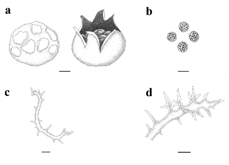
Basidioma 50-60 × 45-50 mm, globose to obovate, slightly depressed at first, later splitting into 6– 9 rays expanding up to 100 mm broad. Exoperidium almost smooth, tough, white to yellowish-white initially, light to dark pinkish-brown later, disappearing finally. Endoperidium grayish brown to brown, smooth, tough, up to 2 mm thick. Gleba firm, white at first, then yellowish-brown to olive-yellowish-brown. Subgleba absent. Spores 9-12 µm,
86
globose and subreticulate. Capillitium 5-15 µm broad and spiny.Ecology: Autumn and spring, terrestrial, solitary to gregarious, in meadows, manured open fields, sandy ground, in hardwood, coniferous, or mixed forest and forest edges (Calonge, 1998; Kujawa et al, 2004; Miller et al, 2005).
Material examined: TURKEY—Muğla: Bodrum, Turgutreis, in meadow, sea level, 37° 01' 12'' N, 27°15' 07'' E, 12.09.2019, ANK Akata & Altuntas 551.
Distribution: Asia (Yemen, Iran, India, China, Mongolia and Turkey), Europea (Poland, The United
Kingdom), America (Uruguay, Mexico, Chile and Argentina, United States and Canada), Africa (Zimbabwe), Caribbean, Australia and New Zealand (Calonge, 1998; Esqueda-Valle et al, 2000; Guzmán, 1969; Hansen and Knudsen, 1997; Homrich and Wright, 1973; Kreisel and Fatimi, 2004; Kujawa et al, 2004; Li et al, 2012; Miller et al, 2005; McNeill, 2013; Morena et al, 2010; Pegler et al, 1995; Pérez-Silva et al, 2015; Saber, 1989; Sharp and Piearce, 1999; Sparling, 2007; Zhishu et al, 1993).
Figure 1. Mycenastrum corium (illustrated by Deniz Altuntaş): a. basidioma (bar: 1 cm), b. basidiospores, c-d. capillitium (bar: 10 µm).
87
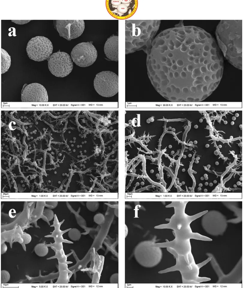
Figure 2. Mycenastrum corium as viewed by a scanning electron microscope (SEM): a-b. basidiospres, c-d. basidiospores and capillitia, e-f. capillitium.
88
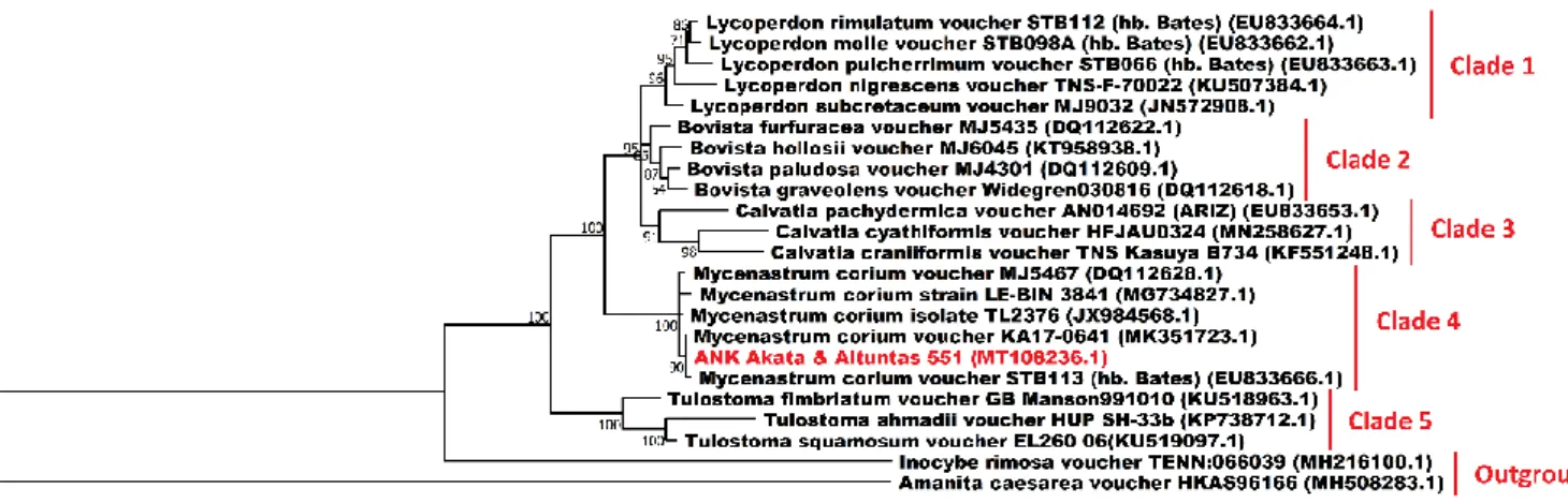
The ITS rDNA sequence of ANK Akata &Altuntas 551 was determined using conventional PCR and subsequent Sanger sequencing and deposited into NCBI GenBank under the accession number of MT108236.1. Regarding the phylogenetic analysis of ANK Akata & Altuntas 551, considering the BLAST search results of the specimens’ ITS sequence, the ITS sequences of the genera Lycoperdon, Bovista, Calvatia, Mycenastrum and Tulostoma, some of the well-known genera of the gasteroid fungi, were selected as ingroup sequences and the ITS sequences of Inocybe rimosa and Amanita caesarea were selected as the outgroup sequences. As a result of the phylogenetic analysis, five distinct clades were revealed along with an outgroup (Figure 3).While the clade 4 contained Mycenastrum species and the specimen Ank Akata & Altunta 551, the Clades 1, 2, 3 and 5 included species from the genera Lycoperdon, Bovista, Calvatia and Tulostoma respectively. On the other hand, Inocybe rimosa and Amanita caesarea were branched far from the rest of the fungi species and formed an outgroup as anticipated. The BLAST analyses carried out with the ITS sequence of Ank Akata & Altuntaş 551 revealed evidence for more than 99.4 % similarities of the specimen with M. corium. The phylogenetic analyses performed based on the ITS sequences of these specimens, further supported the close identity relationship of this specimen with M. corium with a high bootstrap value.
Discussion
M. corium may be confused with other Mycenastrum members in terms of their morphology and ecology. They have globose to obovate basidioma, a double and thick peridium, absence of sungleba powdery gleba, spiny capillitium, globose to subglobose and ornamented basidiospores (Calonge, 1998; Miller et al, 2005).
Mycenastrum catimbauense differs from M. corium by its dark tomentose exoperidium, dark brown endoperidium, dark brown gleba and larger spores (13.3– 15.0 × 11.7–14.5 µm).
M. corium may macroscopically resemble Mycenastrum spinulosum because of their macro-morphology but they have different spore ornamentations. While the former species has subreticulate spores, spore ornamentation of the latter species is minutely verrucose (Gurgel et al, 2017).
Because reliance on the morphological data per se is not always adequate for the precise identification of fungal species, the sequence data from the conserved genomic DNA regions such as ITS, nrSSU and nrLSU are benefited as a suitable tool in molecular taxonomic studies since the last 30 years (Raja et al. 2017). Besides, ITS is one of the most commonly used DNA barcoding markers and thus bestows substantial information for molecular phylogenetic studies. Therefore, we used the ITS region for the molecular identification of Ank Akata & Altuntaş 551. The phylogenetic analyses conducted based on the ITS region revealed the almost 100% genetic identity between the Mycenastrum corium and the specimen (GenBank ID: MT108236.1) (Figure 3).
To the best of our knowledge, this is the first study reporting on the morphological features of a Mycenastrum corium collected from Turkey along with its molecular phylogeny.
Figure 3. The Maximum Likelihood tree exhibiting the phylogenetic relationships of 23 fungi inferred from the ITS region. Percentage bootstrap values obtained from 1000 replicates were given next to the branches. All the sequences used in the phylogenetic analysis were obtained from GenBank except for Ank Akata & Altuntaş 551. Inocybe rimosa and Amanita caesarea were used as the outgroup samples. Accession numbers are indicated in parentheses. The scale bar given at the lower left shows a genetic distance of 0.1.
89
AcknowledgementsAuthors would like to thank Cemile Altuntaş for her valuable contribution during field study. References
Bi, Z., Zheng, G., & Taihui, L. (1993). The macrofungus flora of China's Guangdong Province. Chinese University Press, 1-268.
Calonge, F. D., Gasteromycetes, I., Lycoperdales, N., & Phallales, S. (1998). Tulostomatales. Flora Mycologica Ibérica, 3, 1-271.
Chen, L., Cai, Y., Zhou, G., Shi, X., Su, J., Chen, G., & Lin, K. (2014). Rapid Sanger sequencing of the 16S rRNA gene for identification of some common pathogens. PloS one, 9(2).
Esqueda-Valle, M., Silva, E. P., Herrera, T., Andrade, M. C., & Torres, A. E. (2000). Composición de gasteromicetos en un gradiente de vegetación de Sonora, México. Anales del Instituto de Biología. Serie Botánica, 71(2), 39-62.
Felsenstein, J. (1985). Confidence limits on phylogenies: an approach using the bootstrap. Evolution, 39(4), 783-791. Guzmán, G. (1969). Macromicetos de las zonas áridas de México, II. Gasteromicetos. Anales Inst. Biol. Univ. Nac. Anton.
Mex., 40, 1-92.
Gurgel, R. A. F., Melanda, G. C. S., Ferreira, R. J., Alfredo, D. S., & Baseia, I. G. (2017). Mycenastrum catimbauense (Agaricales, Basidiomycota), a new puffball species from the Brazilian semi–arid region. Stud Fungi, 2(1), 112-118. Hansen, L., & Knudsen, H. (1997). Nordic macromycetes. Vol. 3, Heterobasidioid, Aphyllophoroid and Gastromycetoid
Basidiomycetes. Nordsvamp, 383-620.
Homrich, M. H., & Wright, J. E. (1973). South American Gasteromycetes. The genera Gastropila, Lanopila and Mycenastrum. Mycologia, 65(4), 779-794.
Kaşık, G., & Öztürk, C. (1998). İstanbul’da Görülen Mantar Zehirlenmelerinden Sonra Tesbit Edilen Makrofunguslar. Selçuk Üniversitesi Fen Edebiyat Fakültesi Fen Dergisi, 15, 41-46.
Kreisel, H., & Al‐Fatimi, M. (2004). Basidiomycetes and larger Ascomycetes from Yemen. Feddes Repertorium: Zeitschrift für botanische Taxonomie und Geobotanik, 115(7‐8), 547-561.
Kujawa, A., Bujakiewicz, A. N. N. A., & Karg, J. (2004). Mycenastrum corium (Fungi, Agaricales) in Poland. Polish Bot. J, 49(1), 63-66.
Kumar, S., Stecher, G., Li, M., Knyaz, C., & Tamura, K. (2018). MEGA X: molecular evolutionary genetics analysis across computing platforms. Molecular biology and evolution, 35(6), 1547-1549.
Li, X., Liu, B., Xie, W., Deng, J., & Xu, J. (2012). The contrastive analysis of puffballs produced from Inner Mongolia and Jilin. Chinese Journal of Applied Chemistry, 29(04), 477-482.
McNeil, D. (2013). Some noteworthy British Gasteromycete records. Field Mycology, 1(14), 24-30.
Miller Jr, O. K., Brace, R. L., & Evenson, V. (2005). A new subspecies of Mycenastrum corium from Colorado. Mycologia, 97(2), 530-533.
Moreno, G., Lizárraga, M., Esqueda, M., & Coronado, M. L. (2010). Contribution to the study of gasteroid and secotioid fungi of Chihuahua, Mexico. Mycotaxon, 112(1), 291-315.
Nei, M., & Kumar, S. (2000). Molecular evolution and phylogenetics. Oxford university press.
Pegler, D. N., Læssøe, T., & Spooner, B. (1995). British Puffballs, Earthstars and Stinkhorns: an account of the British gasteroid fungi. Royal Botanic Gardens, Kew, 1-255.
Pérez-Silva, E., Herrera, T., & Medina-Ortiz, A. J. (2015). Mycenastrum corium and gastrointestinal mycetism in Mexico. Mycotaxon, 130(3), 641-645.
Raja, H. A., Miller, A. N., Pearce, C. J., & Oberlies, N. H. (2017). Fungal identification using molecular tools: a primer for the natural products research community. Journal of natural products, 80(3), 756-770.
Rogers, S. O., & Bendich, A. J. (1994). Extraction of total cellular DNA from plants, algae and fungi. In Plant molecular biology manual. Springer, Dordrecht, 183-190.
Stielow, J. B., Levesque, C. A., Seifert, K. A., Meyer, W., Iriny, L., Smits, D., ... & Lomascolo, A. (2015). One fungus, which genes? Development and assessment of universal primers for potential secondary fungal DNA barcodes. Persoonia: Molecular Phylogeny and Evolution of Fungi, 35, 242.
Saber, M. (1989). New records of Aphyllophorales and Gasteromycetes for Iran. Iranian Journal of Plant Pathology, 25: 21-26.
Sharp, C., & Piearce, G. (1999). Some interesting gasteroid fungi from Zimbabwe. Kew bulletin, 739-746. Sparling, J. (2007). Bovista, Mycenastrum, and other puffballs of the Carden Alvar. The Mycophile, 48(1): 1-15.