Journal of the Hellenic Veterinary Medical Society
Vol. 70, 2019
The investigation of xylazine, detomidine, isoflurane and sevoflurane anaesthetic combinations on clinical, laboratory and cardiovascular parameters and on intraocular
pressure in horses
EROL H. Department of Surgery,
Faculty of Veterinary Medicine, The University of Erciyes, Kayseri, Turkey
ARICAN M. Department of Surgery,
Faculty of Veterinary Medicine, The University of Selcuk, Campus, Konya, Turkey
https://doi.org/10.12681/jhvms.20348
Copyright © 2019 H. EROL, M. ARICAN
The investigation of xylazine, detomidine, isoflurane and sevoflurane
anaesthetic combinations on clinical, laboratory and cardiovascular
parameters and on intraocular pressure in horses
H. Erol1, M. Arican21Department of Surgery, Faculty of Veterinary Medicine, The University of Erciyes, Kayseri, Turkey. 2Department of Surgery, Faculty of Veterinary Medicine, The University of Selcuk, Campus, Konya, Turkey.
Corresponding Author:
Hanifi Erol; Department of Surgery, Faculty of Veterinary Medicine, University of Erciyes. 38039, Kayseri, Turkey.
Email address: [email protected]
Date of initial submission: 07-06-2018 Date of revised submission: 07-06-2018 Date of acceptance: 07-06-2018
Ερευνητικό άρθρο
ΠΕΚΕ 2019, 70(1): 1401-1412
ABSTRACT. The aim of this study was to investigate the effects of anaesthetic combinations of xylazine, detomidine, sevoflurane and isoflurane on clinical, laboratory, and cardiovascular parameters as well as their effects on intraocular pres-sure in horses. Twenty-four mixed-breed horses (twelve male and twelve female) were used for this study. The horses were allocated into four groups (six horses in each group): XS (xylazine-sevoflurane), XI (xylazine-isoflurane), DS (detomi-dine-sevoflurane) and DI (detomidine-isoflurane). Clinical evaluations, hematological, biochemical tests and measurement of intraocular pressure were done before (0th), during (5th, 15th and 30th min) and at the end of anaesthesia (60th min). The detected differences were statistically evaluated. In conclusion, this study shows that the anaesthetic combinations of sevoflurane and isoflurane with xylazine and detomidine provided safe and suitable anaesthesia in horses. Our study did not reveal any statistical differences in intraocular pressure measurements. However, it should be noted that intraocular pressures were measured with the animals lying down and our results do not rule out changes in intraocular pressures in a standing position. We concluded that these anaesthesia protocols are suitable for ophthalmic surgery.
1402 H. EROL, M. ARICAN
INTRODUCTION
Α
lpha-2 (α-2) agonists are the greatest improvements in modern equine practice. For treatments and diagnostic procedures, α-2 agonists are widely used in equine medicine and anaesthesia (Mutoh et al., 1997; Guedes et al., 2017). Administration of α-2 agonists also reduces the amount of inhalational anaesthetic needed to maintain general anaesthesia in horses (Nyman et al., 2009; Rohrbach et al., 2009). α-2 Agonists such as detomidine, xylazine, medetomidine and romifidine are used for premedication in horses, and they have similar properties (Arıcan et al., 2015).The inhalation anaesthetic agents such as isoflurane and sevoflurane are used for procedures requiring long-term anaesthesia in horses. Unlike injectable anesthetics, they enter the body through the lungs and move into systemic circulation from the respiratory tract (Hall et al., 2000; Del Barrio et al., 2017). Isoflurane is an inhalant anesthetic agent with a low blood/gas solubility (1.4 in horses) coefficient. The lower blood/gas solubility causes rapid induction and short recovery time from general anaesthesia (Kalchofner et al., 2009; Ida et al., 2013). It leads to dose-dependent cardiac and respiratory depression. The α-adrenergic stimulation associated with isoflurane increases blood flow in skeletal muscles and reduces systemic vascular resistance and arterial blood pressure (Yamashita et al., 2006; Steffey, 2009). Sevoflurane is structurally similar to isoflurane but has lower blood/gas solubility (0.6 in horses). Its effects on the central nervous system (CNS) are fewer than those of isoflurane, but there is still a dose-dependent depression. CNS depression is greater at increasing doses when compared to the other inhalation agents. It also reduces cerebral vascular resistance and metabolism at high doses (Yamanaka et al., 2001; Kronen, 2003).
Intraocular pressure (IOP) is defined as the equilibrium between production and drainage of humor aqueous in the ciliary processes of the eye. The anaesthetic agents may cause changes in IOP (Komáromy et al., 2006; Monk et al., 2017). The aim of this study was to investigate the effects of xylazine, detomidine, isoflurane and sevoflurane anesthetic combinations on clinical, laboratory, and cardiovascular parameters as well as their effects on IOP in horses.
MAŚLANKA T., ZUŚKA-PROT M.
MATERIALS AND METHODS
The current study was performed in the Surgery Department of the Faculty of Veterinary Medicine, University of Selcuk, Turkey and was approved by the Ethics Committee of the Faculty of Veterinary Medicine. Twenty-four mixed-breed horses (twelve male and twelve female) weighing 450 ± 51 kg, 7.6 ± 5 years old were used for this study. The clinical examination and hematological tests were done before starting the study. The horses were allocated into four groups (six horses in each group): XS (xylazine-sevoflurane), XI (xylazine-isoflurane), DS sevoflurane) and DI (detomidine-isoflurane). All horses were fasted for 12 h before premedication; water intake was not restricted. Before premedication, the body weight, respiratory rate, body temperature, and heart rate of the horses were recorded.
Before premedication, a 20-gauge intravenous catheter was placed in the right jugular vein for blood sample collection and administration of anaesthetic drugs. Electrocardiography (ECG) leads (aVL, aVR, aVF) and oxyhaemoglobin saturation (SpO2) were monitored by a BM3 Vet monitor (Bionet, Seoul, Korea) before premedication and during anaesthesia. Intravenous xylazine (1.1 mg/kg b.w., Rompun 2%, Bayer, Mississauga, Canada) was used for premedication in XS and XI groups, and intravenous detomidine (6 µg/kg b.w., Domosedan, Pfizer, New York, NY, USA) was used for premedication in DS and DI groups. Ketamine hydrochloride (2 mg/kg b.w., Ketasol 10%, Richterpharma, Wels, Austria) and midazolam (0.03 mg/kg b.w., Demizolam, Curamed Pharma GmbH. Karlsruhe Germany) were mixed in the same syringe and intravenously administered to induce anesthesia for assisted fall. After induction of anaesthesia, horses were placed in left lateral recumbency and the trachea was intubated with a cuffed endotracheal tube with an internal diameter of 30 mm. The endotracheal tube was attached to a large animal circle breathing system anaesthesia machine (Large animal LSD 3000 anaesthetic machine, Dublin, OH, USA, 12 breath/min), and anaesthesia was maintained with either sevoflurane (Sevorane liquid, Abbott Laboratories, Abbott Park, IL, USA) or isoflurane (Aerrane, Baxter Healthcare, Deerfield, IL,USA) for 60 min. The initial concentration of
sevoflurane was 8% + 4 l O2/min and was reduced to 4%. After 30 min of anaesthesia, the sevoflurane dose was changed to 2% + 4 l O2/min, which was maintained until the end of anaesthesia (60 min). The initial dosage of isoflurane was 5% + 4 l O2/min,
which was reduced to 4% after 15 min. At 30 min of anaesthesia, the isoflurane dose was changed to 2.5 % + 4 l O2/min, which was maintained until the end of anaesthesia (60 min). After 60 min, the administration of inhalation anaesthetics was discontinued, and all animals were supported with O2 for 10 min.
Clinical evaluations
Before pre-anaesthetic administration and during anesthesia (5th, 15th, 30th and 60th min) heart rate (HR), ECG, body temperature (BT), SPO2, respiratory rate (RR), systolic, diastolic and mean arterial pressure were measured and recorded. After initiation of inhalation anaesthesia, palpebral reflex was controlled during and at the end of anesthesia in all groups. Palpebral reflex (sluggish) time (PRs), corneal reflex (strong) (PRS), palpebral reflex (normal) (PRn), spontaneous respiration return time (SRRT) and standing up time (SUT) were recorded on examination papers.
Haematologic evaluations
Before (control) and during anaesthesia (5th, 15th, 30th and 60th min) a 15-ml blood sample was collected from the right jugular vein for hematologic, blood gas, and biochemical evaluations. Red blood count (RBC), white blood count (WBC), blood pH, venous partial carbon dioxide pressure (PvCO2), venous partial oxygen pressure (PvO2), total carbon dioxide (tCO2), hemoglobin level (Hb), hematocrit (Ht), bicarbonate (HCO3-), electrolytes (Na+, K+) and
lactic acid were measured using a Gem Premier 3000 (Biomerieux Diagnostics, Marcy l’Etoile, France) and a Medonic CA 530 hematology analyzer (PZ Cormay, Łomianki, Poland).
Biochemical evaluations
The serum levels of total protein (TP), aspartate transaminase (AST), alanine transaminase (ALT), alkaline phosphatase (ALP), α-glutamyl transferase (γGT), total bilirubin (T-bil) and glucose (Glu) were
measured before and at the end of anaesthesia (60th min) using a VetTest 8008 biochemical analyzer (Idexx Laboratories, Westbrook, ME, USA).
Intraocular pressures measurements
Before pre-anaesthetic administration, eyes examination (palpebral, corneal and pupillary reflexes, conjunctiva) were done in all horses and intraocular pressures were measured using a TonoVet tonometer (Icare, Vantaa, Finland) before administering xylazine and detomidine (control) and at the 5th, 15th, 30th, and 60th min after inducing anaesthesia. The same eyes (right) were evaluated in all horses.
Data and statistical analysis
IBM SPSS 21 statistics programs were used to analyze the data. The Shapiro-Wilk test was used for normality then the one and two-way repeated measure (ANOVA) followed Tukey significance tests to compare intra-group and inter-intra-group values. The level of statistical significance was set at P ˂ 0.05. Results were presented as Mean ± SE (Standard Error).
RESULTS
Clinical evaluation results
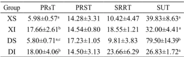
In the current study, suitable and adequate sedation was provided in all groups. The clinical evaluations are shown in Table 1. There were significant differences seen in PRsT and SUT values between groups. The PRsT values of sevoflurane groups were shorter than those of the isoflurane groups (P < 0.05). The SUT values were higher in group DS than in the others (P < 0.05).
The cardiopulmonary findings are shown in Table 2. Increases and decreases were seen in HR, SpO2
Table 1. Clinical Evaluation of groups (Mean ± SE) PRsT;
Palpebral reflex sluggish time, PRST; palpebral reflex strong time, SRRT; spontaneous respiration return time, SUT; standing up time, a,b,c; P ˂ 0,05 between groups. Group PRsT PRST SRRT SUT
XS 5.98±0.57a 14.28±3.31 10.42±4.47 39.83±8.63a
XI 17.66±2.61b 14.54±0.80 18.55±1.21 32.00±4.41a
DS 5.80±0.71a,c 17.23±1.05 9.81±3.83 79.50±14.39b
1404 H. EROL, M. ARICAN
and SP (systolic pressure) values before and during anaesthesia in all groups. The changes detected in these values were not statistically significant. Respiratory rate (RR) increased at 5 min and decreased from 15 min until the end of anaesthesia
Table 2. Cardiopulmonary evaluation of groups (M±SE)
in XI (P < 0.05). Significant differences were seen in diastolic (DP) and mean pressures (MP) between and within groups (P < 0.05). Decreases of DP were recorded at 5th, 15th, 30th, 60th min of anaesthesia in XI, DS and DI. Increases of DP were detected at 15,
Values Groups 0th 5 th 15th 30th 60th Reference ranges
HR XS 57±8.42 47.6±12.04 62.8±13.33 52.1±10.99 51.3±9.24 28-45 XI 54±8.42 68±12.04 55.8±13.33 55±10.09 55.5±9.24 28-45 DS 46.8±8.42 51.6±12.04 56±13.33 34.8±10.99 31.8±9.24 28-45 DI 34.3±8.42 35.6±12.04 49.8±13.33 60.5±10.99 54.3±9.24 28-45 SpO2 % XS 92.3±1.77 95.5±0.77 96.5±0.62 95.3±1.30 95±1.34 85-100 XI 95.6±1.37 97.8±0.77 97.1±0.62 94.6±1.30 94.6±1.34 85-100 DS 96.5±1.77 96.5±0.77 96.1±0.62 95.1±1.30 94.1±1.38 85-100 DI 94.3±1.77 96±0.77 96.5±0.62 95.5±1.30 96±1.34 85-100 RR XS 15.5±4.01 16.6±4.54 19.4±5.74 14.4±3.17 11.4±1.43 8-20 XI 13.1±2.61ab 27.3±9.38a 11.6±2.04ab 7.2±1.20b 7.6±0.98b 8-20 DS 13.1±2.92 9±1.26 10.8±1.55 11.5±1.38 12.8±2.70 8-20 DI 10.4±2.56 8.75±2.78 13.5±4.99 18±7.18 7.6±1.60 8-20 BT XS 36.8±0.09a 36.7±0.08a 35.9±0.12b 35.7±0.18b 35.7±0.2b 37.5-38 XI 37.1±0.21 37±0.22 36.5±0.42 36.3±0.41 36.1±0.30 37.5-38 DS 37±0.26 36.6±0.31 36.5±0.46 36.5±0.40 36.3±0.37 37.5-38 DI 37.1±0.34 36.8±0.34 37.1±0.27 36.9±0.35 36.9±0.40 37.5-38 SP XS 153.3±7.81 105.3±5.57 151.5±10.14 107.8±10.17 87.8±6.22 112±14 XI 161.8±7.8 158.5±5.57 125.6±10.14 118.3±10.17 103.3±6.22 112±14 DS 153.6±7.81 153.6±5.57 122.5±10.14 118.3±10.17 103.3±6.22 112±14 DI 160.1±7.81 155.5±5.57 122.8±10.14 106.6±10.17 94.6±6.22 112±14 DP XS 59.6±10.64*,a 58.3±8.57*,a 121.6±7.3*,b 104.1±5.82*,b 70.8±7.50a 70±14 XI 117±10.64a 107±8.57*,a 80.3±7.36a 61.5±5.82b 47±7.50b 70±14 DS 119.3±10.64a 83±8.57*,b,c 80.3±7.36a,c 61.5±5.82b,c 56.6±7.50b,c 70±14 DI 124.1±10.64a 95.5±8.57*,b 80.3±7.36b 61.5±5.82b 60.6±7.50b 70±14 MP XS 99.5±19.86 77±12.5 132.8±8.14 106.1±6.57* 80.3±4.31 90±14 XI 109.4±21.76a,c 114.4±13.73a 97.2±8.92a 74.6±7.20b,c 68.6±4.72b,c 90±14 DS 98.6±19.86a 111.5±12.54a 91.6±8.14a 72.8±6.57b 67.1±4.31b 90±14 DI 96±19.80a 111.5±12.50a 91.6±8.12a 72.8±6.55b 67.1±4.28b 90±14
HR; heart rate, SpO2; oxygen saturation, RR; respiratory rate, BT; body temperature, SP; systolic pressure, DP; dia-stolic pressure, MP; mean pressure, *,**; P ˂ 0.05 between groups evaluation, a,b,c ; P ˂ 0.05 in groups evaluations. References: Arıcan et al. 2015.
30, 60 min of anaesthesia in XS. In XI, DS and DI the MP increased at 5 min of anaesthesia and decreased until the end of anesthesia. In XS the MP decreased at 5 min and increased at 30 min of anaesthesia. BT decreased from 15 min until the end of anaesthesia in XS. The decrease of BT was statistically significant (P < 0.05).
Haematologic results
The measured hematologic and blood gas parameters are shown in Table 3. In hematologic evaluations, RBC, WBC, Hb, Ht and lactic acid values showed changes. The changes of RBC, WBC, Hb and Ht values were in reference intervals in all groups. The lactic acid levels were lower than reference range before and during anaesthesia in all groups. In DS, there was significant difference recorded in lactic acid levels between 0th min and the other times.
The pH values decreased at 15th min of anaesthesia in DS. This reduction was below the reference range and it was significant (P < 0.05) (Table 3). At 0th, 5th, 15th, 30th, 60th min of anaesthesia, the PvCO2 values were higher in sevoflurane groups
(XS and DS) than in isoflurane groups (XI and DI). The PvCO2 values increased during anaesthesia in sevoflurane groups. After 5 min of anaesthesia, the PvCO2 values started to decrease in isoflurane groups, and the reductions were significantly different (P < 0.05). There were no significant differences seen in PvO2 values between groups at the 0th, 5th, 15th, 30th and 60th min of anaesthesia. Before and during anaesthesia the PvO2 values were lower than reference range in DI (P < 0.05) (Table 3). Differences in tCO2 values were seen in all groups. The tCO2 values were
higher than reference range at 15th, 30th and 60th min of anaesthesia in XS and DS (P < 0.05). The other differences were in reference interval (Table 3). The HCO3 levels of XS and DS were significantly different from XI and DI (P < 0.05).
Biochemical results
Biochemical parameters are shown in Table 4. There were significant differences detected in TP, AST, ALT and ALP values among and within groups. All of the
values were within the normal range (Table 4). T-bil values were higher at 0 and 60th min of anesthesia than the reference range in XS. At the end of the anaesthesia Glu values in XS, XI and DI were higher than XS and reference values (Table 4).
Intraocular pressures results
Intraocular pressures (IOP) were measured before and during anesthesia (5th, 15th, 30th and 60th min) (Table 5). There were no significant differences observed between and within groups (reference range: 13-37 mmHg).
DISCUSSION
Arıcan et al. (2015) compared the clinical effects of sevoflurane, isoflurane, and medetomidine anaesthetic combinations in horses. They found the loss of palpebral reflex, return of palpebral reflex and standing up times to be shorter in a sevoflurane group than in an isoflurane group. They attribute this to the low blood concentration of sevoflurane at a plane of anaesthesia and its relatively rapid elimination from the body. In our study, there were significant differences observed in PRsT and SUT (P < 0.05). Palpebral reflex sluggish time of sevoflurane groups was shorter than those of isoflurane groups which may be supported by the previous finding of low blood concentration and rapid elimination of sevoflurane from the blood. Detomidine binds α-2 adrenoreceptors in the locus coeruleus and medulla spinalis to provide sedative and analgesic effects. It suppresses norepinephrine and dopamine release in CNS. Because of this, detomidine is more potent and has fewer side effects than xylazine. It has been reported that detomidine produces deeper and longer sedation than xylazine (Steffey and Pascoe, 2002; Rochbrach et al., 2009). In this study, SUT was longer in XS and DS than XI and DI. The longest and shortest times were detected in DS and DI. This condition suggested that anaesthetic combinations, anaesthetic doses and animal related factors (age, sex and weight) could influence the post anaesthetic parameters.
Generally cardiac arrhythmias which occur during anaesthesia can cause serious problems, and
1406 H. EROL, M. ARICAN
Values Groups 0th 5th 15th 30th 60th Reference ranges
RBC (x106/mm3) XS 9.8±0.12 8.8±0.63 8.6±0.81 7.7±0.53 7.9±0.48 7-13 XI 10.3±0.43 8.1 ±0.34 7.5±0.28 7.3±0.33 7±0.19 7-13 DS 10.6±0.38 10.8±0.28 10.7±0.18 10.1±0.32 9.3±0.37 7-13 DI 9.7±1.07 10.9±0.87 11.4±0.48 11±0.32 10.6±0.43 7-13 WBC (x103/mm3) XS 9±1.06 8.34±0.93 7.64±0.84 8±1.01 6.96±0.75 6-12.5 XI 10.6±0.74 9.4±0.63 9.2±0.59 8.6±0.66 8.3±0.63 6-12.5 DS 7.8±0.42 8.9±1.23 8.6±1.42 8.65±0.95 7,6±0.60 6.12.5 DI 8±1.20 9.1±1.26 9.2±1.07 9.6±0.95 9.5±0.95 6-12.5 pH XS 7.3±0.02 7.3±0.02 7.3±0.02 7.3±0.02 7.3±0.02 7.36-7.43 XI 7.4±0.001 7.3±0,001 7.4±0.01* 7.4±0.08 7.4±0.03 7.36-7.43 DS 7.4±0.001a 7.3±0.02ab 7.2±0.02*,b 7.2±0.02b 7.2±0.02b 7.36-7.43 DI 7.4±0.01 7.3±0.02 7.3±0.01 7.4±0.03 7.4±0.01 7.36-7.43 PvCO2 (mm/Hg) XS 47.2±1.06*,c 52.6±1.6*,bc 55.8±1.74*,b 59.2±1.24* ,ab 63.4±1.99*,a 38-48 XI 36.9±1.44** 38.2±1.33** 32.9±0.95** 29.6±4.60** 33.3±2.19** 38-48
DS 45.4±0.92*,c 52.8±3.15*,bc 59.4±3.77*,abc 64.5±5.43*,ab 70.2±5.89*,a 38-48
DI 36±0.45**,ab 38.6±1.87**,a 36.4±0.88**,ab 33.8±1.30**,ab 32.8±1.52**,b 38-48
PvO2 (mm/Hg) XS 30.6±1.32 42.8±2.92 48.8±8.5 52.6±7.28 48.2±8.16 37-56 XI 29.6±1.09c 37.5±1.72bc 42.4±1.79ab 43.9±3.21ab 48.7±2.3a 37-56 DS 31.2±2.03bc 28.8±1.35c 42.8±1.80ab 47.5±4.17a 55.2±6.2a 37-56 DI 33.5±3.19ab 29.2±1.56b 33.1±0.90ab 34.3±1.56ab 31.6±2.0b 37-56 tCO2 XS 32.9±0.73* 33.8±1.37 34.1±1.49*,** 35.9±1.04 37.3±0.95* 24-32 XI 27.2±0.83* 26.5±1.20 25.1±1.10* 21.1±4.31 26.6±0.77** 24-32 DS 33.4±0.73**b 33.2±1.16b 34.1±1.08*,**,ab 35.9±1.28ab 39.2±2*,a 24-32 DI 25.9±0.59* 26±1.48 25.4±0.83 24.8±0.54 25.1±1.07** 24-32 Hb (g/dL) XS 14.6±0.30 13.22±1.03 12.56±1.02 11.66±0.94 11.8±0.83 11-19 XI 15.5±0.55a 12.3±0.51b 11.5±0.41b 11±0.46b 10.7±0.2b 11-19 DS 15.3±0.27 15.4±0.51 15.3±0.54 14.4±0.70 13.4±0.39 11-19 DI 13.5±1.18 15.8±0.63 16.2±0.53 15.4±0.36 14.8±0.33 11-19 Ht (%) XS 50.22±1.60 45.16±3.42 44±3.66 39.74±3.21 40.2±2.60 35-52 XI 52.3±1.93 40.5±1.66 37.8±1.39 36.3±1.57 34.8±0.75 35-52 DS 54.8±1.87 55.94±1.88 55.3±1.71 51.4±3.05 47.6±2.30 35-52 DI 44.6±4.61 51.4±2.50 53.6±1 53.1±1.51 51.1±1.40 35-52 HCO3 (mm/L) XS 31.4±0.73* 32.1±1.37 32.4±1.46*,** 34.1±1.04* 35.4±0.94* 22-29 XI 26±0.79** 25.4±1.18 24±1.08* 24.3±1.12** 25.5±0.74** 22-29
DS 32±0.70*,ab 31.6±1.07b 32.3±1**,ab 33.9±1.10*,ab 37±1.83*,b 22-29
DI 24.8±0.59** 24.8±1.43 24.3±0.83* 23.8±0.54** 24±1.04** 22-29 Na (mEq/L) XS 136.4±0.87 136±0.54 137±1.04 136.2±1.06 135.2±0.96 136-142 XI 142.8±0.30 142±1.26 142.6±1.35 127.4±16.46 140.6±1.99 136-142 DS 138±0.83 138.6±0.87 137.2±0.96 137.7±1.25 136.5±0.47 136-142 DI 140±1.06 138±2.25 135±1.92 137±1.44 137.6±1.08 136-142 K (mEq/L) XS 3.5±0.17 3.3±0.20 3.4±0.26 3.6±0.30 3.7±0.28 2.2-4.6 XI 2.9±0.06 2.8±0.11 2.7±0.11 26.4±23.27 2.8±0.16 2.2-4.6 DS 3.5±0.19 3.8±0.23 3.4±0.23 3.7±0.29 3.8±0.36 2.2-4.6 DI 3.2±0.07 3.5±0.09 3.4±0.05 3.4±0.05 3.5±0.08 2.2-4.6 Lactic acid (mg/dL) XS 0.6±0.12 1.1±0.26 1±0.20 0.7±0.04 1±0.16 4-12 XI 0.6±0.25 1.1±0.30 1±0.18 08±0.05 1±0.15 4-12 DS 0.5±0.05b 1.6±0.10a 1.6±0.15a 1.5±0.20a 1.2±0.50a 4-12 DI 0.6±0.25 1.1±0.35 1±0.15 0.8±0.15 1±0.15 4-12
Novakovski et al., 2015) have reported this dose-dependent suppression of the cardiovascular system. Sevoflurane and isoflurane have been reported to be equally arrhythmogenic in animals (Matthews and Hartsfield, 2004). In this study HR, SpO2, BT, RR, SP, DP and MP values were recorded at the beginning of the anaesthesia and SP and MP values were higher than the reference ranges in all groups (Table 2). In this study HR, SpO2, BT, RR, SP, DP and MP values were recorded at the beginning and during of the anaesthesia. DP and MP values showed significant decreases and increases during anesthesia in all groups (P < 0.05). DP values were higher than reference range at 0th 5th and 15th min of anaesthesia in XI, DS and DI. The statistically differences were detected between groups at 0th, 5th and 30th min of anaesthesia in DP values. Intra-groups evaluation of DP values, there were statistically differences recorded at 0th and other times in all groups. In XS, MP value was statistically different at 30th min of anaesthesia than others. Intra-groups evaluation of MP values, the statistically differences were detected between 0th, 5th, 15th and 30th, 60th min of anaesthesia in XI, DS and DI. However, evaluations of ECG traces from all groups did not show atrioventricular blockade, atrial fibrillation, hypoxia-related extra systoles or dysrhythmias. The electrolyte parameters correlated well with our ECG evaluations as there were no significant differences detected in Na and K values. Taylor and Clerk (2007) reported that reduction of BT can occur secondary to variations in skin thickness and environmental temperature in horses. In our study, BT showed decreases during anaesthesia in all groups. However, the decrease was significantly different only in XS.
The horse’s hematologic profile is affected by different factors which are nutritional, environmental, age, sex, performance, and genetics (Padalino et al., 2014). At the same time stress, excitement, fear and the associated catecholamine exchange in blood circulation, as well as monitoring is required (Muir and Hubbell, 2009;
Erol and Arıcan, 2017). Anaesthetic agents cause dose-dependent reductions in arterial blood pressure with myocardial depression and peripheral vasodilatation. Many investigators (Ebert et al., 1995; Aida et al., 1996; Röding et al., 1996;
Duke-RBC; red blood count, WBC; white blood count, PH; blood PvH, PvCO2; venous partial carbon dioxide, PvO2; venous
partial oxygen pressure, tCO2; total carbon dioxide, Hb; hemoglobin level, Ht; hematocrit, HCO3; bicarbonate, *,**; P ˂ 0.05 between groups evaluation, a,b,c; P ˂ 0.05 in groups evaluations. References: Kawamura 2011, Aros et al. 2017, http://cal.vet.upenn.edu/projects/fieldservice/Equine/EQCLPATH.htm
Values Groups 0th 60th Reference ranges TP (g/ dL) XS 7.4±0.27 6.9±0.18 5.6-7.9 XI 7.05±0.18 a 6.2±0.10 b 5.6-7.9 DS 7.07±0.25 a 6.47±0.26 b 5.6-7.9 DI 7.4±0.25 6.9±0.20 5.6-7.9 AST (g/dL) XS 337.66±30.98 320.16±34.3 100-600 XI 591±213.50 345.25±160.08 100-600 DS 410.75±171.55 356.25±146.08 100-600 DI 450±52.23 345±122.05 100-600 ALT (IU/L) XS 25±3.52 28±5.97 5-50 XI 28±8.71 40.75±9.97 5-50 DS 18.50±6.38 30.50±10.41 5-50 DI 25.2±7.28 42.3±6.65 5-50 ALP (IU/L) XS 3.2±0.20 2.86±0.12 1.9-3.2 XI 3.07±0.23 2.65±0.11 1.9-3.2 DS 3.40±0.12 a 2.87±0.08 b 1.9-3.2 DI 3.05±0.23 2.58±0.15 1.9-3.2 T-bil (mg/ dl) XS 4.96±0.98 4.26±0.65 0-3.5 XI 4.15±0.21 3.40±0.44 0-3.5 DS 3.40±0.83 2.95±0.68 0-3.5 DI 4.1±0.65 3.2±0.78 0-3.5 γGT (U/L) XS 15±2.8 15±2.7 0-87 XI 11.25±3.03 18.25±8.08 0-87 DS 14.75±2.46 12.50±3.17 0-87 DI 16±3.05 19±2.18 0-87 Glu (IU/L) XS 105.16±7.53 169.66±2.80 64-150 XI 92.25±1.49a 130.50±5.7b 64-150 DS 88±2.16a 186.75±17.01b 64-150 DI 104.2±8.57 165.6±3.33 64-150
Table 4. Biochemical evaluations of groups (Mean ± SE)
TP; total protein, AST; aspartate transaminase, ALT; alanine transaminase, ALP; alkaline phosphatase, T-bil; total bilirubin, γGT; α-glutamyl transferase, Glu; glu-cose, a,b,c; P ˂ 0.05 in groups evaluations. References: Kawamura 2011, Takasu et al., 2013, Aros et al., 2017, http://cal.vet.upenn.edu/projects/fieldservice/Equine/ EQCLPATH.htm
1408 H. EROL, M. ARICAN
anaesthesia. However, these conditions are not fully observed during anesthesia and may vary depending on the anaesthetic duration (Mutoh et al., 1997; Muir and Hubbell, 2009). The decreases of anaesthetic agent doses during the anaesthetic period and the concurrent ECG and venous blood parameters evaluations support the literature findings (Mutoh et al., 1997; Kronen, 2003; Padalino et al., 2014). Yamashita et al. (2006) emphasized that α-2 agonists and ketamine-midazolam combinations cause to decrease PO2 values but do not significantly change PCO2
and cardiovascular findings. They have also been associated with intrapulmonary shunts and ventilation-perfusion mismatch. Intermittent positive pressure ventilation (IPPV) suppresses the cardiovascular functions during general anaesthesia in horses because the mechanical ventilation increases thoracic pressure and retards venous blood circulation (Taylor and Clerk, 2007). Kushiro et al. (2005) reported that cardiovascular functions are more suppressed than during normal spontaneous breathing during anesthesia in horses due to the increased amount of catecholamine in circulation. There were statistically differences recorded in PvCO2, PvO2, tCO2 and HCO3 values between and among groups. In XS and DS, PvCO2 values were higher than XI and DI at 5, 15, 30 and 60th min of anesthesia. PvO2, tCO2 and HCO3 values of DI were lower than the others at same times. The highest values of PvCO2, PvO2, tCO2
and HCO3 were detected in DS at 60th min of anesthesia. In the current study, the observed decreases and increases in PvCO2, PvO2, tCO2 and HCO3 values in groups were thought to be
due to the changes in the amount of circulating catecholamine and ventilation support, and varied hyperglycemia and hypoxia can all cause changes in
venous blood parameters (Robinson, 2009). These are potential reasons for the changes of RBC, WBC, Hb, Ht and lactic acid at the beginning of and during anaesthesia in the present study.
Analysis of blood gas samples from arterial and venous blood give information about ventilation and fluid-electrolyte balance (Hartsfield et al., 2006; Erol and Arıcan, 2017).In venous blood gas parameters, the significant differences were detected in pH, PvCO2, PvO2, tCO2 and HCO3 values (P < 0.05). The changes in pH values were noted during anesthesia in all groups. Depending on the changes of pH values, effected in PvCO2, PvO2, tCO2 and HCO3- values. These changes were
more evident in sevoflurane groups and related to the compensatory response to respiratory acidosis and metabolic alkalosis. Venous blood values (pH, PvCO2, PvO2, tCO2 and HCO3) are 5-10 mmHg and 1-5 mEq/l higher than arterial values (Steffey et al., 2005; Marcilla et al., 2012).
The α-2 agonists cause cardiac depression and hypoxemia. The resulting cardiac depression and hypoxemia decrease PO2 and increase PCO2 values. In addition, inhalation anaesthetic agents cause respiratory depression and increase in PCO2 values. The level of depression depends on doses of inhalation agents (Kronen, 2003). Detomidine is 10 times more potent and produces deeper and longer sedation than xylazine (Yamashita et al., 2000; Steffey et al., 2005; Padalino et al., 2014). The effects of sevoflurane on CNS are lower than isoflurane and compared to other inhalation anesthetics the suppression effect is greater at increasing doses. Sevoflurane reduces systemic vascular resistance, arterial blood pressure and mean pulmonary arterial pressure during
IOP; intraocular pressure, Reference; Komáromy et al., 2006; Monk et al., 2017
Table 5. IOP evaluation of groups (Mean ± SE)
Groups 0th 5th 15th 30th 60th Reference range
IOP (mm/Hg)
XS 24±0.6 27.8±3.81 38.7±2.95 34.7±2.58 35.4±1.15 13-37 XI 32±1 34.2±0.50 34.2±1.15 33.9±0.57 34.4±0.50 13-37 DS 52.3±4.45 32.7±3.73 50.3±2.71 42.3±2.59 41.5±3.33 13-37 DI 34.4±2.58 25.7±1.78 20.2±0.34 21.1±0.22 20.1±0.48 13-37
In general anaesthesia, anaerobic metabolism begins to produce lactate due to decreased perfusion in tissues and an increase in the amount of lactate during anaesthesia. Along with anaerobic metabolism Glu levels increase due to sympathetic stimulation and lipolysis (Edner et al., 2005). Steffey and Pascoe (2002) emphasized that ɑ-2 agonists increase the serum Glu levels in a dose-dependent manner. In XS, DS and DI the Glu values were greater than reference limits at the end of the anesthesia (Table 4). The increase of Glu values were significant in XI and DS. However, DS Glu values were higher than reference interval. The changes of Lac values were detected in groups but they were in reference limits. The measured Glu values in our study showed changes due to anaerobic metabolism during the anaesthesia. The IOP measurement is part of the routine eye examination in horses with tonometry and has become more widely used in recent years. The reference range for horses using tonometry is reported as 15-37 mmHg (Komáromy et al., 2006; Monk et al., 2017). Several factors such as the time of day, sedation, anesthetic agents and head position influence the IOP (Hall et al., 2000; Stine et al., 2014; Arıcan et al., 2015; Monk et al., 2017). In the present study, there were no statistical differences seen in IOP values at the 0th, 5th, 15th, 30th and 60th minutes of anaesthesia between and within groups. The IOP values of DS were higher at the 0th (Control), 15th, 30th and 60th min of anesthesia. No statistical differences were detected between and within groups. These differences were thought to be related to the anaesthetic agents’ effects on the intracranial and cerebral perfusion pressures. On the other hand, these results suggest that the anaesthetic combinations used in our study can be used safely for eye surgeries in horses.
CONCLUSION
In conclusion, this study shows that the anaesthetic combinations of sevoflurane and isoflurane with xylazine and detomidine provided safe and suitable anaesthesia in horses. It is a comprehensive study on the evaluation of anaesthesia combinations in horses and the transfer of our experience to clinicians. There were statistical differences depending on the anesthetic agents’ doses.
The biochemical values may be different in the pre-anaesthetic, anaesthetic and post-anaesthetic periods due to differences in anaesthetic agents. It is important to have information about liver function before and after anaesthesia. The differences in these values because of detoxification of anaesthetic agents in liver balance the anaesthetic agents’ effects on liver (Steffey, 2009; Hubbell et al., 2011). In the present study, the biochemical parameters’ levels changed after anaesthesia. However, the TP, AST, ALT and ALP values detected, although significantly different, were still within the reference interval (Table 4). T-bil values were higher at 0 and 60th min of anesthesia than reference range in XS. At the end of anaesthesia Glu values in XS, DS and DI were higher than in XI and the reference values. The use of α-2 agonists in horses cause to decrease in TP value and hyperglycaemia (Muir and Hubbell, 2009). In our study TP values decreased in all groups at 60th min. This condition supported the literature information. The changes of AST, ALT and ALP values between 0th and 60th min in all groups were thought due to detoxification of anaesthetic agents in liver. The significant difference of ALP value in DS was more evident than others. However, it was within reference interval. The higher T-bil values reason was thought to be due to age, sex and gender of horses. The increases of Glu values in all groups supported the anaesthetic agents’ effects on liver.
Ketamine is the most commonly used dissociative anaesthetic agent in horses. It has been reported that it increases renal blood flow, mucous secretions of the respiratory tract and the amount of catecholamine in circulation. The fact that ketamine is metabolized in the liver was reported in previous studies (Hall et al., 2000; Rosetti et al., 2008; Levionnois et al., 2010; Oku et al., 2011). Liver function during anaesthesia depends on liver blood flow, metabolites of anaesthetic agents and other drugs. Isoflurane and sevoflurane are less hepatotoxic than other inhalation anaesthetics (Topal et al., 2003). Our study supported the literature’s previously reported information (Hall et al., 2000; Topal et al., 2003; Rosetti et al., 2008; Levionnois et al., 2010; Oku et al., 2011).
1410 H. EROL, M. ARICAN
in hematologic, biochemical and cardiovascular parameters even when these values were found within normal physiological limits. It was determined that both inhalation anaesthetic drugs studied could depress the respiratory system due to dose-dependent effects, and similar effects were observed between sevoflurane and isoflurane anaesthesia on the respiratory and circulatory systems. It has been clinically determined that the choosing of ɑ-2 agonist and their action times are very important in general anaesthesia. The cardiopulmonary changes were more prominent in sevoflurane groups. Our study did not reveal any statistical differences in IOP measurements. However, it should be noted that
intraocular pressures were measured with the animals lying down and our results do not rule out changes in IOP in a standing position. We concluded that these anaesthesia protocols are suitable for ophthalmic surgery.
ACKNOWLEDGEMENT
We would like also to thank for financial support by University of Selcuk Scientific Research Projects (BAP) (Project No. 10401129).
CONFLICT OF INTEREST
The authors declare that they have no conflict of interest.
REFERENCES
Aida H, Mizuno Y, Hobo S, Yoshida K, Fujinaga T (1996) Cardiovascular and pulmonary effects of sevoflurane anaesthesia in horses. Vet Surg 25: 164-170
Arıcan M, Erol H, Esin E (2015) Clinical comparison of medetomidine with isoflurane or sevoflurane for anesthesia in horses. Pak Vet J 35: 474-478
Aros K, Carrosko J, Briones R, Tadich TA (2017) Haematological and serum biochemical reference values for urban-working equines in Chile, Austral J Vet Sci 1: 27-33.
Del Barrio MCN, Bennett RC, Hughes JML (2017) Effect of detomidine or romifidine constant rate infusion on plasma lactate concentration and inhalant requirements during isoflurane anaesthesia in horses. Vet Anaesth Analg 44: 473-482.
Duke-Novakovski TD, Palacios-Jimenez C, Wetzel T, Rymes L, Sanchez-Teran AF (2015) Cardiopulmonary effects of dexmedetomidine and ketamine infusions with either propofol infusion or isoflurane for anesthesia in horses. Vet Anaesth Analg 42: 39-49
Ebert T, Harkin CP, Muzi M (1995) Cardiovascular responses to sevoflu-rane: A review. Anesth Analg 81: 569-575
Edner A, Nyman G, Ess_en-Gustavssen B (2005) The effects of sponta-neous and mechanical ventilation on central cardiovascular function and peripheral perfusion during isoflurane anaesthesia in horses. Vet Anaesth Analg 32: 136-146
Erol H, Arıcan M (2017) A Comparasion of laboratory and cardiopulmo-nary effects of desflurane, detomidine and medetomidine anaesthetic combinations in Horses, Acta Sci Vet 45: 1-9
Guedes AGP, Tearney CC, Cenani A, Aristizabal F, Nicto J (2017) Comparison between the effects of postanesthetic xylazine and dex-medetomidine on characteristics of recovery from sevoflurane anes-thesia in horses. Vet Anaesth Analg 44: 273-280
Hall LW, Clarke KW, Trim CM (2000) Veterinary Anaesthesia. 10th ed, W.B. Saunders, London: pp 247-315.
http://cal.vet.upenn.edu/projects/fieldservice/Equine/EQCLPATH.htm [accessed date: 01.10.2018]
Hubbell JAE, Aaernes TK, Bednarski RM, Lerche P, Muir WM (2011) Effects of 50% and maximal inspired oxygen concentrations on respi-ratory variables in isoflurane-anesthetized horses. BMC Vet Re 7: 1-11 Ida KK, Fantoni DT, Ibiapina BT, Souto MTMR, Zoppa ALV, Silva LC,
Ambrósio AM (2013) Effect of postoperative xylazine administration on cardiopulmonary function and recovery quality after isoflurane anesthesia in horses. Vet Surg 42: 877-884
Kalchofner KS, Picek S, Ringer SK, Jackson M, Hassig M, Bettschart-Wolfensberger R (2009) A study of cardiovascular function under controlled and spontenous ventilation in isoflurane-medetomidine anaesthetized horses. Vet Anaesth Analg 36: 426-35
Kawamura, S (2011) Criteria for clinical biochemistry. In: Textbook of Veterinary Internal Medicine, Large Animal Practice, Bun-Eido Publishing Co, Ltd, Tokyo, pp. 331–336
Komáromy AM, Garg CD, Ying GS, Liu, C (2006) Effect of head position on intraocular pressure in horses. Am J Vet Res 67: 1232-1235 Kronen PW (2003) Anesthetic management of the horse: inhalation
anes-thesia. Available at: International Veterinary Service (www.ivis.org), Ithaca, New York, USA, 1-8. [Accessed November 10, 2017] Kushiro T, Yamashita K, Umar MA, Maehara S, Wakaiki S, Abe R, Seno
T, Tsuzuki K, Izumisawa Y, Muir WM (2005) Anesthesic and cardio-vascular effects of balanced anesthesia using constant rate infusion of midazolam-ketamine-medetomidine with inhalation of oxygen-sevo-flurane (MKM-OS Anesthesia) in horses. J Vet Med Sci 67: 379-384 Levionnois OL, Menge M, Thormann W, Mevissen M, Spadavecchia C
(2010) Effect of ketamine on the limb withdrawal reflex evoked by transcutaneous electrical stimulation in ponies anaesthetised with iso-flurane. Vet J 186: 304-311
Marcilla MG, Schauvliege S, Segaert S, Duchateau L, Gasthuys F (2012) Influence of a constant rate infusion of dexmedetomidine on cardio-pulmonary function and recovery quality in isoflurane anaesthetized horses. Vet Anaesth Analg 39: 49-58
Matthews NS, Hartsfield SM (2004) Arrhythmogenic dose of epinephrine in isoflurane- or sevoflurane-anesthetized horses. J Equine Vet Sci 24: 110-114
Monk CS, Brooks DE, Granone T, Garcia-Pereira FL, Melesko A, Plumber CE (2017) Measurement of intraocular pressure in healty anesthetized horses during hoisting. Vet Anaesth Analg 44: 502-508 Muir WW, Hubbell JAE (2009) History of Equine anaesthesia. In (Muir
W, Hubbell JAE ed): Equine anaesthesia: monitoring and emergency therapy, 2th ed, Saunders Elsevier, Saint Louis: pp 1-10
Mutoh T, Nishimura R, Kim H, Matsunaga S, Sasaki N (1997) Cardiopulmonary effects of sevoflurane, compared with halotane,en-flurane and isoflorane in dogs. Am J Vet Res 58: 885-890
Nyman G, Marntell S, Edner A, Funkquist P, Margan K, Hedenstierna G (2009) Effect of sedation with detomidine and butorphanol on pulmo-nary gas exchange in the horse, Acta Vet Scand 51:1-9
Oku K, Kazıkazı M, Ono K, Ohta M (2011) Clinical evaluation of total intravenous anesthesia using a combination of propofol and medeto-midine following anesthesia induction with medetomedeto-midine, guaifene-sin and propofol for castration in thoroughbred horses. J Vet Med Sci 73: 1639-1643
Padalino B, Rubino G, DBio RL, Petazzi F (2014) Observation on the hematology of the standartbred horses in training and racing in south-ern Italy. Equine Vet J 34: 398-402
Robinson NE (2009) The respiratory system. In (Muir W, Hubbell JAE ed): Equine anaesthesia: monitoring and emergency therapy, 2th ed. Saunders Elsevier, Saint Louis: pp 11-34
Rohrbach H, Korpivaara T, Schatzmann U, Spadavecchia C (2009) Comparasion effects of the alpha-2 agonists detomidine, romifidine and xylazine on nociceptive withdrawal reflex and temporal summa-tion in horses. Vet Anaesth Analg 36: 384-395
Rosetti RB, Cortopassi SRG, Intelizano T, Machado TSL, Da Cruz, RSF (2008) Comparision of ketamine and S(+)-ketamine, with romifidine and diazepam, for total intravenous anesthesia in horses. Vet Anaesth Analg 35: 30–37
Röding G, Keyl C, Wiesner G, Philipp A, Hobbhahn J (1996) Effects of sevoflurane and isoflurane on systemic vascular resistance. Br J Anaesth 76: 9-12
Steffey EP (2009) Inhalation anesthetics and gases. In (Muir W, Hubbell JAE ed): Equine anaesthesia: monitor¬ing and emergency therapy, 2th ed. Saunders Elsevier, Saint Louis: pp 288-314
Steffey EP, Mama KR, Galey FD, Puschner B, Woliner MJ (2005) Effects of sevoflurane dose and mode of ventilation on cardiopulmonary function and blood biochemical variables in horses. Am J Vet Res 66: 606-614
Steffey EP, Pascoe PJ (2002) Detomidine reduce isoflurane anesthetic requirement (MAC) in horses. Vet Anaesth Analg 29: 223-227 Stine JM, Michau TM, Williams MK, Kuebelbeck KL, Stengard ME
(2014) The effects of intravenous romifidine on intraocular pressure in clinically normal horses and horses with incidental ophthalmic find-ings. Vet Ophthal 17: 134-139
Takasu M, Nagatani N, Tozaki T, Kakoi H, Maeda M, Murase M, Mukoyama H (2013) Hematological and Biochemical Reference Values for he Endargered Kiso Horse, J Equine Sci 24: 75-88 Taylor PM, Clerk KW (2007) Handbook of equine anaesthesia, 2th ed,
Saunders Elsevier, Philadelphia: pp 1-50.
Topal A, Gül N, İlçöl Y, Görgül OS (2003) Hepatic effects of halothane, isoflurane or sevoflurane anaesthesia in Dogs. J Vet Med A 50: 530-533
1412 H. EROL, M. ARICAN
medetomidine, detomidine and xylazine in horses. J Vet Med Sci 62: 1025-1032
Yamashita K, Wijayathilaka TP, Kushiro T, Umar MA, Taguchi K, Muir WW (2006) Anesthetic and cardiopulmonary effects of total intrave-nous anesthesia using a midazolam, ketamine and medetomidine drug combination in horses, J Vet Med Sci 69: 7-13
Walton RM (2013) Clinical laboratory data. In: Practical Guide to Equine Colic. 1st ed, Willey-Blackwell, Oxford: pp 78-86
Yamanaka T, Oku K, Koyama H, Mizuno Y (2001) Time-related changes of cardiovascular system during maintanence anesthesia with sevoflu-rane and isoflusevoflu-rane in horses. J Vet Med Sci 63: 527-532
Yamashita K, Tsubakishita S, Futaoka S, Ueda I, Hamaguci H, Seno T, Katoh S, Uzimasawa Y, Muir WW (2000) Cardiovascular effects of