After respiratory infections, inflammation of the middle ear is the most prevalent disease of childhood. The other areas of the temporal bone which can be contiguous with the middle ear, including the mastoid, petrousus apex and perilabyrinthine air cells, may also be involved. Otitis media can be further divided into acute otitis media (AOM) without effusion, otitis media with effusion (OME), chronic suppurative otitis media (COM) with or without cholesteatoma, and atelectasis of the tympanic membrane, middle ear or mastoid. Acute otitis media is usually suppurative or purulent, but serous effusions may also have an acute onset (1).
Epidemiology and Pathogenesis:
The peak age for attacks is between 6 and 18 months. The incidence is 10-20 % for each year of life up to 6 years of age and then decreases dramatically to less than 1 % by the age of 12. Till 3 years of age almost two thirds of children have
had at least one episode of AOM, and up to one half have recurrent AOM after 3 years old. The risk factors for AOM is as follows (1-4 ):
1. Sex (Males have more middle ear disease than females.)
2. Occurrence of the first episode age (If the first episode occurs in early ages the recurrence rate is higher.)
3. History of severe or recurrent AOM in patients or one of twins. (Suggesting a genetic basis for the disease)
4. Environmental factors a. Allergy
b. Exposure to smoke,
c. Breast-feeding (infants who are breast fed for as little as 3 months have less disease in the first year of life than children who are not breast-fed) d. Season
Yavuz KÖKSAL*, İsmail REİSLİ*
ACUTE OTITIS MEDIA IN CHILDREN
–––––––––––––––––––––––––
* Sel uk University, School of Medicine, Department of Pediatrics, Konya
–––––––––––––––––––––––––––––––––––––––––––––––––––––––––––––––––––––––––––––––––––––––––––––––––––– Received: Nov. 19, 2001 Accepted: Feb 04, 2002
SUMMARY
Otitis media is the most common outpatient diagnosis with a bacterial etiology made in pediatric practice. The incidence is 10-20 %for each year of life up to 6 years of age and then decreases dramatically to less than 1 % by the age of 12. By definition, these patients require antimicrobial
therapy and therefore, this diagnosis accounts for the greatest proportion of antimicrobial prescriptions written.
Key Words: Acute Otitis Media, Children
ÖZET
Çocuklarda Akut Otitis Media
Otitis media, poliklini e ba vuran pediatrik hastalarda en s k kar la lan ve etiyolojisinde bakterilerin yer ald hastal klardan biridir. nsidans ilk 6 ya a kadar her y l i in %10-20 iken, 12 ya ndan itibaren bu oran dramatik olarak %1 in alt na d mektedir. Bu hastalar n tedavisinde antimikrobiyal i eren re etelerin en b y k b l m n olu turmaktad r.
Several factors contribute tothe predisposition of infants to middle ear infection. The eustachian tube is short, the orifice patulous and easily comprosed, and its horizontal position hinders drainege.
The route of infection is presumed to be through the eustachian tube in most cases, although hematogenus infection is conceivable. During the act of swalling, fluid in the nasopharynx may be propelled up the eustachian tube to the middle ear.
Adenoidal hypertrophy, nasopharygeal tumors, malocclusion, developmental defects such as cleft palate, connective tissue disease, barotrauma and allergy are the primary factors underlying recurrent or chronic middle ear disease.
Etiology:
Streptococcus pneumoniae, H. influenzae
and M. catarrhalis are the most responsible microorganisms in AOM (Table 1) (2-5). At least one middle ear fluid sample was taken from 772 AOM cases; S. pneumoniae, H. influenzae and
M. catarrhalis were isolated in 201 (26%), 174
(23%), 177 (23%) children respectively. The incidence of S. pneumoniae in AOM peaked at 12 months, whereas the incidence of M.
catarrhalis in AOM showed the first peak at 6
months and H. influenzae in AOM at 20 months.
Streptococcus pneumoniae AOM relation with
season has lesser than with other two agents.
Haemophilus influenzae rarely caused the first 2
AOM episodes (13%) but became increasingly common after the third episode (approximately 32%) (5).
Signs and Symptoms:
Acute otitis media is the inflammation of the area behind the tympanic membrane (TM, eardrum). The middle ear contains air and is placed behind the eardrum. When the eardrum vibrates, tiny bones within the middle ear transmit the sound signals to the inner ear. In the inner ear, nerves are stimulated in order to relay the sound signals to the brain. The eustachian tube, which connects the middle ear to the nose, normally ventilates and equalizes pressure of the middle ear. When child’s ears “pop” while yawning or swallowing, the eustachian tube adjusts the air pressure in the middle ear (1, 3, 6).
Acute otitis media is an infection that produces pus, fluid, and inflammation in the middle ear. Older children often complain about ear pain, ear fullness, or hearing loss. Irritability, fussiness or difficulty in sleeping, feeding or hearing may be seen in infants. Fever may be present in a child of any age (1, 3).
These symptoms are frequently associated with signs of upper respiratory infection, such as a runny or stuffy nose or a cough. Severe ear infections may cause TM rupture. If TM rupture occurs the pus then starts to drain through out of the middle ear and into the ear canal. The hole in the eardrum from the rupture usually heals after medical treatment (1, 3).
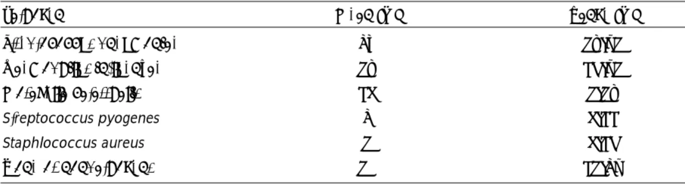
Table 1: Bacterial pathogens isolated from middle ear fluids in children with AOM
Pathogen Mean (%) Range (%)
Streptococcus pneumoniae 39 27-52 Haemophilus influenzae 27 16-52 Moraxella catarrhalis 10 2-27 Streptococcus pyogenes 3 0-11 Staphlococcus aureus 2 0-16 None or nonpathogens 28 12-35
Although the incubation period is variable, otitis media is usually develops 4 to 7 days after upper respiratory tract infection (1).
Diagnosis:
The tympanic membrane can not be adequately seen while partially occluded by cerumen. (7-10) Cerumen removal by curette is essential and should be regularly used. If the wax is dry or deep in the auditory canal then cerumenolytics and/or warm water irrigation may be necessary.
Pneumatic otoscopy has been advocated as an important adjunct to assist in diagnostic accuracy of AOM (9, 10) yet most physicians find it inconvenient or remain unconvinced. Four characteristics of the TM should be evaluated and described in every examination (position, mobility, colour, degree of translucency). The normal TM is in the neutral position (neither retracted nor bulging), pearly gray, translucent and responds briskly to positive and negative pressure, indicating an air filled space. The abnormal TM may be retracted or bulging, and immobile or poorly mobile to positive and/or negative air pressure. The colour of the eardrum is of minor importance although patients with AOM more often have a red TM. The key differentiating features of AOM compared to OME on physical exam relate to TM position. In AOM the TM almost always is bulging and in OME it is usually retracted or, occasionally, it is in neutral position. The TM is thickened in both AOM and OME, thereby reducing visibility through it. Sometimes a yellow or grayish middle ear effusion can be seen behind the TM in either condition.
Tympanometry and acoustic reflectometry each have attributes which make them of value in providing information about the possible presence of a middle ear effusion (1, 3, 8-10). The sensitivity, specificity, positive predictive value and negative predictive value of the two instruments has been assessed in comparison with pneumatic otoscopy, audiometry and tympanocentesis findings (11, 12). As a result
both of them have some limitations. Acoustic reflectometry has the advantage of not requiring a seal within the canal which improves its usefulness in the crying child because a reading can be obtained when a child stops crying to take a breath. Tympanometry provides additional information about actual pressures within the middle ear space (13).
Myringotomy is not necessary rountinely for the patient with intense pain and imminent rupture of the membarane it provides dramatic relief. (14-17). Selective use of tympanosynthesis may improve diagnostic accuracy because it validates or refutes the physicians’ impression after visual examination. Certainly proper restraint of the patient and excellent visualisation of the TM are essential; mild sedation may also be helpful in some cases. Tympanosynthesis should be performed and it is beneficial (14).
Radiography:
Roentgenographic evaluation of the temporal bone is indicated when complications or sequelae of otitis media are suspected or present. Plain radiographs are of limited value in the diagnosis of mastoiditis or cholesteatoma; computed tomography and magnetic resonance imaging are more precise and should be obtained if a suppurative intratemporal or intracranial complication is suspected (3).
Complications and sequelae:
Suppurative complications of acute infection of the middle ear are now uncommon in areas where children have access to medical care. However contiguous spread of infection may be responsible for mastoiditis, petrositis, labyrinthitis, brain abscess and meningitis. Impairment of hearing associated with fluid in the middle ear. Sensorineural hearing loss is uncommonly associated with otitis media (1, 3).
The most common etiologic factors of serous otitis media are untreated or deficiently treated AOM. Serous effusions of the middle are believed
to originate as a phsical phenomenon secondary to blockage of the eustachian tube and negative pressure in the middle ear cavity. The inciting cause of the obstructing edema or lymphoid hyperplasia may be nasopharyngeal inflamation, allergy or barotrauma. The increasing recognition of serous otitis media in the antibiotic era suggests that some cases represent incompletely resolved bacterial infections of the middle ear, but proof of this hypothesis is lacking (1-4).
Treatment:
Antimicrobial therapy is one of the cornerstones in the management of AOM but some studies have suggested that its routine use is not indicated (17-19). As the most cases of AOM resolve spontaneously (20), antimicrobial therapy is not always necessary. Nonetheless, complications such as mastoiditis may be developed, so that (16); routine use of antibiotics were recommended. It is probably not possible to determine a priori which cases of AOM will result in suppurative complications, it is likewise not possible to determine which cases require antimicrobial therapy and which will resolve spontaneously. Therefore, it appears prudent to consider all cases of AOM candidates for antimicrobial therapy in order to minimize the likelihood of complications. Some authors recommend watchful waiting for 48 to 72 hours before initiating antibiotic therapy (16). This approach may be feasible in children over two years of age if good follow-up can be assured; therefore, decisions about whether to withhold antibiotics therapy initially must be made on a patient-by-patient basis.
The most frequent etiological bacterial agents are S. pneumoniae, H. influenzae, M.catarrhalis, group A streptococcus and S. aureus. Viruses continue to cause a substantial minority of cases (1, 3-5), and antibiotic therapy would not be expected to affect the outcome. With the increasing prevalence of beta-lactamase-producing (penicillin-resistant) strains of H
influenzae and M catarrhalis, alarms have been
sounded about the wisdom of routinely using aminopenicillins (such as amoxicillin) as the standard first-line antimicrobial for uncomplicated AOM. Despite theoretical concerns about the diminishing usefulness of amoxicillin, it continues to be as effective as any other oral antimicrobial agent for childhood AOM. Most comparative trials of antimicrobial therapy in AOM have failed to demonstrate a difference in effectiveness between amoxicillin and any other agent. Their use may be associated with relatively high rates of side effects and may increase the pressure for selection of multiple antibiotic-resistant strains of bacteria (Table 2). Therefore, because of its excellent track record’ (for infections due to penicillin-susceptible and-resistant bacteria), low cost, safety and acceptability to patients, amoxicillin remains the drug of choice for uncomplicated AOM.
What is a reasonable expectation for response to therapy?
One can reasonably expect that the symptoms of AOM (fever, irritability and ear pain) will resolve within 72 hr. of initiation of antimicrobial therapy. If symptoms persist after this length of therapy, the child should be re-evaluated to determine if the infection is persisting or has evolved into one of the suppurative complications. If the patient has complied with the prescribed therapy and the symptoms, such as pain and fever, have persisted, a change in antimicrobial regimen is appropriate. The different agents from which to choose are listed in Table 2 (3). Whereas the symptoms of AOM listed above should resolve promptly with antimicrobial therapy, the middle ear effusion may persist for up to three months despite bacteriological cure (15). Therefore, persistence of middle ear fluid after a full course of antibiotic therapy of AOM is not an indication for therapy continuation or for institution of treatment with different drugs.
The role of parenteral therapy in AOM With the advent of extended spectrum
cephalosporins with prolonged half-life (eg, ceftriaxone), the option of parenteral therapy with a single dose has become feasible (21). Today there is, at present, little published evidence that parenteral therapy provides any advantage to the conventional 10-day oral therapy. Furthermore, the use of such broad-spectrum agents may hasten the emergence of antibiotic-resistant organisms. Except in extraordinary situations, parenteral therapy should not be employed for simple uncomplicated childhood AOM. If a child appears to be too ill instend of the standard oral therapy, a diagnosis other than AOM should be entertained and admission to hospital should be considered.
What is the optimal antimicrobial management of treatment failures?
As stated above, the symptoms of AOM (fever, irritability and otalgia) should resolve within 72 hr. of initiating antimicrobial therapy. Failure of symptomatic response to appropriate therapy (with evidence of compliance) constitutes a
treatment failure. The optimal management of such patients is controversial and various approaches have been advocated (22). A tympanosynthesis should be considered for both therapeutic (relief of pressure and pain) and for diagnostic (recovery of the etiologic agent) purposes. If a tympanosynthesis is not practical, consideration should be given to adding amoxicillin-clavulanate or selecting one of the alternative agents from Table 2. If a tympanosynthesis is performed, the antibiotic choice should ultimately be guided by the etiological agent and antimicrobial susceptibility (1-5).
Supportive therapy:
Supportive therapy, including analgesics, antipyretics and local heat, is usually helpful. An oral decongestant may relieve some nasal congestion and antihistamines may help patients with known or suspected nasal allergy. Antihistamines, decongests and corticosteroids are not effective in the actual treatment of AOM, however (1).
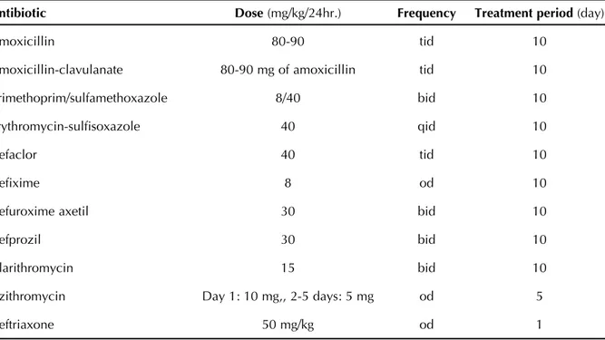
Table 2: Antibiotics for therapy of child with acute otitis media (3)
Antibiotic Dose (mg/kg/24hr.) Frequency Treatment period (day)
Amoxicillin 80-90 tid 10
Amoxicillin-clavulanate 80-90 mg of amoxicillin tid 10
Trimethoprim/sulfamethoxazole 8/40 bid 10
Erythromycin-sulfisoxazole 40 qid 10
Cefaclor 40 tid 10
Cefixime 8 od 10
Cefuroxime axetil 30 bid 10
Cefprozil 30 bid 10
Clarithromycin 15 bid 10
Azithromycin Day 1: 10 mg,, 2-5 days: 5 mg od 5
1. Kenna M. Otitis media and its complications. In: Behrman RE, Kliegman RM and Jenson HB, eds. Nelson Textbook of Pediatris. 16th ed. Philadelphia, W.B. Saunders, 2000: 1950-9 2. FadenH. The microbiologic and immunologic
basis for recurrent otitis media in children. Eur J Pediatr 2001; 160(7): 407-13
3. Klein JO. Otitis media. In: Katz SL, Gershon AA and Hotez PJ. eds. Krugman’s Infectiious Diseases of Chikdren. 10th ed. Missouri, Mosby 1997: 302-10
4. Demireller A. Acute otistis media.In: Topcu AW, Söyletir G and Doğan M, eds. Infeksiyon Hastalıkları 1st ed. Ankara: Nobel Tıp Kitapevi 1996: 345-6
5. Kilpi T, Herva E, Kaijalainen T, Syrjanen R, Takala AK. Bacteriology of acute otitis media in a cohort of Finnish children followed for the first two years of life. Pediatr Infect Dis J 2001 Jul; 20(7): 654-62
6. McGahren ED and Wilson WG. In: McGahren
ED and Wilson WG, eds. Otitis media. Pediatrics Recall. 1st ed. Baltimore, Williams & Wilkins, 1997: 389-94
7. Barriga F, Schwartz RH, Hayden GF. Adequate
illumination for otoscopy: variations due to power source, bulb, and head and speculum design. Am J Dis Child 1986; 140: 1237-1240.
8. Schwartz RH, Stool SE, Rodriguez WJ,
Grundfast KM. Acute otitis media: towards a more precise definition. Clin Pediatr (Phila) 1981; 20: 549-554.
9. Kaleida PH, Stool SE. Assessment of
otoscopists’ accuracy regarding middle-ear effusion. Am J Dis Child 1992; 146: 433-435.
10. Cavanaugh RM Jr. Pediatrician and the
pneumatic otoscope: are we playing it by ear? Pediatrics 1989; 84: 362-364.
11. Barnett ED, Klein JO, Hawkins KA, Cabral HJ, Kenna M, Healy G. Comparison of spectral gradient acoustic reflectometry and other diagnostic techniques for detection of middle ear effusion in children with middle ear disease. Pediatr Infect Dis J 1998; 17: 556-559.
12. Block SL, Mandel E, McLinn S, Pichichero ME, Bernstein S, Kimball S, Kozikowski J. Spectral gradient acoustic reflectometry for the detection of middle ear effusion by pediatricians and parents. Pediatr Infect Dis J 1998; 17: 560-564.
13. Brookhouser PE. Use of tympanometry in office practice for diagnosis of otitis media. Pediatr Infect Dis J 1998; 17: 544-551.
14. Pichichero ME. Changing the treatment
paradigm for acute otitis media in children. JAMA 1998; 279: 1748-1750.
15. Klein JO. Otitis media. Clin Infect Dis 1994; 19: 823-833.
16. Berman S. Otitis media in children. N Engl J Med 1995; 332: 1560-1565.
17. Van Buchem FL, Dunk JH, van’t Hof MA. Therapy of acute otitis media: Myringotomy, antibiotics, or neither? A double-blind study in children. Lancet 1981; ii: 883-7
18. Mygind N, Meistrup-Larsen KI, Thomsen J, et al. Penicillin in acute otitis media: A double-blind placebo-controlled trial. Clin Otolaryngol 1981; 6: 5-13
19. Van Buchem FL, Peeters MF, van’t Hof MA. Acute otitis media: A new treatment strategy. Br Med J 1985; 290: 1033-7
20. Rosenfeld RM, Vertrees JE, Carr J, et al. Clinical efficacy of antimicrobial drugs for acute otitis media: Metaanalysis of 5400 children from thirty-three randomized trials. J Pediatr 1994; 124: 355-67.
21. Heikkinen T, Saeed KA, McCormick DP,
Baldwin C, Reisner BS, Chonmaitree T. A single intramuscular dose of ceftriaxone changes nasopharyngeal bacterial flora in children with acute otitis media. Acta Paediatr 2000 89(11): 1316-21
22. Klein JO, Bluestone CD. Management of otitis media in the era of managed care. Adv Pediatr Infect Dis 1996; 12: 351-86