© AME Publishing Company. All rights reserved. www.amepc.org/qims Quant Imaging Med Surg 2015;5(4):626-627 A 25-year-old male patient with thalassemia major
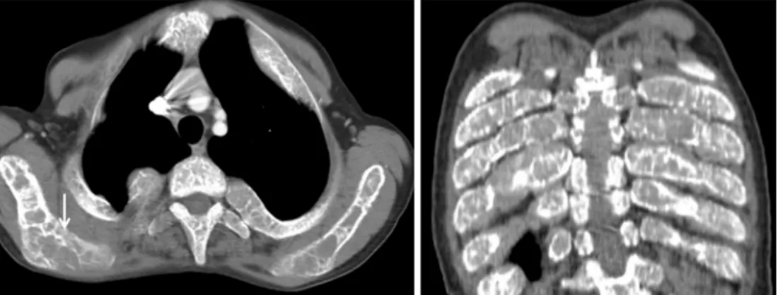
presented at the polyclinic for a routine examination. On the pulmonary radiograph, mass lesions were observed superimposed on the parenchyma of both lungs (Figure 1). In the bone structures, scattered expansile and lytic areas were determined. On the CT examination applied because of this, expansile lytic expansions were determined in all the ribs bilaterally, in the sternum, in both scapula and in the vertebrae (Figure 2). There were scattered losses of height in the vertebrae corpuses. All the bone changes were
evaluated as secondary to excessive erythropoiesis. On the thoracic CT, mass lesions were observed associated with foci of extramedullary hematopoiesis in the paravertebral areas (Figure 3). After informing the patient of the condition, rather than the routine follow-up tests, blood transfusion was administered.
Skeletal changes in patients with untreated thalassemia originate from ineffective erythropoiesis and result in expansion of the bone marrow. The whole skeletal system may be affected. In addition, ineffective erythropoiesis can Images of the Issue
Changes in the skeletal system and extramedullary hematopoiesis
in a patient with thalassemia
Nesat Cullu1, Mehmet Deveer1, Onder Yeniceri2, Serdar Kalemci3
1Department of Radiology, Mugla Sitki Kocman University, Faculty of Medicine, Mugla, Turkey; 2Department of Radiology, Yücelen Hospital, Mugla, Turkey; 3Department of Pulmonology, Mugla Sitki Kocman University, Faculty of Medicine, Mugla, Turkey
Correspondence to: Assist. Prof. Dr. Nesat Cullu. Mugla Sitki Kocman School of Medicine, Department of Radiology, Central Campus, 48000, Mugla, Turkey. Email: [email protected].
Submitted Jun 30, 2014. Accepted for publication Jul 03, 2014. doi: 10.3978/j.issn.2223-4292.2014.07.12
View this article at: http://dx.doi.org/10.3978/j.issn.2223-4292.2014.07.12
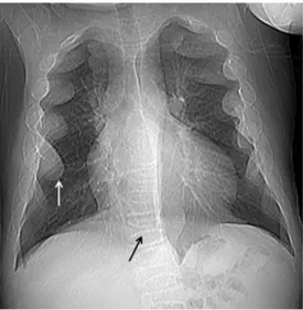
Figure 1 Expansion in the ribs forming the suspected mass on both sides (white arrow) and scattered losses of height in the vertebrae (black arrow) observed on the posterior anterior (PA) pulmonary radiograph.
627 Quantitative Imaging in Medicine and Surgery, Vol 5, No 4 August 2015
© AME Publishing Company. All rights reserved. www.amepc.org/qims Quant Imaging Med Surg 2015;5(4):626-627 develop in paravertebral areas forming the appearance of a
mass in extramedullary areas (1-3).
Acknowledgements
None.
Footnote
Conflicts of Interest: The authors have no conflicts of interest to declare.
References
1. Tyler PA, Madani G, Chaudhuri R, Wilson LF, Dick EA. The radiological appearances of thalassaemia. Clin Radiol 2006;61:40-52.
2. Tunaci M, Tunaci A, Engin G, Ozkorkmaz B, Dinçol G, Acunaş G, Acunaş B. Imaging features of thalassemia. Eur Radiol 1999;9:1804-9.
3. Chan YL, Li CK, Pang LM, Chik KW. Desferrioxamine-induced long bone changes in thalassaemic patients - radiographic features, prevalence and relations with growth. Clin Radiol 2000;55:610-4.
Cite this article as: Cullu N, Deveer M, Yeniceri O, Kalemci S. Changes in the skeletal system and extramedullary hematopoiesis in a patient with thalassemia. Quant Imaging Med Surg 2015;5(4):626-627. doi: 10.3978/j.issn.2223-4292.2014.07.12 Figure 3 Extramedullary hematopoiesis areas observed in the left paravertebral region on axial thorax CT image.