DOI: 10.5455/annalsmedres.2019.05.256 2019;26(8):1588-93
Non-invasive prediction of fibrosis in chronic hepatitis B
Mustafa Salih Akin1, Abdullah Ozgur Yeniova2, Osman Demir3
1Medipol University, Faculty of Medicine Department of Gastroenterology, Istanbul Turkey
2Tokat Gaziosmanpasa University, Faculty of Medicine Department of Gastroenterology, Tokat, Turkey 3Tokat Gaziosmanpaşa University, Faculty of Medicine, Department of Bioistatistics, Tokat, Turkey Copyright © 2019 by authors and Annals of Medical Research Publishing Inc.
Abstract
Aim: To date, the main diagnostic tool for assessing the liver fibrosis stage has been liver biopsy. Despite the advantages in patient
prognosis prediction and disease management, the application of liver biopsy is hampered by its limitations. The limitations of liver biopsy have led to a need for the development of a non- invasive scoring system that is simple to use, reliable, and cheap. The aim of this study was to evaluate the accuracy of APRI (AST to Platelet Ratio Index) and FIB-4 (Fibrosis-4) scores in the prediction of liver fibrosis in Chronic Hepatitis B patients.
Material and Methods: A retrospective assessment was made of the electronic medical records to identify patients who underwent
liver biopsy for evaluation of fibrosis related to CHB (Chronic Hepatitis B). A total of 352 patients were included in the study. Laboratory parameters that were measured within 7 days of the liver biopsy day were considered suitable for the calculation of the formulae. The APRI and FIB-4 scores was calculated. Liver biopsy reports, which were eligible for inclusion in the study, were assessed retrospectively. The fibrosis stage and histological acitvity index were used as previously described by Ishak. Analyses were performed using SPSS 22 (IBM SPSS Statistics 22, SPSS inc., an IBM Co., Somers, NY).
Results: APRI was well correlated with fibrosis and HAI score (r=0.280, p<0.05; r=0.22, p<0.05) respectively. The FIB-4 correlation
between fibrosis and HAI was significant (r=0.433, p<0.05; r=0.34, p<0.05) respectively.
Conclusion: Inexpensive, non-invasive, and reliable fibrosis assessement models for CHC should be applied in clinical practice.
Keywords: Chronic hepatitis B; liver fibrosis; APRI, FIB-4.
Received: 10.05.2019 Accepted: 02.07.2019 Available online: 28.08.2019
Corresponding Author: Mustafa Salih Akin, Medipol University, Faculty of Medicine Department of Gastroenterology, Istanbul Turkey E-mail: [email protected]
INTRODUCTION
Chronic Hepatits B (CHB) is one of the major public health concern that affects approximately two billion people, 350 million of whom are chronically infected worldwide (1). CHB related mortality and morbidity depends on the liver fibrosis (2). Cirrhosis, the last stage of the liver damage, is major cause of hepatocellular carcinoma (HCC) also, cirrhosis related complications increase risk of mortality. Thus determining the liver fibrosis will not also guide the treatment but also predict the patient’s prognosis.
Prevention from development of fibrosis will allow the chronic hepatitis B patient to live normal lifespan by decreasing the rate of HCC and cirrhosis complications. Antiviral therapy have shown to be effective against the disease progression (3). Main question is when to initiate the antiviral therapy? Because the natural course of the chronic hepatitis B is variable and patient’s fibrosis stage can pass through the forward stage or backward stage,
disease progression can be defined as a dynamic state (4).
Until today, the main diagnostic tool for assessing the liver fibrosis stage is liver biopsy. Many staging systems have been using for scoring the liver fibrosis. Knodell established a scoring system for description and classification necroinflammation and fibrosis stages of liver (5). Other comparable systems based on definition of necroinflammation and fibrosis stage (Metavir, Batts and Ludwig, Scheuer) followed this system (6-8). Modified HAI system combined firstly described histology activity index established by Knodell and Ishak’s scoring system which is commonly using system with METAVIR in most of the countries (9).
Despite its advantages for the patient’s prognosis prediction and disease management, performing liver biopsy is hampered by its limitations. Rare but sometimes life threatining complications and invasiveness of the
procedure that cause patient discomfort are the main limitations of the liver biopsy. Sampling errors may not allow the appropriate assessment of the fibrosis because of the heterogenity of liver paranchyme in the mean of fibrosis. Also inter observer variability may also cause confusion (10).
Limitations of liver biopsy lead the necessarity for development of non-invasive scoring systems that must be easily used, reliable, and cheap. Methods based on routine laboratory tests, ultrasound based methods, laboratory tests that are not routinely available have been proposed in the last decades. Fibroscan and acoustic radiation force impulse (ARFI) which are ultrasound-based methods have been tried in CHB amd chronic hepatitis C (CHC) patients. There are promising results but cost and operator dependence of the methods limits their ability (11,12).
Routinely measured biochemical and complete blood count indices considered as possible alternatives for fibrosis definition as they are easy to use, inexpensive and reliable. Prompt and accurate diagnosis of fibrosis may be achieved by combination of these indices. Aspartate aminotransferase (AST) to alanine aminotransferase ratio (AAR), APRI and Fibrosis 4 score (FIB-4) which are scoring systems using ALT, AST, age and platalet count were established in the past decades (13). CHC results were promising although the data about CHB is insufficient. Red cell distribution width to platelet ratio another system using complete blood count indices determined as effective for fibrosis and cirrhosis prediction in CHB patients (14). Fibrotest are more complex systems that requires laboratory tests that cannot be found easily on daily clinical practice thus this serum marker panel cannot find a place in non-invasive assessment of liver fibrosis (15).
The aim of this study is to evaluate the accuracy of APRI and FIB-4 scores in prediction of liver fibrosis in CHB patients.
MATERIAL and METHODS
Electronic medical records were assessed retrospectively. Patients who underwent liver biopsy for assessment of fibrosis related to CHB between 2010 and 2014 were enrolled into the study. Written informed consent was obtained from all patients. Ethics Committee Approval received for this study. Patients of whose surface B antigen (Hbs antigen) were positive for over 6 months were accepted as CHB. Patients with hepatitis C, D and HIV co-infection, alcohol abuse over 20 gr/day, suffering from other liver diseases (autoimmun hepatitis, metabolic liver diseases, toxic hepatitis), of whose laboratory indices for calculating APRI and FIB-4 scores were not available were excluded from the study.
APRI score was calculated as AST (U/L)/AST (Upper Limit Normal [ULN])/platelet count 109/L. FIB-4 scores were calculated as age (years) X AST/ platelet count 109/Lx ALT (U/L) ½ as previosly described. Laboratory parameters
that were measured in seven days of liver biopsy day were considered as available for calculating formulas. When records revealed that there were multiple laboratory parameters, closest time to the biopsy was taken into account. Age was defined as the age in which the patient were underwent liver biopsy. All laboratory parameters were measured according to the routinely using methods. Liver biopsy reports, which were eligible for enrolling into the study, were assessed retrospectively. Biopsies that were described detailed and were determined as enough for iagnosis by pathologist were enrolled into the statistical analysis. Fibrosis stage and histological acitvity index were used as described by Ishak previously (9). Seven levels of fibrosis have been described; F0 (no fibrosis), F1 (fibrous expansion of some portal areas), F2 (fibrous expansion of most portal areas), F3 (portal to portal bridging), F4 (marked birdging), F5 (markes bridging with occasional nodules, incomplete cirrhosis), F6 (cirrhosis). Cutoff fibrosis stage for initiate treatment was F2 so patients between F2 and F6 was described as significant fibrosis (16). Also histologic acitivity index (HAI) over six was considered as significant fibrosis. Because of the retrospective characteristic of study and potential heterogenity of the data, to identify best cutoff value and test its adequate power, we obtained optimal values in a training group then validate these parameters in an independent group and entire cohort. Patients were randomly divided into training set and validation set. Obtained parameters of training set then applied into validation set and entire cohort. The ratio of the training set and validation set was 2:1.
Analyses were performed using SPSS 22 (IBM SPSS Statistics 22, SPSS inc., an IBM Co., Somers, NY). Descriptive analyses were performed to provide information on general characteristics of the study population. Distribution was analyzed by Kolmogrov-Smirnov and histograms. Continous variables were given as mean ± standard deviation (SD) or median (25th- 75th percentiles) where appropriate. Categorical variables were given as ratios. Two independent sample t test or Mann Whitney U test were used to compare the continuous data between training set and validation set. Pearson correlation was the preferred method for correlations. Receiver operating characteristic (ROC) curves were applied to determine the power of APRI and FIB-4 in predicting significant fibrosis in each group (training set, validation set and entire cohort). Optimal cutoff value was defined as maximum sum of the (sensivity+[1+1-specifity]). Cutoff value obtained from training group in each fibrosis stage and HAI classification variable were applied for validation and entire cohort. Sensitivity, specificity, and accuracy of the test as well as the positive and negative predictive values were calculated for Toradol - change in NIF. Comparison of ROC curves of APRI and FIB-4 for each group was analyzed by method of DeLong test. A p-value <0.05 was considered significant.
RESULTS
A total of 352 patients were enrolled into the study. 139 (39.5%) of them were female while 213 (60.5%) of them were male. Median age was 45 (35-55). Minimum age was 16 while maximum age was 73.283 (80%) of the patients were HbeAg negative while 69 (19.6%) of them were HbeAg positive. Distribution of the patients according to the fibrosis stages were F0 (34 patients, 9.7%), F1 (80 patients, 22.7%) F2 (80 patients, 22.7%), F3 (73 patients 20.7%), F4 (35 patients 9.9%), F5 (26 patients, 7.4%), F6 (24 patients, 6.8%). 238 of the patients’ (67.6%) fibrosis stage were over 2. 243 (69%) of the patients’ HAI score were over 6.
Demographic and the clinical characteristics of the training group and validation group can be seen in Table 1. There was no significant differences between groups. APRI was well correlated with fibrosis and HAI score (r=0.280, p<0.05; r=0.22, p<0.05) respectively. Also FIB-4 correlation between fibrosis and HAI is significant (r=0.433, p<0.05; r=0.34, p<0.05) respectively.
Area under the receiving operating characteristics (AUROC) curve of APRI for predicting significant fibrosis in the mean of fibrosis variable was 0.825 (95% CI, 0.769-0.870) in training group. AUROC of FIB-4 was 0.750 (% 95 CI, 0.690-0.804). When AUROC curves of APRI and
FIB-4 were compared, it was demonstrated that APRI had significantly higher AUROC than FIB-4 (p=0.017) (Figure1). Optimal cutoff values of APRI was 0.55 (sensivity 63.4%, specifity 93.8%). Optimal cutoff value of FIB-4 was 1.28 (sensivity 58.8%, specifity 82.7%) (Table 2).
In the validation group, AUROC of APRI was 0.856 (95% CI, 0.780-0.914). AUROC of FIB-4 was 0.725 (95% CI, 0.635-0.803). APRI had significant higher AUROC than FIB-4 (p=0.0066) (Figure1). Sensivity, specivity, PPV, NPV and accuracy values can be seen in Table 2.
In the entire cohort AUROC of APRI and FIB-4 were 0.835 (95% CI, 0.793-0.873) and 0.742 (95% CI, 0.693-0.787) respecitvely. APRI’s AUROC curve was significantly higher than FIB-4 (p=0.0003) (Figure 1). Sensivity, specivity, PPV, NPV and accuracy values can be seen in Table 2.
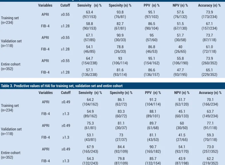
For predicting significant fibrosis determined as patients’ with HAI score over six AUROC’s and predictive values were similar with fibrosis variable. In the training set AUROC of APRI was 0.793 (95% CI, 0.735-0.843). AUROC of FIB-4 was 0.733 (95% CI, 0.672-0.789). APRI can predict significant fibrosis more accurate than FIB-4 (p=0.075) (Figure 2) Optimal cutoff values obtained from training set was 0.49 for APRI and 1.4 for FIB-4. Sensivity, specivity, PPV, NPV and accuracy values can be seen for HAI variable in Table 3.
Table 1. Demographic characteristics of training and validation set
Variables Group p Training Validation (n=234) (n=118) Age (years) 44.32±13.51 44.46±13.46 0.931 ALT (U/L) 72.2±79.82 91.3±98.83 0.070 AST (U/L) 51.91±56.76 60.05±53.62 0.197 Platelet/mm3 196.99±56.1 204.6±59.42 0.239 APRI 0.84±1.74 0.87±0.88 0.855 FIB-4 1.65±1.9 1.59±1.24 0.774 HBsAg 2315.87±1534.17 2233.33±1548.09 0.635 HBVDNA (copies/ml) 169250 [14240-4579000] 591100 [18880-11650000] 0.138 HAI 8.25±4.27 7.86±3.71 0.397 Fibrosis 2.5±1.73 2.44±1.52 0.752 Gender Female 90 (38.5) 49 (41.5) 0.579 Male 144 (61.5) 69 (58.5) Fibrosisgorup ≥F2 153 (65.4) 85 (72) 0.208 <F2 81 (34.6) 33 (28) HBeAg Negative 192 (82.1) 91 (77.1) 0.271 Positive 42 (17.9) 27 (22.9)
HAI GROUP ≥HAI 6 162 (69.2) 81 (68.6) 0.911 <HAI 72 (30.8) 37 (31.4)
Table 2. Predictive values of fibrosis for training set, validation set and entire cohort
Variables Cutoff Sensivity (n) % Specivity (n) % PPV (n) % NPV (n) % Accuracy (n) %
Training set (n=234) APRI ≥0.55 (97/153)63.4 (76/81)93.8 (97/102)95.1 (76/132)57.6 (173/234)73.9 FIB-4 ≥1.28 (90/153)58.8 (67/81)82.7 (90/104)86.5 (67/130)51.5 (157/234)67.1 Validation set (n=118) APRI ≥0.55 (57/85)67.1 (30/33)90.9 (57/60)95 (30/58)51.7 (87/118)73.7 FIB-4 ≥1.28 (46/85)54.1 (26/33)78.8 (46/53)86.8 (26/65)40 (72/118)61.0 Entire cohort (n=352) APRI ≥0.55 (154/238)64.7 (106/114)93 (154/162)95.1 (106/190)55.8 (260/352)73.9 FIB-4 ≥1.28 (136/238)57.1 (93/114)81.6 (136/157)86.6 (93/195)47.7 (229/352)65.1
Table 3. Predictive values of HAI for training set, validation set and entire cohort
Variables Cutoff Sensivity (n) % Specivity (n) % PPV (n) % NPV (n) % Accuracy (n) %
Training set (n=234) APRI ≥0.49 (104/162)64.2 (62/72)86.1 (104/114)91.2 (62/120)51.7 (166/234)70.1 FIB-4 ≥1.3 (89/162)54.9 (60/72)83.3 (89/101)88.1 (60/133)45.1 (149/234)63.7 Validation set (n=118) APRI ≥0.49 (61/81)75.3 (30/37)81.1 (61/68)89.7 (30/50)60 (91/118)77.1 FIB-4 ≥1.3 (43/81)53.1 (27/37)73 (43/53)81.1 (27/65)41.5 (70/118)59.3 Entire cohort (n=352) APRI ≥0.49 (165/243)67.9 (92/109)84.4 (165/182)90.7 (92/170)54.1 (257/352)73.0 FIB-4 ≥1.3 (132/243)54.3 (87/109)79.8 (132/154)85.7 (87/198)43.9 (219/352)62.2
In the validation group AUROC of APRI was 0.821 (95% CI, 0.740-0.886). AUROC of FIB-4 was 0.680 (95% CI, 0.587-0.762).
When AUROC curves were compared with similar results obtained. APRI can significantly predict more accurate than FIB-4 (p=0.0013) (Figure 2). Sensivity, specivity, PPV,
NPV and accuracy values of validaiton set can be seen for HAI variable in Table 3.
In the entire group, AUROC of APRI and FIB-4 were 0.806 (95% CI, 0.760-0.846) and 0.715 (95% CI, 0.665-0.762) respectively. APRI’s prediction power is significantly higher than FIB-4 again (p=0.0005) (Figure 2).
Figure 1. Comparison of ROC curves of APRI and FIB-4 for predicting fibrosis stage. a) training set b) validaiton set c) entire set
Figure 2. Comparison of ROC curves of APRI and FIB-4 for predicting HAI over 6. a) training set b) validaiton set c) entire set
DISCUSSION
In this study, we aimed to identify best cutoff values for predicting significant fibrosis in CHB patients. Developing noninvasive predictive models become popular in the last decade because of the limitations of liver biopsy, which is considered as gold standard for fibrosis assessment. Until today, no current approach have been standardized and validated for this aim. First predictive models were developed for CHC patients. APRI and FIB-4 score systems can be considered as practical because they can be calculated from routine laboratoty parameters.
For minimizing the bias and variance; we randomized the entire cohort to training and validation set (17). There were no significant differences between training and validation group in the mean of demographic characteristics. In addition, ratios of patients with or without siginificant fibrosis, ratio of HbeAg positive and negative patients and other caharcteristics related to chronic hepatits B did not differ between two groups. Cutoff values obtained from training group were applied to validation set and entire cohort. In all three groups, the results were similar. For predicting fibrosis score ≥ 2 AUROCs of APRI were over 0.8. AUROCs of FIB-4 were between 0.7 and 0.8. In all three group APRI predict fibrosis more accurate than FIB-4. When HAI ≥6 was considered as significant fibrosis, all AUROCs of APRI were over 8 except one. AUROC of APRI in the training set was 0.793 but also this is near to 0.8. AUROCs of FIB-4 were bewteen 0.7 and 0.8 except AUROC of FIB-4 in validation set which was found 0.680. Again all APRI scores were significantly higher than FIB-4 scores in training, validation set and entire cohort.
Sensivity, specivity, PPV and PPV values were found consistent in each group again. Sensivity values were approximately between 55%-65%. Specifivity values were approximately between 85%- 95%. NPV values and PPV values were between 40-60% and 85-95% respectively. Our findings were similar with previous studies. One study that aimed to find AUROCs for APRI and FIB-4 and identify their diagnositc values for predicting fibrosis used also similar method and enrolled patients into training and validation set (18). AUROCs of APRI was 0.693, 0.728 and 0.704 in the training set, validation set and entire cohort
respectively. AUROCs of FIB-4 was 0.766 and 0.775 in the training and validation set respectively.
Another study that used training and validation set reiterated these findings. AUROC of APRI and FIB-4 for predicting METAVIR score 2 and over were 0.81 and 0.86 respectively in the training set. In the validaiton set, the AUROC of FIB-4 was lower than the training set which was 0.71 (19).
Two studies did not used this validation method and assessed ROC for entire cohort. AUROC of APRI and FIB-4 were 0.662 and 0.687 respectively (20). Other study found that AUROC of APRI and FIB-4 were 0.670 and 0.701 respectively (21). Sensivity and specivity values changes according to the cutoff values. None of these two studies compared APRI with FIB-4.
One meta-analysis that aimed to determine the diagnostic value of FIB-4 enrolled 12 studies. Hierarchical summary receiver operating curve (AUHRSOC) which is a model for calculating summary of curves was found as 0.78 (95% CI=0.74-0.81) which can be considered as similar but also a little bit higher than our study’s result (22). Another meta analysis that assessed APRI and FIB-4 together for predicting significant fibrosis found that summary AUROC of APRI and FIB-4 were 0.7407 and 0.7844 respectively (23). In this meta-analysis, cutoff point 0.5 for APRI which is near to our cutoff point 0.55; the summary of sensitivities and specificities were 70.0% (range, 35.0%-97.0%) and 60.0% (range, 34.0%-86.7%). For FIB-4 at cutoff value of 1.45 (also which can be considered as near to cutoff value obtained from our study 1.28); the summary sensitivities and specificities were 70.0% (range, 35.0%-97.0%) and 60.0% (range, 34.0%-86.7%).
Most of the studies mentioned above also applied ROC for predicting cirrhosis but also none of them reach to the values 0.9 and over. Although histopathologic assessment of liver fibrosis scores were comparable, in this study; the aim is to predict Ishak score ≥ 2 and HAI ≥ 6 because this cutoff point is described as histologic indication of treatment and our country’s national health insurance have this score as an indication for treatment payment (16). Most of the studies were designed to predict the METAVIR score ≥ 2. Main difference of our study can be stated as
APRI is more valuable than FIB-4 for predicting fibrosis. This difference may be explained by methodological differences between other studies mentioned above. Also age which is the main difference between two scoring system may explain this controversial result of our study but two studies that compared APRI and FIB-4 score. Our study reported the same median or mean age which was 45 (18,19). But one study that applied meta analysis for comparing efficacy between APRI and FIB-4 found that there is no statistically significant difference between APRI and FIB-4 (Z score=1.59, p=0.06) (23).
There is no study that aimed to predict HAI ≥ 6 in the literature. Our study revealed the same results for Ishak ≥ 2 and HAI ≥ 6.
This study has limitations. First retrospective characteristics of study can be a limitation. We collect the data of liver fibrosis assessment from the initial reports. İn addition, appropriate reportings according to the guidelines were considered for enrolling into the study, this situation could be determined as weakness of the study as compared with prospective studies.
AUROC value 1 classified as perfect. Results between 1 and 0.9 has been interpreted as excellent, values between 0.9 and 0.8 were good. Values between 0.8 and 0.7 were fair and results less than 0.7 was poor. None of the studies mentioned above reached the excellent study but some of them can be classified as good. Our results also reiterated previous findings. Natural history of CHB is different from CHC. Fibrosis development in CHC can be defined as slowly progressive and stable characteristic but CHB can show alleviating of inflammation. APRI and FIB-4 were derived scoring scores from CHC studies so moderate accuracy and sensivity may be explained by these natural history differences of two chronic viral hepatitis.
CONCLUSION
In conclusion; inexpensive, non-invasive, and reliable fibrosis assessement models for CHC must be applied for clinical practice.
Mustafa Salih Akin ORCID: 0000-0002-1309-6588 Abdullah Ozgur Yeniova {ORCID: 0000-0003-1681-364X Osman Demir ORCID: 0000-0002-1322-2716
Competing interests: The authors declare that they have no competing interest.
Financial Disclosure: There are no financial supports
Ethical approval: The research was approved by the Istanbul Medipol University Ethics Committee.
REFERENCES
1. Lavanchy D. Hepatitis B virus epidemiology, disease burden, treatment, and current and emerging prevention and control measures. J Viral Hepat 2004;11:97-107.
2. Lok AS, Hepatitis B: liver fibrosis and hepatocellular carcinoma. Gastroenterol Clin Biol 2009;33:911-5.
3. EASL clinical practice guidelines: Management of chronic hepatitis B virus infection. J Hepatol 2012;57:167-85. 4. Rotman Y, Brown TA, Hoofnagle JH. Evaluation of the patient
with hepatitis B. Hepatology 2009;49:22-7.
5. Knodell RG, Ishak KG, Black WC, et al. Formulation and application of a numerical scoring system for assessing histological activity in asymptomatic chronic active hepatitis. Hepatology 1981;1:431-5.
6. Batts KP, Ludwig J. Chronic hepatitis. An update on terminology and reporting. Am J Surg Pathol 1995;19:1409-17.
7. Scheuer PJ. Classification of chronic viral hepatitis: a need for reassessment. J Hepatol 1991;13:372-4.
8. Bedossa P, Poynard T. An algorithm for the grading of activity in chronic hepatitis C. The METAVIR Cooperative Study Group. Hepatology 1996;24:289-93.
9. Ishak K, Baptista A, Bianchi L, et al. Histological grading and staging of chronic hepatitis. J Hepatol 1995;22:696-9. 10. Regev A, Berho M, Jeffers LJ. et al., Sampling error and
intraobserver variation in liver biopsy in patients with chronic HCV infection. Am J Gastroenterol 2002;97:2614-8. 11. Foucher J, Chanteloup E, Vergniol J, et al. Diagnosis
of cirrhosis by transient elastography (FibroScan): a prospective study. Gut 2006;55:403-8.
12. Cassinotto C, Lapuyade B, Aït-Ali A et al. Liver fibrosis: noninvasive assessment with acoustic radiation force impulse elastography-comparison with FibroScan M and XL probes and FibroTest in patients with chronic liver disease. Radiology 2013;269:283-92.
13. Ma J, Jiang Y, Gong G. Evaluation of seven noninvasive models in staging liver fibrosis in patients with chronic hepatitis B virus infection. Eur J Gastroenterol Hepatol 2013;25:428-34.
14. Chen B, Ye B, Zhang J, et al. RDW to platelet ratio: a novel noninvasive index for predicting hepatic fibrosis and cirrhosis in chronic hepatitis B. PLoS One 2013;8:e68780. 15. Rossi E, Adams L, Prins A, et al. Validation of the FibroTest
biochemical markers score in assessing liver fibrosis in hepatitis C patients. Clin Chem 2003;49:450-4.
16. Papatheodoridis GV, Manesis EK, Manolakopoulos S, et al, Is there a meaningful serum hepatitis B virus DNA cutoff level for therapeutic decisions in hepatitis B e antigen-negative chronic hepatitis B virus infection? Hepatology 2008;48:1451-9.
17. Adams ST, Leveson SH. Clinical prediction rules. BMJ 2012;344:8312.
18. Lin CL, Liu CH, Wang CC, et al. Serum biomarkers predictive of significant fibrosis and cirrhosis in chronic hepatitis B. J Clin Gastroenterol 2015;49:705-13.
19. Li J, Gordon SC, Rupp LB, et al. The validity of serum markers for fibrosis staging in chronic hepatitis B and C. J Viral Hepat 2014;21:930-7.
20. Ucar F, Sezer S, Ginis Z, et al. APRI, the FIB-4 score, and Forn’s index have noninvasive diagnostic value for liver fibrosis in patients with chronic hepatitis B. Eur J Gastroenterol Hepatol 2013;25:1076-81.
21. Erdogan S, Dogan HO, Sezer S, et al. The diagnostic value of non-invasive tests for the evaluation of liver fibrosis in chronic hepatitis B patients. Scand J Clin Lab Invest 2013;73:300-8.
22. Wang F, Gatsonis CA. Hierarchical models for ROC curve summary measures: design and analysis of multi-reader, multi-modality studies of medical tests. Stat Med 2008;27:243-56.
23. Xiao G, Yang J, Yan L. Comparison of diagnostic accuracy of aspartate aminotransferase to platelet ratio index and fibrosis-4 index for detecting liver fibrosis in adult patients with chronic hepatitis B virus infection: a systemic review and meta-analysis. Hepatology 2015;61:292-302.