International Journal of Surgical Pathology 2016, Vol. 24(7) 607 –613
© The Author(s) 2016 Reprints and permissions: sagepub.com/journalsPermissions.nav DOI: 10.1177/1066896916653211 ijs.sagepub.com
Original Article
Introduction
Breast cancer is the second most common cause of cancer-related death.1 Therefore, the studies are being carried out to develop effective treatments targeting some molecules involved in the pathogenesis and the prognosis of breast cancer seriously. In addition, some clinical, genetic, histo-pathological, and immunohistochemical findings that might be associated with prognosis are being investigated.
One of the well-known major characteristics of malig-nant tumors is the high mitotic rate.2,3 Ki67 is a protein expressed by proliferating cells.2,4 High Ki67 index has been observed to be associated with poor prognosis in breast cancer and it has been suggested to have indepen-dent prognostic significance in some studies.2,5,6
RacGAP1, is a Rac guanosin triphosphatase (GTPase) activating protein, located at the metaphase of mitotic
spindle of the normal cell cycle and is necessary for cyto-kinesis.2,7 RacGAP1 upregulation has been demonstrated in some tumors such as hepatocellular carcinoma, ovarian cancer, and bladder cancer.2,8-10 Also, some studies have suggested that overexpression of RacGAP1 is associated with poor prognosis in breast cancer patients.2,7,11
Topoisomerase 2 alpha (TOP2a) is an important nuclear DNA-binding protein and its expression level is highest in growing cells in the G2/M phase.2,12,13 TOP2a expression
1Bozok University School of Medicine, Yozgat, Turkey
2Gazi University School of Medicine, Ankara, Turkey
3Istanbul Medipol University School of Medicine, Istanbul, Turkey
Corresponding Author:
Sevinç Şahin, Department of Pathology, Bozok University School of Medicine, 66100, Yozgat, Turkey.
Email: [email protected]
Clinicopathological Significance of
the Proliferation Markers Ki67,
RacGAP1, and Topoisomerase
2 Alpha in Breast Cancer
Sevinç Şahin, MD
1, İpek Işık Gönül, MD
2, Aslı Çakır, MD
3, Selda Seçkin, MD
1,
and Ömer Uluoğlu, MD
2Abstract
Objectives. The aims of this study are to evaluate expressions of Ki67, RacGAP1 (MgcRacGAP) and topoisomerase 2 alpha (TOP2a), the markers related with cell proliferation that have been proposed to affect the prognosis in the literature and correlate the results with clinicopathological parameters of breast cancer patients. Methods. Ki67, RacGAP1, and TOP2a antibodies were applied immunohistochemically to the tissue micrarray blocks of 457 female breast cancer patients. The results were correlated with clinical, prognostic, histopathological features, and other immunohistochemical findings (estrogen receptor [ER], progesterone receptor [PR], HER2, cytokeratin [CK]5/6, CK14, epidermal growth factor receptor [EGFR] and vimentin), statistically. Results. Ki67 expression demonstrated direct correlation with TOP2a expression, mitotic count, tumor grade, geographic necrosis, basal-like phenotype. RacGAP1 expression was directly correlated with TOP2a expression, nipple invasion, and number of metastatic lymph nodes, and it was inversely correlated with PR expression. TOP2a expression was directly correlated with vimentin and Ki67 expressions, mitotic count, tumor grade, and geographic necrosis, and nipple invasion, and negatively correlated with ER and PR expressions. Higher TOP2a and Ki67 expressions were correlated with shorter overall survival. Higher TOP2a expression and RacGAP1 positivity were directly correlated with shorter disease-free survival. Conclusion. This study showed that the overexpressions of Ki67, RacGAP1, and TOP2a affect the prognosis adversely, thus to develop target therapies against RacGAP1 and TOP2a as well as using Ki67 as a part of routine pathology practice might be beneficial in breast cancer therapy and prediction of prognosis.
Keywords
is used as an indicator of susceptibility to the anthracycline neoadjuvant therapy of breast cancer in some studies.2,14,15 TOP2a overexpression has been reported to be associated with shorter disease-free survival and overall survival in some studies, however there are controversial results in the literature on this issue.2,12,14,16
The goals of this study are to find out the expressions of Ki67, RacGAP1, and TOP2a that are thought to play a role in the breast cancer prognosis and treatment with some conflicting data in the literature by immunohistochemistry, and investigate the association with the prognostic and his-topathological features, and other immunohistochemical antibodies (estrogen receptor [ER], progesterone receptor [PR], HER2, cytokeratin [CK]5/6, CK14, epidermal growth factor receptor [EGFR], and vimentin) performed previously for a thesis.
Materials and Methods
After obtaining informed consents and ethic committee approval, 457 cases of breast cancer diagnosed at Department of Pathology, Gazi University School of Medicine between 2006 and 2010 were included. The tissue microarray paraffin blocks containing 4 samples about 0.1 cm in diameter (about filling the objective of 20× of the light microscope, Olympus BX53F, Tokyo, Japan) from each case previously prepared for a thesis were used in the study. The antibodies of Ki67 (7 mL RTU, mouse anti-human monoclonal antibody, clone K2, Leica Biosystems, Danvers, MA, USA), RacGAP1 (1: 500, rabbit polyclonal antibody, Abcam, Cambridge, MA, USA) and TOP2a (1:150, mouse monoclonal antibody, Abcam, USA) were applied to the 4-µm thick sections pre-pared from tissue microaaray blocks by immunohistochem-istry at the Department of Pathology, Bozok University School of Medicine. The immunohistochemical staining was evaluated by a pathologist under a light microscope (Olympus BX53F, Tokyo, Japan). Nuclear staining for TOP2a and Ki67, and cytoplasmic staining for RacGAP1 were considered as positive. The extent of TOP2a and RacGAP1 staining (score 0 [negative], ≤10%; score 1, 11% to 80%; score 2, 81% to 100%) were evaluated. Ki67 expres-sion was evaluated by counting the number of the nuclei showing positivity in the 4 tissue microarray samples each filling the objective of 20× of the light microscope. The num-ber of Ki67-positive nuclei were scored as follows: score 0, negative; score 1, ≤10 nucleus/nuclei; score 2, 11 to 50 nuclei; score 3, 51 to 100 nuclei; score 4, 101 to 200 nuclei; score 5, 201 to 400 nuclei; score 6, 401 to 600 nuclei; score 7, 601 to 1000 nuclei; score 8, >1001 nuclei. The immunos-taining results were correlated statistically with the clinico-pathological features and the expressions of other immunohistochemical antibodies (ER, PR, HER2, CK5/6, CK14, EGFR, and vimentin) performed previously for a the-sis. Membranous staining for EGFR; cytoplasmic staining
for vimentin, CK5/6 and CK14 had been considered as posi-tive. The cases that showed no staining had been considered as negative. Nuclear staining more than 1% for ER and PR had been considered as positive. HER2 status had been scored using the system as scores 0 to 3.1,17 The HER2 status of the cases that had showed score 2 by immunohistochem-istry had been evaluated by florescent in situ hibridization.
Statistical Analysis
All data were analyzed using PASW Statistics version 18. The demographic variables were detected using descrip-tive statistics. The chi-square test, Fisher’s exact test, Pearson and Spearman’s rho correlation analysis were used for investigating the association between immunoex-pressions of antibodies and the clinicopathological param-eters. Kaplan-Meier method was used for survival analysis. The effects of associated variables were studied by multi-ple linear regression analysis using the backward method. P < .05 was considered as significant.
Results
All 457 patients included in the study were female. The mean (±SD) age of the patients was 53.3 ± 12.8 years (range 19-86 years). The operation material was mastec-tomy in 420 patients, and lumpecmastec-tomy in 37 patients. The tumor size ranged from 0.4 to 20 cm (mean 2.7 ± 1.6 cm). Lymph node metastases were found in 250 of 447 cases performed lymph node dissection. The mean number of metastatic lymph nodes was 2.71 ± 5.09. Nipple involve-ment was detected in 32 tumors, skin involveinvolve-ment was present in 14, and fascia involvement was found in 38 tumors. There were 371 invasive ductal carcinomas, 22 invasive lobular carcinomas, 11 mixed carcinomas, 11 mucinous carcinomas, 8 metaplastic carcinomas, 5 papil-lary carcinomas, 5 medulpapil-lary carcinomas, 1 atypical med-ullary carcinoma, 5 tubular carcinomas, 4 apocrine carcinomas, 4 micropapillary carcinomas, 4 signet ring cell carcinomas, 3 pleomorphic carcinomas, 2 cribriform carcinomas, and 1 neuroendocrine carcinoma. Geographic necrosis was observed in 66 cases. The mean number of mitosis in 10 high-power fields was calculated as 11.3 ± 9. Disease-free survival and overall survival were evaluated in 254 patients. Disease-free survival ranged from 5 to 84 months (mean 42.58 ± 14.92 months), and overall survival ranged from 13 to 84 months (mean 44.59 ± 14.38 months). Among 254 patients in whom current status was achieved, 239 were dead, and 15 were alive. Pathological tumor stage was pT1 in 186 cases, pT2 in 249 cases, and pT3 in 22 cases. Pathological lymph node stage was pN0 in 197 cases, pN1 in 152 cases, and pN2 in 67 cases. Distant organ metastasis was detected in 31 of 254 patients. According to modified Bloom-Richardson classification,
130 patients had grade 1, 166 had grade 2, 161 had grade 3 tumors.18 Clinicopathological features of the patients are presented in Table 1. The tumor was accompanied by
carcinoma in situ in 347 cases. ER-positivity was present in 355 cases, PR-positivity was present in 339 cases, HER2 positivity was present in 159 cases. There were 248 cases of luminal A (ER+, PR+, HER2−), 125 cases of luminal B (ER+, PR+, HER2+), 34 cases of HER2+ (ER−, PR−, HER2+), 50 cases of basal-like (ER−, PR−, HER2−, CK5/6+, and/or EGFR+) phenotype according to St Gallen International Breast Cancer Conference.19 Sixty-six patients were positive for EGFR, 235 were positive for CK 5/6, 43 cases were positive for CK14, and 67 case were positive for vimentin.
Eight cases showed score 0, 85 cases showed score 1, and 348 cases showed score 2 for RacGAP1. Seventy-four cases exhibited score 0, 211 cases exhibited score 1, and 148 cases exhibited score 2 for TOP2a. There were 233 cases of score 0, 58 cases of score 1, 47 cases of score 2, 32 cases of score 3, 30 cases of score 4, 19 cases of score 5, 9 cases of score 6, 9 cases of score 7, and 1 case of score 8 for Ki67.
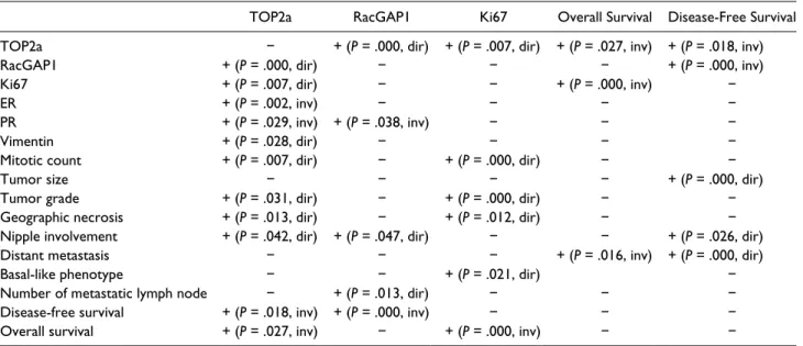
The data about the immunohistochemical, clinicopath-ological and prognostic parameters that showed statisti-cally significant correlation according to univariate analysis are given in Table 2. TOP2a (Figure 1A and B) expression showed direct correlation with RacGAP1 (Figure 1C and D) (P = .000), vimentin (P = .028) and increased Ki67 (Figure 1E and F) proliferation index (P = .007), number of mitosis (P = .007), high tumor grade (P = .031), geographic necrosis (P = .013), and nipple involvement (P = .042); however, there were inverse cor-relation with ER (P = .002) and PR (P = .029) positivity. It was detected that when TOP2a expression increased, dis-ease-free survival (P = .018) and overall survival (P = .027) tended to decrease statistically. There were direct correla-tion between RacGAP1 expression and TOP2a expression (P = .000), nipple involvement (P = .047), and the number of metastatic lymph nodes (P = .013), but there was inverse correlation between PR expression (P = .038). It was detected that RacGAP1 negative (≤10% expression) cases tended to have longer disease-free survival; however, shorter disease-free survival was detected in RacGAP1-positive (>10% expression) cases (P = .000). Ki67 expres-sion showed direct correlation with TOP2a expresexpres-sion (P = .007), number of mitosis (P = .000), tumor grade (P = .000), and geographic necrosis (P = .012). In addition, higher Ki67 expression was observed in the tumors of basal-like phenotype (P = .021). The overall survival of the patients were found to be shorter in the cases that showed higher Ki67 proliferation index and distant metas-tasis (P = .000 and P = .016, respectively). There were direct correlation between disease-free survival and nipple involvement (P = .026), distant metastases (P = .000), and tumor size (P = .000).
According to multiple linear regression analysis, over-all survival was found to be inversely correlated with Table 1. Clinicopathologic Features (n = 457).
Age, years, mean ± SD (range) 53.3 ± 12.8 (19-86)
Gender (female/male) 457/0
Operation materials, n
Mastectomy 420
Lumpectomy 37
Tumor size, cm, mean ± SD (range) 2.7 ± 1.6 (0.4-20)
Tumor types, n
Invasive ductal carcinoma 371
Invasive lobular carcinoma 22
Mixed carcinoma 11 Mucinous carcinoma 11 Metaplastic carcinoma 8 Medullary carcinoma 6 Tubular carcinoma 5 Papillary carcinoma 5 Apocrine carcinoma 4 Micropapillary carcinoma 4
Signet ring cell carcinoma 4
Pleomorphic carcinoma 3 Cribriform carcinoma 2 Neuroendocrine carcinoma 1 Nipple involvement, n Present 32 Absent 387 Unknown 38 Skin involvement, n Present 14 Absent 443 Fascia involvement, n Present 38 Absent 392 Unknown 27
Metastatic lymph nodes, n
Present 250 Absent 197 Unknown 10 Distant metastasis, n Present 31 Absent 223 Unknown 203 Geographic necrosis, n Present 66 Absent 391
The number of mitosis/10 high-power fields,
mean ± SD (range) 11.36 ± 9.04 (1-60)
Tumor grade, n
Grade 1 130
Grade 2 166
Grade 3 161
Overall survival, months, mean ± SD (range) 44.59 ± 14.38 (13-84)
Disease-free survival, months, mean ± SD (range) 42.58 ± 14.92 (5-84)
The current status of patients, n
Alive 239
Dead 15
higher TOP2a expression (P = .000, R2 = 0.14, β = 0.19). Disease-free survival was detected to be inversely corre-lated with higher TOP2a expression (P = .000, R2 = 0.29, β = 0.29) and presence of distant metastasis (P = .000, R2 = 0.29, β = 0.40). The other associated parameters in univari-ate analysis did not show any significant correlation by multiple linear regression analysis.
Discussion
High proliferation rate is one of the major characteristics of malignancy, however prognostic gene signatures about proliferation are not the same in different types of can-cer.2,20 The cell proliferation in breast cancer has been sug-gested to vary according to the molecular subtypes of the tumor.2 In our study, the expressions of three different pro-liferation markers as Ki67, RacGAP1, and TOP2a in breast cancer patients were evaluated by immunohistochemical methods and compared with the clinicopathological parameters associated with prognosis.
Ki67 is a nuclear nonhistone protein first defined in 1991.2,4 It is strongly expressed in all phases of the cell cycle of proliferating cells, but not present in nondividing cells.2 In the literature, some studies have reported that high Ki67 expression is related with poorer prognosis in breast cancer; however, most of other studies have stated that it is not an independent prognostic factor.2,5,6 Also, some stud-ies have suggested that high Ki67 expression is associated with chemotherapy response.2,21 Many studies have pro-posed that the proliferation markers are important prognos-tic factors related with disease-free survival and overall survival, particularly for ER+ tumors.2 Nevertheless, the
assess of proliferation markers has not been widely used yet in routine practice.2 Using time consuming methods, the standardization challenges and problems, the difficul-ties in reproducibility of the results of those studies are among the reasons for inability to use them routinely.2,22 The St Gallen guidelines published in 2011 have suggested a cutoff value of 14% for Ki67 proliferation index immuno-histochemically for the molecular subtypes of luminal A and luminal B of breast cancer.2,19 Romero et al23,24 have proposed a 20% cutoff value for Ki67 expression.25 However, it is stated that those cutoff values for Ki67 immunohistohemistry are not reliable for routine clinical use due to the intra- and interobserver variability.2 In our study, Ki67 expression was assessed by counting the num-ber of stained nuclei using tissue microarray method that was thought to be a more objective method rather than the other studies that counted the percentage of staining. In the present study, high Ki67 expression was found in the tumors exhibiting high mitotic count, geographic necrosis, and high histological grade indicating poorer prognosis. Similarly, high Ki67 expression was observed in the tumors with basal-like phenotype implying poorer prognosis. In addition, the tumors with high expression of Ki67 were demonstrated to have statistically significant shorter over-all survival and higher TOP2a expression.
TOP2a shows high expression in growing cells in the G2/M phase and overexpression of TOP2a has been reported to be associated with shorter disease-free sur-vival and overall sursur-vival in breast cancer in some stud-ies.2 TOP2a is located on the same chromosome as HER2-neu and often amplified with this receptor.2 TOP2a is suggested as a target receptor for anthracycline. Table 2. Statistically Significant Associations Between Immunohistochemical and Clinicopathologic Characteristics According to
Univariate Analysis (P < .05).
TOP2a RacGAP1 Ki67 Overall Survival Disease-Free Survival TOP2a − + (P = .000, dir) + (P = .007, dir) + (P = .027, inv) + (P = .018, inv)
RacGAP1 + (P = .000, dir) − − − + (P = .000, inv)
Ki67 + (P = .007, dir) − − + (P = .000, inv) −
ER + (P = .002, inv) − − − −
PR + (P = .029, inv) + (P = .038, inv) − − −
Vimentin + (P = .028, dir) − − − −
Mitotic count + (P = .007, dir) − + (P = .000, dir) − −
Tumor size − − − − + (P = .000, dir)
Tumor grade + (P = .031, dir) − + (P = .000, dir) − −
Geographic necrosis + (P = .013, dir) − + (P = .012, dir) − −
Nipple involvement + (P = .042, dir) + (P = .047, dir) − − + (P = .026, dir)
Distant metastasis − − − + (P = .016, inv) + (P = .000, dir)
Basal-like phenotype − − + (P = .021, dir) −
Number of metastatic lymph node − + (P = .013, dir) − − −
Disease-free survival + (P = .018, inv) + (P = .000, inv) − − −
Overall survival + (P = .027, inv) − + (P = .000, inv) − −
Therefore, amplification of TOP2a is considered to be an indicator of sensitivity to anthracycline therapy.2 In the literature, high TOP2a RNA levels have been reported to be related with more short-term metastasis-free survival in breast cancer patients without lymph node metasta-sis.2,26 Besides, patients treated with anthracycline have been detected to show high rate of pathological complete response.2,26,27 Chen et al16 have investigated the predic-tive and prognostic value of TOP2a expression in the pri-mary tumors as well as the residual tumors sampled after the treatment of the patients that had locally advanced breast cancer and recieved neoadjuvant chemotherapy with anthracycline. In the literature, most of the studies
have advocated that TOP2a expression is a predictive parameter of longer disease-free survival and overall sur-vival of early breast cancer patients received anthracy-cline therapy; however, there are some other studies with contradictory results that have stated no association on that issue.2,12-14,16
The tumors of the patients who did not receive neoadju-vant therapy were evaluated in our study and high expres-sion of TOP2a was detected in the high-grade tumors with high mitotic count, geographic necrosis, nipple involve-ment, indicating poor prognosis. Similarly, high TOP2a expressions were found in the tumors showing ER nega-tivity, PR neganega-tivity, and vimentin positivity28-32 implying Figure 1. Photomicrographs of immunostaining of breast cancer samples. (A, B) TOP2a positivity (avidin-biotin-peroxidase
method, 100×, 400×, respectively). (C, D) RacGAP1 positivity (avidin-biotin-peroxidase method, 100×, 400×, respectively). (E, F) Ki67 positivity (avidin-biotin-peroxidase method, 200×, 400×, respectively).
poorer prognosis in the present study. In addition, high expression of the TOP2a was directly correlated with high RacGAP1 and Ki67 expression, and shorter disease-free survival and overall survival in our study, as previously mentioned.
RacGAP1 is a protein that participates in mitosis.2,33 It is phosphorylated by Aurora B that is necessary for RhoA GAP.2,34 Additionally, it plays a role as a nuclear chaper-one and participates in nuclear transportation for the STAT transcription factors.2,35 Milde-Langosch et al2 have pro-posed RacGAP1 as a marker indicating poor prognosis in ER+ (luminal) breast tumors in contrast to HER2+ and triple negative tumors. They have demonstrated that the prognostic value of RacGAP1 in luminal tumor is inde-pendent from histological grade, clinical stage, and lymph node involvement.2 RacGAP1 expression was demon-strated to be correlated with both TOP2a and Ki67 expres-sions; however, the prognostic impact of their expressions were detected to vary between different subtypes of breast cancer.2 Pliarchopoulou et al7 investigated the prognostic importance of RacGAP1 mRNA expression on the overall and disease-free survival in high-risk early breast cancer patients and compared with Ki67 expression. The cases with high RacGAP1 expression were found to show higher histological grade and higher Ki67 expression.7 High RacGAP1 mRNA expression was observed to be related with both shorter disease-free survival and overall sur-vival, also suggested to be an independent prognostic fac-tor.7 Similar to that study, RacGAP1 positive cases were found to show shorter disease-free survival statistically significantly in our study. Additionally, as favouring poor prognosis, the tumors with nipple and lymph node involve-ment, PR expression, and high TOP2a expression16,30-32 were demonstrated to exhibit higher RacGAP1 expression in the present study.
Similar to our study, Milde-Langosch et al2 studied Ki67, TOP2a, and RacGAP1 expression in different molecular subtypes of breast cancer. Only Ki67 was sug-gested to be a statistically significant independent prog-nostic marker for triple negative tumors; however, none of these markers was found to have prognostic significance in HER2+ patients.2 They advocated that mRNA analysis of RacGAP1 was superior to Ki67 and TOP2a to predict the prognosis of luminal tumors.2 The effect of prolifera-tion markers was observed to vary in luminal tumors depending on the method of therapy used.2 Ki67, TOP2a, and RacGAP1 have been demonstrated to be statistically significant independent prognostic markers among untreated patients.2 In addition, overexpressions of these 3 markers were determined to be predictive for early recur-rence for the patients treated with chemotherapy.2 RacGAP1 was advocated to be the only predictive prolif-eration marker in the patients treated with endocrine ther-apy.2 In our study, significant prognostic significance was
detected only for high Ki67 expression in basal-like tumors, there was no relationship between the other molec-ular subgroups and the expressions of Ki67, RacGAP1, and TOP2a.
In conclusion, our study has demonstrated that overex-pressions of Ki67, RacGAP1, and TOP2a have been asso-ciated with poorer prognosis in breast cancer, also the effect of TOP2a was supported by multivariate analysis. Thus, performing RacGAP1, TOP2a, and particularly Ki67 by immunohistochemistry in routine practice of pathology while diagnosing breast cancer is strongly advised in order to determine the therapy and predict the prognosis. It is also claimed that comprehensive studies conducted with multivariate analysis are crucial to clarify-ing the effect of those markers on pathogenesis and prog-nosis of breast cancer.
Declaration of Conflicting Interests
The author(s) declared no potential conflicts of interest with respect to the research, authorship, and/or publication of this article.
Funding
The author(s) disclosed receipt of the following financial support for the research, authorship, and/or publication of this article: This study was supported financially by Bozok University Scientific Research Projects Unit (Project No. 2014TF/A92).
References
1. Cakir A, Gonul II, Uluoglu O. A comprehensive morpholog-ical study for basal-like breast carcinomas with comparison to nonbasal-like carcinomas. Diagn Pathol. 2012;20:145. 2. Milde-Langosch K, Karn T, Müller V, et al. Validity of
the proliferation markers Ki67, TOP2A, and RacGAP1 in molecular subgroups of breast cancer. Breast Cancer Res
Treat. 2013;137:57-67.
3. Hanahan D, Weinberg RA. The hallmarks of cancer. Cell. 2000;100:57-70.
4. Gerdes J, Li L, Schlueter C, et al. Immunobio-chemical and molecular biologic characterization of the cellproliferation-associated nuclear antigen that is defined bymonoclonal antibody Ki-67. Am J Pathol. 1991;138:867-873.
5. Stuart-Harris R, Caldas C, Pinder SE, et al. Proliferation markers and survival in early breast cancer: a systematic review and meta-analysis of 85 studies in 32,825 patients.
Breast. 2008;17:323-334.
6. Urruticoechea A, Smith IE, Dowsett M. Proliferation marker Ki-67 in early breast cancer. J Clin Oncol. 2005;23:7212-7220. 7. Pliarchopoulou K, Kalogeras KT, Kronenwett R, et al.
Prognostic significance of RACGAP1 mRNA expression in high-risk early breast cancer: a study in primary tumors of breast cancer patients participating in a randomized Hellenic Cooperative Oncology Group trial. Cancer Chemother
Pharmacol. 2013;71:245-255.
8. Lu KH, Patterson AP, Wang L, et al. Selection of potential markers for epithelial ovarian cancer with gene xpression
arrays and recursive descent partitionanalysis. Clin Cancer
Res. 2004;10:3291-3300.
9. Stone R 2nd, Sabichi AL, Gill J, et al. Identification of genes correlated with early-stage bladder cancer progression.
Cancer Prev Res (Phila). 2010;3:776-786.
10. Wang SM, Ooi LL, Hui KM. Upregulation of Rac GTPase-activating protein 1 is significantly associated with the early-recurrence of human hepatocellular carcinoma. Clin Cancer
Res. 2011;17:6040-6051.
11. Fritz G, Brachetti C, Bahlmann F, et al. Rho GTPases in human breast tumors: expression and mutation analy-ses and correlation with clinical parameters. Br J Cancer. 2002;87:635-644.
12. Rody A, Karn T, Ruckhaberle E, et al. Gene expression of topoisomerase II alpha (TOP2A) by microarray analysis is highly prognostic in estrogen receptor (ER) positive breast cancer. Breast Cancer Res Treat. 2009;113:457-466. 13. Colozza M, Azambuja E, Cardoso F, et al. Proliferative
markers as prognostic and predictive tools in early breast cancer: where are we now? Ann Oncol. 2005;16:1723-1739. 14. O’Malley FP, Chia S, Tu D, et al. Topoisomerase II alpha
and responsiveness of breast cancer to adjuvant chemother-apy. J Natl Cancer Inst. 2009;10:644-650.
15. Nielsen KV, Ejlertsen B, Moller S, et al. The value of TOP2A gene copy number variation as a biomarker in breast cancer: update of DBCG trial 89D. Acta Oncol. 2008;47:725-734. 16. Chen S, Huang L, Liu Y, et al. The predictive and
prog-nostic significance of pre- and post-treatment topoi-somerase IIα in anthracycline-based neoadjuvant chemotherapy for local advanced breast cancer. Eur J Surg
Oncol. 2013;39:619-626.
17. Wolff AC, Hammond ME, Schwartz JN, et al. American Society of Clinical Oncology/College of American Pathologists guideline recommendations for human epider-mal growth factor receptor 2 testing in breast cancer. J Clin
Oncol. 2007;25:118-145.
18. Elston CW, Ellis IO. Pathological prognostic factors in breast cancer. I. The value of histological grade in breast cancer: experience from a large study with long-term follow-up. Histopathology. 1991;19:403-410.
19. Goldhirsch A, Wood WC, Coates AS, et al. Strategies for subtypes-dealing with the diversity of breast cancer: high-lights of the St. Gallen International Expert Consensus on the Primary Therapy of Early Breast Cancer. Ann Oncol. 2011;22:1736-1747.
20. Wirapati P, Sotiriou C, Kunkel S, et al. Meta-analysis of gene expression profiles in breast cancer: toward a unified understanding of breast cancer subtyping and prognosis sig-natures. Breast Cancer Res. 2008;10:65.
21. Yerushalmi R, Woods R, Ravdin PM, et al. Ki67 in breast cancer: prognostic and predictive potential. Lancet Oncol. 2010;11:174-183.
22. Dowsett M, Nielsen TO, A’Hern R, et al. Assessment of Ki67 in breast cancer: recommendations from the International Ki67 in Breast Cancer Working Group. J Natl Cancer Inst. 2011;103:1656-1664.
23. Romero Q, Bendahl PO, Fernö M, et al. A novel model for Ki67 assessment in breast cancer. Diagn Pathol. 2014;9:118. 24. Romero Q, Bendahl PO, Klintman M, et al. Ki67 prolif-eration in core biopsies versus surgical samples - a model for neo-adjuvant breast cancer studies. BMC Cancer. 2011;11:341.
25. Stathopoulos GP, Malamos NA, Markopoulos C, et al. The role of Ki-67 in the proliferation and prognosis of breast cancer molecular classification subtypes. Anticancer Drugs. 2014;25:950-957.
26. Brase JC, Schmidt M, Fischbach T, et al. ERBB2 and TOP2A in breast cancer: a comprehensive analysis of gene amplification, RNA levels, and protein expression and their influence on prognosis and prediction. Clin Cancer Res. 2010;16:2391-2401.
27. Park K, Kim J, Lim S, Han S. Topoisomerase II-alpha (topoII) and HER2 amplification in breast cancers and response to preoperative doxorubicin chemotherapy. Eur J
Cancer. 2003;39:631-634.
28. Karihtala P, Auvinen P, Kauppila S, et al. Vimentin, zeb1 and Sip1 are up-regulated in triple-negative and basal-like breast cancers: association with an aggressive tumour phe-notype. Breast Cancer Res Treat. 2013;138:81-90.
29. Yamashita N, Tokunaga E, Kitao H, et al. Vimentin as a poor prognostic factor for triple-negative breast cancer. J
Cancer Res Clin Oncol. 2013;139:739-746.
30. Mohammed H, Russell IA, Stark R, et al. Progesterone receptor modulates ERalpha action in breast cancer. Nature. 2015;523:313-317.
31. Prat A, Cheang MC, Martin M, et al. Prognostic significance of progesterone receptor-positive tumor cells within immu-nohistochemically defined luminal A breast cancer. J Clin
Oncol. 2013;31:203-209.
32. Purdie CA, Quinlan P, Jordan LB, et al. Progesterone recep-tor expression is an independent prognostic variable in early breast cancer: a population-based study. Br J Cancer. 2014;110:565-572.
33. Hirose K, Kawashima T, Iwamoto I, Nosaka T, Kitamura T. MgcRacGAP is involved in cytokinesis through asso-ciating with mitotic spindle and midbody. J Biol Chem. 2001;276:5821-5828.
34. Minoshima Y, Kawashima T, Hirose K, et al. Phosphorylation by aurora B converts MgcRacGAP to a RhoGAP during cytokinesis. Dev Cell. 2003;4:549-560.
35. Kawashima T, Bao YC, Minoshima Y, et al. A Rac GTPase-activating protein, MgcRacGAP, is a nuclear localizing sig-nal-containing nuclear chaperone in the activation of STAT transcription factors. Mol Cell Biol. 2009;29:1796-1813.