© Springer Nature Singapore Pte Ltd. 2017 61 A. Badnjevic (ed.), CMBEBIH 2017,
IFMBE Proceedings 62,
DOI: 10.1007/978-981-10-4166-2_10
Multi-Regional Adaptive Image Compression (AIC) for Hip Fractures
in Pelvis Radiography
Huseyin Nasifoglu1,*, Osman Erogul1, Gokce Kaan Atac2, and Galip Ozdemir1 1 Department of Biomedical Engineering, TOBB University of Economics and Technology,
Ankara, Turkey
{hnasifoglu, erogul, g.ozdemir}@etu.edu.tr
2 Department of Radiology – Faculty of Medicine, Ufuk University, Ankara, Turkey [email protected]
Abstract. High resolution digital medical images are stored in DICOM (Digital Imaging and Communications in Medi-cine) format that requires high storage space in database.
Therefore reducing the image size while maintaining diagnostic quality can increase the memory usage efficiency
in PACS. In this study, diagnostic regions of interest (ROI) of pelvis radiographs marked by the radiologist are
seg-mented and adaptively compressed by using image processing algorithms. There are three ROIs marked by red,
blue and green in every image. ROI contoured by red is defined as the most significant region in the image and compressed by lossless JPEG algorithm. Blue and green regions have less importance than the red region but still contain diagnostic data compared to the rest of the image. Therefore, these regions are compressed by lossy JPEG algorithm with higher quality factor than rest of the image. Non-contoured region is compressed by low quality factor which does not have any diagnostic information about the patient. Several compression ratios are used to determine sufficient quality and appropriate compression level. Compression ratio (CR), peak signal to noise ratio (PSNR), bits per pixel (BPP) and signal to noise ratio (SNR) values are calculated for objective evaluation of image quality.
Experimental results show that original images can approximately be compressed six times without losing any
diagnostic data. In pelvis radiographs marking multiple regions of interest and adaptive compression of more than one ROI is a new approach. It is believed that this method will improve database management efficiency of PACS while preserving diagnostic image content.
Keywords: Adaptive compression, DICOM, JPEG, loss-less, lossy, medical image, radiography, region of interest (ROI)
INTRODUCTION
In medical centers and hospitals, high amount of medical imaging data are generated by various imaging modalities such as: Magnetic Resonance Imaging (MRI), Computed Radiography (CR), Computed Tomography (CT), Positron Emission Tomography (PET), Ultrasonography, Single Photon Emission Computerized Tomography (SPECT), Digital Fluorography etc. This data needs to be stored and transmitted to another medical modality or facility if needed by a standardized Picture Archiving and Communication System (PACS). Handling of this data via PACS is critical and requires not only high storage capacity but also high transmission bandwidth. Medical image compression is an essential image processing technique in this manner. The aim of image compression is to reduce the number of bits required to represent an image while preserving highest image quality as possible. However, medical images contain significant diagnostic data which can not be compromised. Due to this fact, lossless compression techniques are com-monly used for decreasing the required storage size of a medical image while preserving 100% of the image data.
The downside of lossless compression is that the obtained compression ratio is not sufficient to achieve the storage
space of high amount of data. On the contrary, lossy com-pression techniques can reach to higher comcom-pression ratios when some content of the image data is lost forever which is affecting diagnostic accuracy in further examinations. Adaptive Image Compression (AIC) technique is a hybrid modality utilizing both lossless and lossy compression algo-rithms [1-2]. To be able to use this technique, first region of interest (ROI), containing significant diagnostic infor-mation, should be determined precisely. By applying loss-less compression algorithms, the size of the information in this region can be reduced where later it will be possible to
62 H. Nasifoglu et al.
IFMBE Proceedings Vol. 62
reconstruct the image perfectly. The remaining part of the image can be considered as less significant and the diagnos-tic information belongs to this region will be compressed in lossy manner. In reconstruction, some of the image data will obviously be lost however the image must still provide suf-ficient diagnostic information.
A lossless compression achieves low compression ratios typically 1.5:1 to 3:1 but allows an exact reconstruction of the original image. Some of the common algorithms are Run-Length Encoding (RLE), Huffman Encoding, Lempel-Ziw-Welch (LZW) [3-4]. These methods are generally preferred for compression of graphical and text data. Due to the fact that the image should be recovered exactly, the number of developed lossless compression techniques are limited. On the other hand, a lossy compression can achieve high compression ratios up to 100:1 with the consequence of loss of data or in this case, image distortion. Some of the most popular lossy compression approaches are wavelet transform [5-6], lossy JPEG and JPEG-2000 [7-8]. Dodig M., Grgic S. and Dodig S. compared the quality of X-ray images with lung tuberculosis (TB) by using JPEG and JPEG2000 compression algorithms [9]. According to the results, JPEG performed better at lower compression ratios and the authors mentioned that both algorithms can be used for store-and-forward chest X-ray teleradiology. Selvi G. and Nadarajan R. obtained high compression ratio and PSNR for lossy compression of CT, MRI and US images by applying Bilinear Interpolation [10]. In another study, Sophia P. and Anitha J. researched on implementation of ROI based medical images [11]. ROI layer is compressed by lossless algorithm while the Non-ROI layer is comressed by lossy. According to the results, good compression ratio of about 4.2 with PSNR 20.75 values are obtained during the study. Genetic algorithm is also used for lossless compression of DICOM images [12]. The extracted region is compressed by using Huffman encoding and genetic algorithm for the enhancement of compression ratio. Yulianti L. and Mengko T.R proposed a hybrid method to enhance the performance of Fractal lmage Compression (FIC) technique by FIC (lossy compression) and Huffman coding [13]. Raja J. A., Raja G. and Khan K., have made a resarch on multiple regions of interest for medical images [7]. The regions are encoded seperately by JPEG-XR and JPEG2000 standards. Since JPEG-XR is computationally less intensive, it is found to be more suitable for embedded applications or for the systems with limited memory and computation. In this paper, marking multiple regions of interest and adaptive compression of more than one ROI in pelvis radiography is investigated as a new approach.
METHODOLOGY
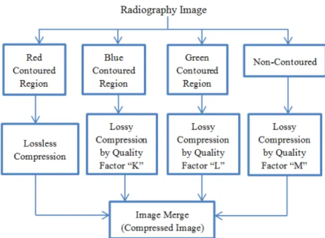
There are two main methodologies in this study: detec-tion of regions of interest (ROI) and adaptive compression of images. ROIs marked by radiologist with red, blue and green colors specify the significance order of these regions. In other words, red is described as the most significant re-gion of a possible hip fracture in the image, blue and green represent second and third most significant regions respec-tively. Remaining part of the image is considered as not containing any diagnostic information. Indeed, expert radi-ologist does not expect to see any hip fracture out of the marked regions. In adaptive compression both lossy and lossless algorithms are implemented. Red marked region is compressed by lossless compression algorithm. The blue region is compressed without compromising any diagnostic data by using an appropriate quality factor. Similarly, lower quality factors are selected for lossy compression of green region and remaining part of the image respectively. Figure 1 shows the general block diagram of the methodology.
Fig. 1. Regions of interest based compression methodology
1
Specifying Regions of Interest
In this study 10 pelvis radiographs obtained from Ufuk University Dr. Ridvan Ege Health Research and Application Hospital PACS database are used. All images are in DICOM format that contains hip fracture. Information such as name, surname, test date etc. are anonymized to preserve classified personal data. Expert radiologist is asked to mark three ROI depending on the location of fracture in the order of importance by red, blue and green respectively. Red