SUMMARY – Intracranial lipomas (ICLs) are rare lesions, the vast majority encountered as in-cidental findings on imaging studies. ICLs are generally pericallosal midline lesions and thought to be asymptomatic and can be accompanied by additional intracranial congenital malformations. We describe a 17-year old male with an unusual case of ICL on the frontal lobe associated with cortical dysplasia and abnormal vasculature mimicking arteriovenous malformation on magnetic resonance images.
Frontal Lobe Lipoma Associated with Cortical
Dysplasia and Abnormal Vasculature
OZDIL BASKAN, SERDAR GEYIK
Department of Radiology, Istanbul Medipol University School of Medicine; Istanbul, Turkey
Key words: cortical dysplasia, lipoma, magnetic resonance imaging, magnetic resonance angiography, vasculature
Introduction
Intracranial lipomas (ICLs) are very rare congenital malformative lesions accounting for about 0.1-1.7% of all intracranial tumours 1,2.
The majority of the ICL occur at or near the midline, mostly in the callosal cisterns 1. Other
involved locations include the quadrigeminal-ambient cistern, suprasellar, interpeduncular, cerebellopontine and sylvian cisterns 2.
Lipo-mas on the cerebral surface are extremely rare
2-7. A variety of brain malformations have been
reported to be associated with ICLs, mostly agenesis or dysgenesis of the adjacent struc-tures with the lesions located at midline 8 and
cortical dysplasia with lesions located at the cerebral surface 2.
Herein we describe a rare case of cerebral surface lipoma with an unusual location in the inferior medial part of the right frontal lobe in association with cortical dysplasia and abnor-mal vasculature mimicking an arteriovenous malformation (AVM) on initial magnetic reso-nance (MR) scan.
Case Report
A 17-year-old male patient was admitted to our hospital with a prior diagnosis of cerebral AVM. He had a history of syncope after a head trauma two months previously while playing
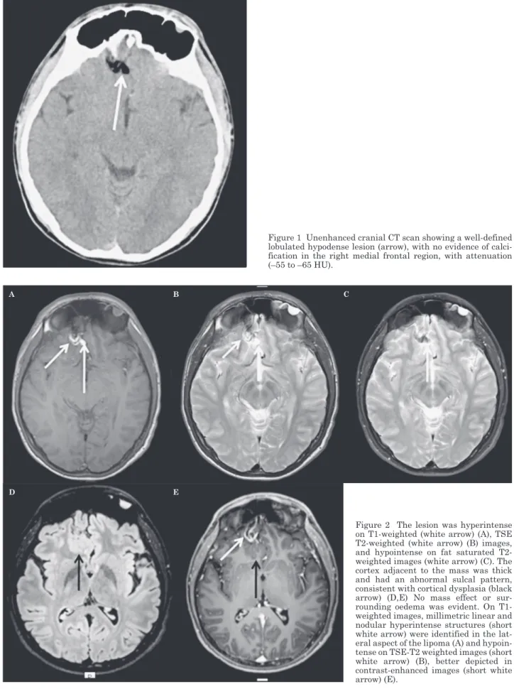
soccer. Unenhanced cranial computed tomog-raphy (CT) showed a hypodense lesion outside the cortex in the right medial frontal region, with attenuation (–55 - –65 HU) characteristics of adipose tissue without calcification. No as-sociated haemorrhage was detected (Figure 1). A lesion in the right inferior frontal region was detected on MR scan performed immedi-ately after the trauma in emergency conditions and considered an AVM. The patient had no episodes of epileptic seizures. The physical and neurologic examinations were unremarkable. We re-evaluated the patient for the purposes of differential diagnosis.
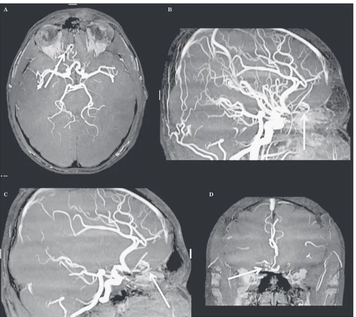
Cranial magnetic resonance imaging (MRI) was performed to evaluate the lesion and ad-jacent cerebral parenchyma. MRI revealed an extra-axial lesion which was hyperintense on T1-weighted (Figure 2A), T2-weighted (turbo spin-echo, TSE) (Figure 2B) images. Because of the signal intensity of the lesion on the T1 and T2-weighted images we performed fat-sat-urated T2-weighted images to rule out fat pres-entation. On fat-saturated T2-weighted, images suppression of the lesion was detected (Figure 2C). The CT and MRI characteristics of the le-sion were compatible with lipoma. The lipoma adhered to the medial inferior aspect of the gyrus rectus and medial orbital gyrus. Thick-ness of the cortex adjacent to the mass was increased and had an abnormal sulcal pattern consistent with cortical dysplasia (Figure 2D).
Figure 1 Unenhanced cranial CT scan showing a well-defined lobulated hypodense lesion (arrow), with no evidence of calci-fication in the right medial frontal region, with attenuation (–55 to –65 HU).
Figure 2 The lesion was hyperintense on T1-weighted (white arrow) (A), TSE T2-weighted (white arrow) (B) images, and hypointense on fat saturated T2-weighted images (white arrow) (C). The cortex adjacent to the mass was thick and had an abnormal sulcal pattern, consistent with cortical dysplasia (black arrow) (D,E) No mass effect or sur-rounding oedema was evident. On T1-weighted images, millimetric linear and nodular hyperintense structures (short white arrow) were identified in the lat-eral aspect of the lipoma (A) and hypoin-tense on TSE-T2 weighted images (short white arrow) (B), better depicted in contrast-enhanced images (short white arrow) (E).
D E
an enlarged drainage vein. Nonetheless, there was no evidence of an AVM on TOF MRA.
Considering the limitations of TOF MRA, a conventional cerebral angiogram was performed to rule out a vascular malformation. The angio-gram revealed an enlarged polar frontal branch of the right anterior cerebral artery feeding the region of the cortical dysplasia and lipoma seen on the MRI (Figure 4). There was no evidence of an early draining vein or any venous anom-aly. In the absence of a vascular malformation or arteriovenous fistula, these findings were considered suggestive of an arterial dysplasia. Additionally, millimetric linear and nodular
hyperintense structures were identified in the lateral aspect of the lipoma (Figure 2A) on T1-weighted images, which were hypointense on TSE-T2-weighted images (Figure 2B). These were better seen in contrast-enhanced images (Figure 2E).
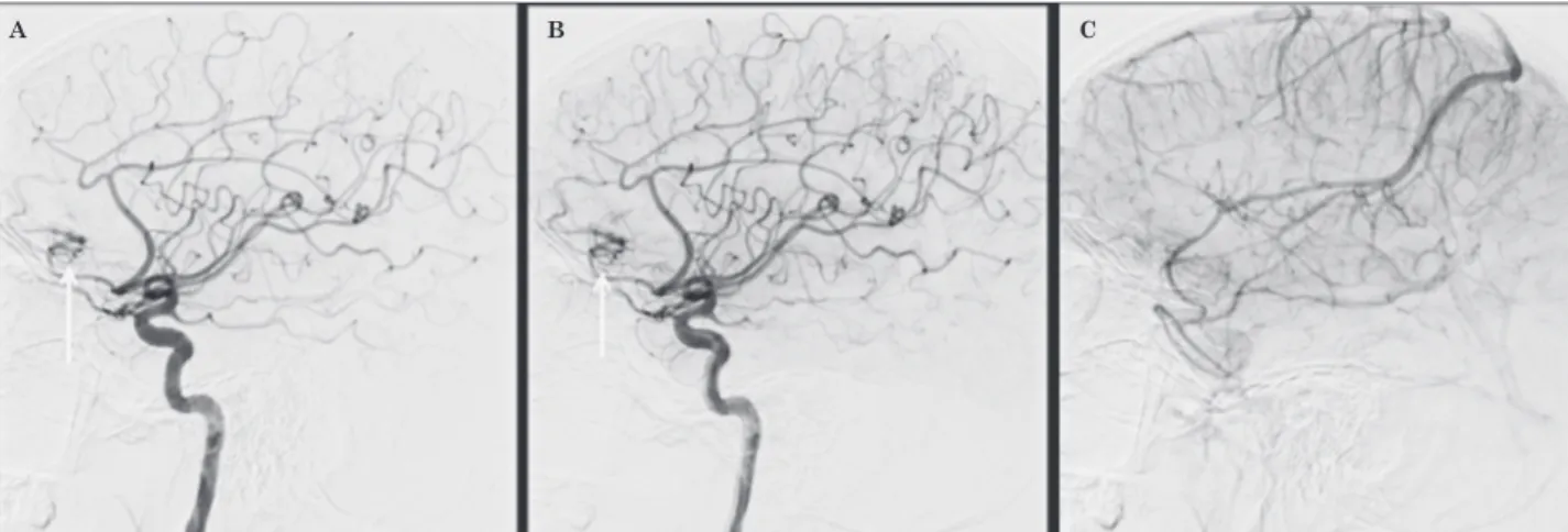
Magnetic resonance angiography (MRA) performed using the time-of-flight (TOF) tech-nique confirmed the presence of abnormal ar-terial branches originating from the anterior cerebral artery (Figure 3). No abnormally large vessel was detected that could represent
Figure 3 TOF MRA. Abnormal arterial branches (arrows) originating from the anterior cerebral artery in axial (A), sagittal (B,C), coronal (D) images.
A B
tal malformations 8. Various brain
malforma-tions are often seen in association with ICLs, including dysplasia or agenesis of the corpus callosum, absence of the septum pellucidum, spina bifida, encephalocele, myelomeningocele, malformation of the cortex and abnormal in-tracranial vessels 8,2. There are a few cases of
cortical malformations associated with ICLs in the literature, including pachygyria-like abnor-malities and subcortical nodular heterotopia 10,
an abnormal sulcal pattern-thickened cortex
4. Various vascular anomalies have also been
described in association with lipomas such as venous angioma, dilatation and prominent tor-tuous arteries, and saccular aneurysm 4,10,11.
In our case, the lipoma under the frontal lobe was also associated with cortical dysplasia and abnormal vasculature. To our knowledge, only four cases of ICLs on the cerebral surface have been reported with cortical dysplasia and ab-normal vasculature in or around them 2-5. It is
not clear whether cortical dysplasia and lipoma arise together or not and if abnormal vascula-ture is caused by lipoma or cortical dysplasia. There have been two ideas for the combination of lipoma and anomaly, one is that an anomaly arises primarily whether with a lipoma or not, and the other is the anomaly occurs secondary to the preexistence of a lipoma 2,8. Truwit et al. 8 suggested that ICLs are the result of
abnor-mal persistence and abnor-maldifferentiation of the meninx primitiva which as a rule dissolves to form the primitive subarachnoid space by the end of eighth gestational week. Most cortical dysplasias are considered to result from inju-ries in the first half of the second trimester 2.
Discussion
The first descriptions of intracranial lipomas (ICLs) were mainly through incidental findings at autopsy. ICL was first described in 1818 by Meckel, who found a chiasmatic lipoma, while in 1856 Rokitansky first described a perical-losal lipoma associated with corpus callosum agenesis 9.
ICLs are rare lesions. The majority of the le-sions are interhemispheric and more than 50% are located in the callosal cisterns 1. Other
typi-cal lotypi-calizations are the quadrigeminal plate (25%), suprasellar-interpeduncular cisterns (14%), and the cerebellopontine angle (9%). The sylvian cistern is a rare location for lipo-mas having a reported incidence of 3.4% 1, and
5% 8. Cerebral hemispheric lipomas located on
the cortical surface are extremely rare 2,6,7. In
our case, the lesion was located superficially, overlying the right inferior medial frontal lobe.
ICLs are mostly asymptomatic, the vast ma-jority of them appearing as incidental findings on imaging studies. However, they can cause symptoms differing according to their localiza-tion and comprising seizures, headache, pa-resis and cranial nerve impairments 8.
Hemi-spheric lipomas are more likely to be sympto-matic than lipomas in the midline 6.
ICLs are neither hamartomatous nor neo-plastic lesions. These are assumed to result from abnormal persistence and maldifferentia-tion of the meninx primitiva, the mesenchymal precursor of the leptomeninges, during the de-velopment of the subarachnoid cisterns 8. These
lesions should be accepted as being
congeni-Figure 4 Cerebral angiography. Lateral projection of right internal carotid artery injection showing the abnormal vasculature (ar-rows) (A,B). There is no evidence of a draining vein (C).
tics similar to adipose tissue (–50 - –100 HU) on CT scan. MRI is sufficient to establish the diag-nosis. On MRI, ICLs demonstrate a fat signal with short T1 and T2 relaxation times, i.e., a bright homogeneous signal on T1-weighted im-ages and an intermediate signal on T2-weighted images. Moreover, fat may appear isointense or hyperintense on TSE T2 sequences. Also with MRI, fatty tissue has become easily detectable with the fat saturation techniques. MRI find-ings can be useful for differential diagnosis and detecting associated malformations.
ICls are rare benign malformations and primarily detected incidentally during radio-logical examinations but sometimes they can cause symptoms. They are easily diagnosed using CT or MR. Cortical and vascular malfor-mations can be associated with ICLs located in the cortical sulci, therefore, a careful radiologic evaluation for any associated anomalies is nec-essary. Differential diagnosis should be made especially for vascular malformations that may need interventional procedures.
These concepts and the abnormal vasculariza-tion around the lesions support the idea that ICLs arise first, physical interruption and focal perfusion insufficiency due to preexistence of the lipomas might contribute to the formation of the focal cortical dysplasia 2.
In this case, we observed abnormal sulcal pattern and thickened cortex, consistent with
focal cortical dysplasia (Figure 2D), and tortuous and prominent vascular structures within and adjacent to the lipoma (Figures 2E and 3). There was no evidence of a feeding ar-tery, early draining vein or any venous anom-aly associated with an AVM. In the absence of a vascular malformation or arteriovenous fis-tula, these findings were considered suggestive of an arterial dysplasia. The other rare pos-sibility of a spontaneously thrombosed AVM could be considered. However, there were no signs of arteriovenous shunt in the angiogram.
On CT and MR images, lipomas show a typi-cal appearance. ICL appears as a homogenous low-density mass with attenuation
characteris-References
1 Maiuri F, Crillo S, Simonetti L. Lipoma of the slyvian region. Clin Neurol Neurosurg. 1989; 91: 321-323. doi: 10.1016/0303-8467(89)90008-5.
2 Sasaki H, Yoshida K, Wakamoto H, et al. Lipomas of the frontal lobes. Clin Neurol Neurosurg. 1996; 98: 27-31. doi: 10.1016/0303-8467(95)00073-9.
3 Britt PM, Bindel AK, Balko MG, et al. Lipoma of the cerebral cortex. Case report. Acta Neurochir. 1993; 121: 88-92. doi: 10.1007/BF01405190.
4 Saatci I, Aslan C, Renda Y, et al. Parietal lipoma as-sociated with cortical dysplasia and abnormal vascu-lature: case report and review of the literature. Am J Neuroradiol. 2000; 21: 1718-1721.
5 Thakur S, Sood RG, Jhobta A, et al. Sylvian fissure lipoma with angiomatous component and associated brain malformation: A case report. Iran J Neurol. 2013; 12 (4): 169-171.
6 Fujii Y, Konishi Y, Kriyama M, et al. Lipoma on sur-face of centroparietal lobes. Pediatr Neurol. 1993; 9: 144-6. doi: 10.1016/0887-8994(93)90052-E.
7 Devasia Bobby, Samuel Annie S, Kesavadas C. Lipo-matous cortical dysplasia with callosal lipoma: a rare association. Pediatr Radiol. 2006; 36: 83.
8 Truwit CL, Barkovich AJ. Pathogenesis of intracranial lipoma: An MR study in 42 patients. Am J Roentgenol. 1990; 155: 855-865. doi: 10.2214/ajr.155.4.2119122. 9 Rokitansky C. Lehrbuch der Pathlogischen anatomie.
Vol 2. Vienna; Braumuller. 1856; p. 33.
10 Guye M, Gastaut JL, Bartolomei F. Epilepsy and peri-sylvian lipoma/cortical dysplasia complex. Epileptic Disord. 1999; 1: 69-73.
11 Futami K, Kimura A, Yamashita J. Intracranial li-poma associated with cerebral saccular aneurysm. J Neurosurg. 1992; 77: 640-642. doi: 10.3171/jns.1992.77. 4.0640.
Ozdil Baskan, MD Department of Radiology
Istanbul Medipol University School of Medicine Istanbul; Turkey