ABSTRACT
Background: This study was designed to investigate the anatomical relationship of the different levels of aortic root. Materials and Methods: The morphological features of the aortic root were examined using of 12 adult hearts from fixed male
cadavers who had expired due to noncardiac causes by magnetic resonance imaging and applied mathematical analyses to the results. The measurements of the aortic root were done at four levels: at the ventriculoarterial junction (annulus), at the largest level of the Valsalva sinuses (sinus), at the level of commissures (sinotubular junction [STJ]), and at 1 cm above the STJ (aorta ascendens). We derived an equation that allows calculation of the appropriate diameter of the aortic root from four levels. Statistical analysis among the variation of the diameters at the four levels of aortic root was achieved using test one‑way analysis of variance.
Results: The data showed a geometric pattern of the aortic root. The comparison of the values from four levels showed that
the narrowest at the sinotubular junctional level and the widest at the sinus level.
Conclusion: The analysis of our data shows that the aortic root has a consistent shape with varying size and that is a definable
mathematical relationship between root diameter.
Key words: Aortic root, cardiac anatomy, mathematical modeling
The Geometrical Modeling of Aortic Root Complex
Murat Ugurlucan, Metin Onur Beyaz
1, Didem Melis Oztas
2, Adnan Ozturk
3, Kayihan Sahinoglu
3,
Ufuk Alpagut
4, Nilgun Bozbuga
4Department of Cardiovascular Surgery, Istanbul Medipol University, 1Cardiovascular Surgery Clinic, Sultanbeyli State Hospital, 2Cardiovascular Surgery Clinic, Bagcilar Training and Research Hospital, 3Department of Anatomy, Istanbul University, Istanbul
Medical Faculty, 4Department of Cardiovascular Surgery, Istanbul University, Istanbul Medical Faculty, Istanbul, Turkey
Original Article
INTRODUCTION
T
he functional anatomy and the geometry ofthe left ventricle are so important to attain the proper mechanical pump function of the heart. Mathematical modeling method could usefully be applied to the design of geometric structure and biomechanics of the heart. The obtained mathematical modeling from three-dimensional imaging of the heart could be adapted for corrective procedures of cardiovascular anomalies. Evaluation and analysis of the geometric structure and anatomical features of the heart using mathematical modeling methods have been developed as an adjuvant
method for clinical experiments.[1,2]
The aortic valve does not present as an active subvalvular apparatus with a real surgical annulus
component.[3] The aortic root is complex, having
surgical annulus which extends from ventriculoarterial to sinotubular junction and consists of cusps and the
Valsalva sinuses of the aortic valve.[4] The aortic wall
which is located posteriorly of each cusp expands for construction of the Valsalva sinus. The aortic root is either anatomical or physiological junctional structure instead of a strait cylinder.[5]
The aim of this study is to obtain not only further understanding and knowledge about normal aortic root anatomy and geometry but also more criteria about structural features of the left ventricular outflow region.
MATERIALS AND METHODS
The mathematical modeling of anatomic structures and their relationship in the left ventricle were performed at
Address for correspondence: Prof. Nilgun Bozbuga,
Department of Cardiovascular Surgery, Istanbul University Istanbul Medical Faculty, Millet Caddesi, Capa/Fatih, 34390, Istanbul, Turkey.
E‑mail: [email protected]
Access this article online
Quick Response Code:
Website: www.heartviews.org DOI:
10.4103/HEARTVIEWS. HEARTVIEWS_115_18
How to cite this article: Uġurlucan M, Beyaz MO, Oztas DM, Ozturk A, Sahinoġlu K, Alpaġut U, et al. The geometrical modeling of aortic root complex. Heart Views 2019;20:6-10.
This is an open access journal, and articles are distributed under the terms of the Creative Commons Attribution‑NonCommercial‑ShareAlike 4.0 License, which allows others to remix, tweak, and build upon the work non‑commercially, as long as appropriate credit is given and the new creations are licensed under the identical terms.
the Department of Anatomy of İstanbul Medical Faculty. The morphological effects of geometry and dimensions of the left ventricle were examined using 12 young adult hearts (all males) from fixed cadavers who had expired due to noncardiac causes. A detailed dissection of the anatomic structure of the hearts was done. The hearts were prepared for magnetic resonance imaging (MRI) by trimming the ascending aorta to 1 cm above the sinotubular junction (STJ).
The present study was designed to answer two questions: first does the human aortic root have a standard shape with varying size and second is there a constant definable relationship between the root and clinically measurable aortic valve annulus?
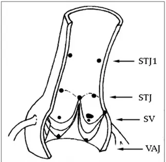
The cadaveric hearts were analyzed by MRI. The aortic root and valve geometry were extracted from three-dimensional MRI imaging and imported into the software program. The sequential cross-sectional slices of MRI were processed and converted the mathematical data using IRIX 6.5 software (Silicon Graphics - SGI Computer Systems, CA, US). The measurements of the aortic root were done at four levels: at the ventriculoarterial junction (VAJ) (annulus), at the largest level of the Valsalva sinuses (sinus), at the level of commissures (STJ), and at 1 cm above the STJ (aorta ascendens). A better understanding of the aortic root shape and the measurement levels are shown graphically [Figures 1 and 2]. Our purpose in calculating aortic annulus dimensions was to identify predictors of root size.
Mathematic curve fitting (polynomial) was done, and regression analysis was performed to determine the level correction. We derived an equation that allows
calculation of the appropriate diameter of the aortic root from four levels.
Statistical analysis among the variation of the diameters at the four levels of aortic root was achieved using one-way analysis of variance (ANOVA) test.
RESULTS
A geometric pattern of the aortic root was attained to the data. The comparison of the related diameters from four levels showed that in terms of related diameter, the aortic root was narrowest at the STJ level and widest at the sinus Valsalva level (one-way ANOVA, P < 0.05). The mean diameters of the aortic root and the mean interlevel distance are listed in Table 1.
The fraction of diameter of the sinus Valsalva level to the VAJ and to the STJ was found 1.1 and 0.8, respectively. The fraction of diameter of STJ to 1 cm above the STJ was 0.9.
One of the root dimensions at each of the four levels was plotted against one of the averaged root dimensions, and curves were fit to the data. In all specimens, the relationship is linear, with correlation coefficients ranging from 0.86 to 0.94. To determine the level of correlation, the measured root dimensions were plotted versus calculated diameter analog. Regression analysis showed the relationship of the measured root dimensions to the diameter analog to be linear, with a correlation coefficient of 0.90. If the average measured root dimension is plotted versus average diameter analog for each specimen, better correlation is demonstrated, with correlation coefficient of 0.96. To verify that the new derived measurement (diameter analog) could be used as a predictor of root size. Regression analysis showed the relationship to
Figure 1: Schematic presentation of four levels of aortic root
longitudinally where measurements were taken. STJ1: 1 cm above the sinotubular junction, STJ: Sinotubular junction, SV: Sinus of Valsalva, VAJ: Ventriculoarterial junction
Figure 2: Schematic presentation of four levels of aortic root
from transverse section. STJ1: 1 cm above the sinotubular junction, STJ: Sinotubular junction, SV: Sinus of Valsalva, VAJ: Ventriculoarterial junction
be linear, with correlation coefficients ranging from 0.88 to 0.93.
DISCUSSION
Better knowledge of the opening and closure of the aortic valve has clearly shown that the aortic valve is complex which includes the left ventricular outflow tract, the aortic annulus, (both fibrous trigones) the three leaflets and sinuses of Valsalva, and the coronary
ostia.[4] The importance of the sinuses and their role
in the creation of fluid eddy currents was recognized
as early as 1513 by Leonardo da Vinci.[6] The aortic
valve has been extensively studied over the last few decades.[7-10]
However, with increasing surgical sophistication in aortic root surgery, including valve-sparing root replacements, the simplistic assumption that the valve consists of leaflets that open and close due to
pressure differentials on either side is inadequate.[11-14]
Sophisticated analysis of the complex interplay between the aortic root and the leaflets, as well as the development of stresses and strains in the leaflet during opening and
closing is necessary for diagnostic techniques.[15]
A sophisticated dynamic analysis may aid in a better understanding of the dynamics of the aortic valve complex and hopefully will help in designing better substitutes for replacing the diseased aortic valve. More importantly, these might prove invaluable in predicting patterns of failure of replacement devices or repair techniques in the workbench setting rather than experiencing it in the clinical situation, with unfortunate consequences.
Mathematics supplies not only the common structural framework on which each of the many
components can be suspended but also represents the only manner in which to adequately model the complex interactions among these various components.
It is also the mathematics that is already allowing the development of a surgeon-friendly, interactive framework that will allow real‑time modification of the various model components, as well as the descriptions of their interrelationships. This is the key to the model’s potential as a clinical tool. It will allow us to enter clinical problem‑specific or patient‑specific variables and then test the various therapeutic options that are available to us.
Mathematical theory and applied mathematics display the answers to the inquiries of the cardiac model in the interactive, three‑dimensional visual field
of the computer workstation.[1,2,16] Display it in three
dimensions using variable degrees of transparency, with a dynamic, rotatable display of the contracting and relaxing ventricles with their opening and closing valves and tensing and relaxing papillary muscles. Geometrical model of the aortic valve and root is a significant advance in modeling and allows a closer simulation of valvular function.
Historically, surgical treatment of valvular heart disease has been based on designing a strategy or a valve substitute, applying it in a clinical setting, and then waiting (sometimes for years) to see if it will work. With increasing sophistication in the surgical treatment of aortic root diseases, including in the application of valve-sparing root replacements and aortic valve repairs, there is clearly a need for a precise understanding of the complex interplay of the various
components of the aortic root.[17-22]
Some of the earlier failures of replacement devices could have been predicted if sophisticated techniques
to test them had been available.[23] With the widespread
availability of powerful computers and supportive software, precise mathematical modeling using techniques make it possible to model and study the aortic valve in a dynamic state. These techniques also allow one to observe the most minute changes taking place in the leaflets and root, as well as the stresses developing in the leaflets in a manner that is not possible using animal or other models. The use of these techniques will enable the testing of newer devices, development of new leaflet materials, as well as newer types of stents for stent-mounted valves and repair techniques at the computer workbench before clinical application so that better long-term results can be expected.
A compliant aortic root contributes substantially to smooth and symmetrical leaflet opening with minimal
gradients.[2,11,13] The sinuses of Valsalva play a very
important role in minimizing stress in the leaflets.[4]
The changes in leaflet stress, strain, and coaptation indicate a departure from the normal aortic root–valve
Table 1: The mean aortic root measurements and the normalized the values as fraction of the diameter for annular level (ventriculoarterial) and the sinus level (Valsalva sinuses)
Level Interlevel distance (mm) Diameter (mm)
Annulus 23.3±2.6 9.4±0.7 Sinus 24.2±1.2 6.6±0.1 STJ 19.1±2.0 4.7±0.05 STJ1 21.6±2.3 Level Diameter Annulus 100 Sinus 104 STJ 82 STJ1 91 Level Diameter Annulus 96 Sinus 100 STJ 79 STJ1 87 STJ: Sinotubular junction, STJ1: 1 cm above the sinotubular junction
relationship, in that the definitive shape of the valve‑root
sinus functional unit has been changed.[9,24] Each leaflet
and corresponding sinus wall comprise a cylindrically shaped functional “unit” with continuity between the leaflets and root to distribute the diastolic pressure
load.[12] In this manner, the aortic valve transfers the
high attachment edge stresses from the leaflet to the lower-stiffness root wall.
This stress transfer appeared to be reversed in the graft models, where high stresses in the grafts were transferred to the valve, thereby increasing the overall leaflet stresses.[13] In addition, the functional unit
represented by the sinus wall and leaflet together in these models was not cylindrically shaped. The stress and velocity changes to the root–valve relationship in the pseudosinus model may be more physiological as that in the cylindrical and tailored models. Normalized leaflet stresses in the clinical setting with geometrical modeling may result in improved longevity of the spared
valve or replaced xenoprosthesis.[25]
The compliance also contributes much to the ability of the normal aortic valve to increase its effective valve
orifice in response to physiologic demands of exercise.[2]
The best hemodynamics in terms of gradients and valve areas during exercise are in normal aortic valves, pulmonary autografts, and cryopreserved homografts, followed by stentless valves.[7,19,23] This effect is strikingly
absent in stiff roots. The least efficient valves in terms of exercise gradients are stented valves.
The implication of these model results is that patients with advancing root dilatation may have aortic valve leaflets that are no longer normal; as such, these patients might not benefit as much as desired from a valve‑sparing procedure. Although the valve leaflets may enlarge in response to the early stages of root dilatation, it has been suggested that there is a critical ratio of total leaflet area to root cross‑sectional area equal to 1.5:1 below which the leaflet can no longer maintain the same degree of coaptation and loss of
coaptation ensues.[24]
The compliant tissue properties and rounded shape of the native aortic root promote the normal function of valve, aortic root replacement with a cylindrically shaped, and relatively stiffer polyester graft would affect the resulting levels of stress and strain
in the spared aortic valve leaflets.[14] Despite different
trimming techniques in valve-sparing operations to provide the graft with a rounded sinus-like shape, all these methods are applied to the same class
of polyester material grafts.[25] Because the elastic
modulus of prosthetic graft material is an order of magnitude greater than that of the normal aortic root, concern has been expressed that the use of this material might transfer high stresses to the spared aortic valve, perhaps reducing the durability of a
surgical valve repair.[25] Further research regarding
the timing of tissue remodeling would be helpful to surgeons in determining the optimal time for a valve-sparing replacement of the dilated aortic root.
CONCLUSION
The analysis of our data shows that the aortic root has a consistent shape with varying size and that is a definable of mathematical relationship between root diameter and clinically measured aortic valve annulus. The obtaining geometrical modeling from this research will be based on to determine computed data and be adapted to the corrective procedures of aortic root pathologies and be accepted reference for reconstructing of left ventricular outflow configuration.
The application of these results will allow prosthetic graft design to more closely resemble the native aorta. This should improve physiologic function of the aortic root, reduce annular stress, and increase the long-term durability. The results of mathematical modeling implied that the success of the surgical correction of the various aortic root pathologies depends on resembling of procedure to the anatomic and geometric design of the exact aortic root.
Financial support and sponsorship Nil.
Conflicts of interest
There are no conflicts of interest.
REFERENCES
1. Ranga A, Mongrain R, Mendes Galaz R, Biadillah Y, Cartier R. Large‑displacement 3D structural analysis of an aortic valve model with nonlinear material properties. J Med Eng Technol 2004;28:95‑103.
2. Redaelli A, Di Martino E, Gamba A, Procopio AM, Fumero R. Assessment of the influence of the compliant aortic root on aortic valve mechanics by means of a geometrical model. Med Eng Phys 1997;19:696‑710.
3. Choo SJ, McRae G, Olomon JP, St. George G, Davis W, Burleson‑Bowles CL, et al. Aortic root geometry: Pattern of
differences between leaflets and sinuses of valsalva. J Heart Valve Dis 1999;8:407‑15.
4. Sutton JP 3rd, Ho SY, Anderson RH. The forgotten interleaflet
triangles: A review of the surgical anatomy of the aortic valve. Ann Thorac Surg 1995;59:419‑27.
5. Yacoub MH, Kilner PJ, Birks EJ, Misfeld M. The aortic outflow and root: A tale of dynamism and crosstalk. Ann Thorac Surg 1999;68:S37‑43.
6. Bozbuğa N, Yakut C. The cardiac anatomy and Leonardo da Vinci. Koşuyolu Heart J 1996;2:95‑8.
7. Lockie KJ, Butterfield M, Fisher J, Juster NP, Watterson K, Davies GA, et al. Geometry of homograft valve leaflets: Effect
of dilation of the aorta and the aortic root. Ann Thorac Surg 1993;56:125‑30.
8. Kunzelman KS, Grande KJ, David TE, Cochran RP, Verrier ED. Aortic root and valve relationships. Impact on surgical repair.
J Thorac Cardiovasc Surg 1994;107:162‑70.
9. Grande KJ, Cochran RP, Reinhall PG, Kunzelman KS. Mechanisms of aortic valve incompetence in aging: A finite element model. J Heart Valve Dis 1999;8:149‑56.
10. Grande KJ, Cochran RP, Reinhall PG, Kunzelman KS. Stress variations in the human aortic root and valve: The role of anatomic asymmetry. Ann Biomed Eng 1998;26:534‑45.
11. Nicosia MA, Cochran RP, Einstein DR, Rutland CJ, Kunzelman KS. A coupled fluid‑structure finite element model of the aortic valve and root. J Heart Valve Dis 2003;12:781‑9.
12. De Hart J, Peters GW, Schreurs PJ, Baaijens FP. A three‑dimensional computational analysis of fluid‑structure interaction in the aortic valve. J Biomech 2003;36:103‑12.
13. Sripathi VC, Kumar RK, Balakrishnan KR. Further insights into normal aortic valve function: Role of a compliant aortic root on leaflet opening and valve orifice area. Ann Thorac Surg 2004;77:844‑51.
14. Beck A, Thubrikar MJ, Robicsek F. Stress analysis of the aortic valve with and without the sinuses of valsalva. J Heart Valve Dis 2001;10:1‑11.
15. Gnyaneshwar R, Kumar RK, Balakrishnan KR. Dynamic analysis of the aortic valve using a finite element model. Ann Thorac Surg 2002;73:1122‑9.
16. Weston MW, LaBorde DV, Yoganathan AP. Estimation of the shear stress on the surface of an aortic valve leaflet. Ann Biomed Eng 1999;27:572‑9.
17. Sarsam MA, Yacoub M. Remodeling of the aortic valve anulus. J Thorac Cardiovasc Surg 1993;105:435‑8.
18. Bhatnagar G, Christakis GT, Murphy PM, Oxorn D, Goldman BS. Technique for reconstruction of the sinotubular junction. Ann Thorac Surg 1997;63:559‑60.
19. van Son JA, Battellini R, Mierzwa M, Walther T, Autschbach R, Mohr FW, et al. Aortic root reconstruction with preservation of
native aortic valve and sinuses in aortic root dilatation with aortic regurgitation. J Thorac Cardiovasc Surg 1999;117:1151‑6. 20. David TE, Armstrong S, Ivanov J, Webb GD. Aortic valve sparing
operations: An update. Ann Thorac Surg 1999;67:1840‑2. 21. Yakut C. A new modified bentall procedure: The flanged technique.
Ann Thorac Surg 2001;71:2050‑2.
22. Kirali K, Mansuroğlu D, Omeroğlu SN, Erentuğ V, Mataraci I, Ipek G, et al. Five‑year experience in aortic root replacement with
the flanged composite graft. Ann Thorac Surg 2002;73:1130‑7. 23. Gallo R, Kumar N, al Halees Z, Duran C. Early failure of aortic
valve conservation in aortic root aneurysm. J Thorac Cardiovasc Surg 1995;109:1011‑2.
24. Grande‑Allen KJ, Cochran RP, Reinhall PG, Kunzelman KS. Mechanisms of aortic valve incompetence: Finite‑element modeling of marfan syndrome. J Thorac Cardiovasc Surg 2001;122:946‑54.
25. Grande KJ, Cochran RP, Reinhall PG, Kunzelman KS. Mechanisms of aortic valve incompetence: Finite element modeling of aortic root dilatation. Ann Thorac Surg 2000;69:1851‑7.