See discussions, stats, and author profiles for this publication at: https://www.researchgate.net/publication/11421250
The use of stentless valves for root replacement during repair of ascending
aortic aneurysms with aortic valve regurgitation
Article in The Heart Surgery Forum · January 2002 Source: PubMed CITATIONS 7 READS 70 8 authors, including: Saide Aytekin
Koc University Hospital & American Hospital 126PUBLICATIONS 799CITATIONS
SEE PROFILE
Ertan Sagbas
Istanbul Bilim University 54PUBLICATIONS 685CITATIONS
A B S T R AC T
Background: Early and mid-term results of stentless
valves for the treatment of ascending aortic aneurysm (AAA) were evaluated in a retrospective manner.
Material and Methods: During a four-year period,
26 patients with ascending aortic aneurysms and aortic valve insufficiency underwent a total root replacement procedure using a stentless “Freestyle” valve (Medtronic Inc., Min-neapolis MN). Mean age was 71 ± 4 years (range 66 to 79 years). Eight patients were in NYHA Class 2, 13 in Class 3, and 5 in Class 4. Cardiopulmonary bypass (CPB) was begun with femoral artery-right atrium (two-stage) cannula-tion in all cases but four, in which the right axillary artery was used. Myocardial protection was established by retrograde, cold-blood cardioplegia and direct antegrade blood cardio-plegia from the right coronary ostium. The left ventricle outflow tract was constructed by using 2-0 ticron sutures and incorporating a pericardial strip in between. Coro-nary buttons were sewn to the xenograft with 6-0 polypro-pylene sutures. Meanwhile, the patient was cooled down to 18 degrees nasopharyngeal temperature and the distal anasto-mosis with the proximal arch was performed with a Dacron graft under total circulatory arrest (TCA), using 4-0 polypro-pylene sutures. During rewarming, the connection between the Freestyle valve and the Dacron graft was performed.
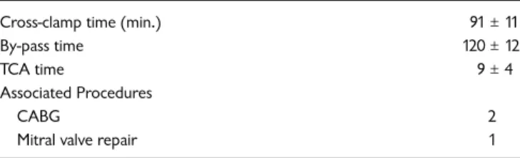
Results: Ischemic time was 91 ± 11 minutes and TCA time
was 9 ± 4 minutes. Operative mortality was zero, and there was one 30-day mortality (3.8%). At discharge, all 25 patients had a functional valve with low transvalvular gradients. Patients were followed for a mean period of 15 months, with one patient being lost to follow-up and one patient dying of non-cardiac causes. Follow-up was 97% complete, and echocardiographic control during the follow-up period
revealed competent valves with gradients comparable to those at discharge. Two patients were screened with electron beam tomography (EBT) three years after the operation and there was no sign of wall or leaflet calcification. At the end of the 15 months (mean) follow-up, the functional capacity of the patients had improved significantly (p <0.05).
Conclusions: Our early results suggest that use of the
Freestyle valve in conjunction with a Dacron tube graft can be a good alternative for patients over 65 years of age who present with ascending aortic aneurysm with aortic valve insufficiency.
B AC KG R O U N D
Ascending aortic aneurysm (AAA) with aortic valve insuffi-ciency is a frequently encountered pathology. Total root replacement with a conduit consisting of a mechanical valve and a Dacron tube as described by Bentall and DeBono is still accepted as the gold standard of treatment for these patients [Bentall 1968]. However, the necessity of anticoagulation therapy and the high risk for thromboembolic events and bleeding complications make this treatment far from ideal, especially in the elderly population [Akpinar 1999]. We describe a group of patients with AAA and aortic regurgita-tion who underwent total root replacement using a stentless “Freestyle” valve (Medtronic Inc., Minneapolis MN), with continuity to the proximal arch established by a Dacron tube graft interposition.
M AT E R I A L S A N D M E T H O D S
Between January 1996 and September 2000, 26 patients with AAA and aortic valve insufficiency underwent total root replacement with the Freestyle valve. There were 19 male and 7 female patients having a mean age of 71 ± 4 years. The mean aneurysm diameter was 5.8 ± 0.6 cm. There was one patient with a chronic Stanford Type A dissection (Table 1 ).
Selection criteria for this method were: (1) age greater than 65 years; (2) contraindication to oral anticoagulants; (3) deliberate request for a biological valve by the patient. Two patients were below 65 years of age but they could not use oral anticoagulants due to previous gastrointestinal bleeding.
The Use of Stentless Valves for Root Replacement During
Repair of Ascending Aortic Aneurysms with Aortic Valve
Regurgitation
(#2001-40913 … August 23, 2001)
Belhhan Akpinar, MD,
1Mustafa Güden, MD,
1Saide Aytekin, MD,
2Ilhan Sanisoglu, MD,
1Ertan Sagbas, MD,
1Ugur Özbek, MD,
3Baris Çaynak, MD,
1Z. Bayramoglu, MD
1Kadir Has University Medical Faculty, Florence Nightingale Hospital, Istanbul, Turkey,
Departments of Cardiac Surgery,1Cardiology,2and Anesthesia.3
Dr. Akpinar
Submitted August 17, 2001; accepted August 23, 2001.
Address correspondence and reprint requests to: Belhhan Akpinar, MD, Associate Professor, Department of Cardiac Surgery, Florence Nightingale Hospital, Abide’i Hurriyet Cad. No:280, Sisli, Istanbul, Turkey, Phone: 090 212 2398790, Fax: 090 212 2398791, Email: [email protected]
The Use of Stentless Valves for Root Replacement During Repair of Ascending Aortic Aneurysms with Aortic Valve Regurgitation—Akpinar et al.
Operative Technique
The right femoral artery was exposed and cannulated for arterial return in 22 patients. In four patients with severe ath-erosclerotic disease of the femoral vessels, the right axillary artery was prepared. An 8 mm Goretex graft was anasto-mosed to the axillary artery, and the arterial cannula was introduced through the graft. In all cases CPB was estab-lished with a two-stage venous cannula in the right atrium, and the left ventricle was vented via the right superior pul-monary vein. While the patient was cooled down to 28°C, the aorta was clamped and cardioplegia administered. Retro-grade blood cardioplegia in combination with direct ante-grade cardioplegia from the right coronary ostium was used for myocardial protection. The aorta was transected, and both coronary ostia were mobilized on generous buttons of aortic wall. The remaining tissue of the valsalva was excised and the diseased aortic wall was removed. While the Freestyle valve was being washed, we developed inflow sutures using 2-0 ticron sutures (Davis Geck, Danbury, CT). We then trimmed the porcine valve to create openings for the mobilized coronary ostia, removing generous buttons of porcine aortic tissue from the left and right coronary sinuses indicated on the valve. A thin strip of pericardium was incor-porated in the inflow suture line for hemostasis. Continuous 6-0 polypropylene sutures were used to reimplant each coro-nary ostium by means of its button of aortic wall in its corre-sponding sinus of valsalva. During this time, the patient was cooled down to 18°C nasopharyngeal temperature. A Dacron tube graft one size smaller than the Freestyle valve was
anasto-mosed to the proximal arch using 4-0 polypropylene sutures and by incorporating a pericardial strip between the suture lines under total circulatory arrest (TCA) (Figure 1, ). After this anastomosis was completed, the Dacron graft was clamped and circulation restarted. The connection between the Freestyle valve and the Dacron tube was established dur-ing rewarmdur-ing also usdur-ing 4-0 polypropylene sutures. We used four 25 mm, thirteen 27 mm, and nine 29 mm Freestyle valves. De-airing was performed using standard methods under transesophageal echocardiography (TEE) monitoring.
Two patients had a concomitant CABG procedure while one patient underwent a mitral valve repair.
Follow-Up Methods
Patients were followed for a mean period of 15 months (minimum 6 mo., maximum 40 mo.). Echocardiographic examination was performed at discharge and 15 months (mean) postoperatively.
R E S U LT S
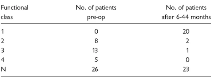
All patients were weaned from CPB with moderate inotrop-ic support. The mean cross-clamp time was 91 ± 11 minutes and mean CPB time was 120 ± 12 minutes, while TCA time was 9 ± 4 minutes (Table 2, ). Operative mortality was zero and there was one 30-day mortality (3.8%). The patient who died had been readmitted to the hospital with a deep sternal wound infection five days after he was discharged. The infec-tion did not respond to treatment and the patient died of sep-sis 22 days after the operation. One patient was reoperated upon for bleeding and one patient developed a minor neuro-logical deficit that resolved before discharge. Another patient had a prolonged ventilation period (Table 3, ). Mean hospi-tal stay was 9 ± 2 days. At discharge all patients had a compe-tent valve with low transvalvular gradients. There was a slight decrease in transvalvular gradients during follow-up but this was not significant (Table 4, ). One patient died of an unknown cause six months after the operation, and one patient was lost to follow-up. A patient who had mild mitral insufficiency during her first operation returned with severe mitral regurgitation one year later and underwent a mitral valve replacement. In the remaining patients, the mean func-tional class (NYHA) dropped significantly at the end of the 15-month follow-up period (Table 5, ). During this time, no valve failure was detected. Electron beam tomography (EBT) of a patient 34 months after the operation showed no sign of wall calcification of the Freestyle valve (Figure 2, ).
Table 1. Demographic Data of Patients
Age (mean) 71 ± 4 (range 61-78)
Gender (male/female) 19/7
Aneurysm diameter (cm) (mean) 5.8 ± 0.6 (range 4.9-7)
Aortic dissection (type A) 1
Figure 1.
Table 2. Operative Data
Cross-clamp time (min.) 91 ± 11
By-pass time 120 ± 12
TCA time 9 ± 4
Associated Procedures
CABG 2
Mitral valve repair 1
Statistical Analysis
All data are represented as mean ± standard deviation. One-way analysis of variants (ANOVA) was used to test the signifi-cance of changes in measurements over the follow-up time.
D I S C U S S I O N
Over the years, many surgical methods have been devel-oped to treat AAA with aortic regurgitation [Mingke 1998, Shapira 1999]. The operation described by Bentall and DeBono continues to be the gold standard therapy for this pathology [Bentall 1968]. However, when it comes to the choice of the valve being used, there is no perfect device for this operation. Mechanical valves present the risk of throm-boembolism, the need for anticoagulation, and bleeding complications. Stented biological valves are another option, but reoperation can be difficult in case of late degeneration since it is usually necessary to remove the entire conduit [Myken 1995, Akpinar 1999, Cartier 1999, Peterseim 1999]. Homografts may offer the ideal solution but they are not readily available, and reports of early wall calcification are disturbing [David 1994, Grodd 1995, Chon 1996, Cartier 1999]. Stentless xenografts are the porcine equivalent of the aortic homograft, with virtually identical flow characteristics [Westaby 1998c, Westaby 2000]. The concept of aortic valve replacement (AVR) with a stentless porcine valve originated with the pioneering work of O’Brien in the mid-1960s [O’Brien 1967]. Observation of optimal hemodynamics and hopes of longer durability due to absence of stents has revived attention for these valves [O’Brien 1967, Sidiropou-los 1997, Peterseim 1999]. Early and mid-term reports of ideal hemodynamic function comparable to homografts and the advantage of availability in all sizes have made the less valves highly attractive. The major advantages of stent-less valves are better hemodynamics, fewer thromboembolic events, absence of dramatic failure, and increased durability [Westaby 1998a, Cartier 1999].
The combination of aortic root replacement with a xenograft and replacement of the ascending aorta with a Dacron tube is one of the proposed treatment options for this
pathology. However, bleeding at the suture line between the two components can be troublesome, especially if the anasto-mosis is under tension [Urbanski 1999]. Urbanski and Hack-er explained a method in which a valved composite graft con-sisting of a stentless valve and a Dacron tube prosthesis was prepared in the operating room after the annulus was meas-ured. The stentless valve was sewn to the aortic annulus together with the tube graft using 4-0 polypropylene suture passing through both the vascular tube and the valve. The authors had to abandon this technique because of bleeding problems, and in later attempts they first sewed the valve to the tube graft and then anastomosed the tube graft to the aortic annulus with interrupted pledgeted sutures [Urbanski 1999, 2000]. The drawback to this option is that the assembly of the composite graft during surgery prolongs the ischemic arrest time and makes the procedure more complicated.
In our experience the major advantage of the Freestyle valve is its versatility. It can be used as a whole root or a mini-root, or a subcoronary implantation can be performed during ordinary AVR [Kon 1995]. An account of a root inclusion pro-cedure with this valve for the treatment of an acutely dissected root in an elderly patient was published by Westaby [Westaby 1998b]. Excellent early and mid-term results (7 years) have Table 3. Complications
Bleeding 1
Pulmonary 1
Neurological 1
Infection 1
Table 4. Echocardiographic Results
Valve size Gradient (discharge) Mean 13 months AI
25 8 ± 1.1 7 ± 1.8 0
27 6 ± 1.5 5.0 0
29 4.0 3.2 0
AI = aortic insufficiency
Table 5. Improvement in Functional Class (NYHA)
Functional No. of patients No. of patients
class pre-op after 6-44 months
1 0 20
2 8 2
3 13 1
4 5 0
N 26 23
NYHA = New York Heart Association
The Use of Stentless Valves for Root Replacement During Repair of Ascending Aortic Aneurysms with Aortic Valve Regurgitation—Akpinar et al.
made this valve our choice for patients undergoing AVR who are over 65 years of age [Cartier 1999]. This valve was an appealing option for our patients in this series because most of them were elderly and two could not use anticoagulants.
Replacement of the aortic valve with a stentless xenograft is a more demanding surgical procedure, and attention to detail is essential. There are some technical points that have to be mentioned. Myocardial protection with retrograde blood car-dioplegia and direct carcar-dioplegia from the right coronary ostia is very important, especially for right ventricle protection. Taking deep sutures from the annulus and incorporating a strip of pericardium between the Freestyle valve and the annulus is very helpful for hemostasis. The coronary ostia have to be freed completely and the corresponding openings on the Freestyle valve have to be generous in order to prevent tension. We advise the enlargement of the openings on the Freestyle valve to prevent any kinking of the coronary ostia and to permit a tension-free anastomosis. Finally, the length of the tube graft has to be measured correctly. A graft that is too short will cause tension on the anastomosis and may lead to bleeding and dehiscence, which has been reported by some authors [Urbanski 1999]. On the other hand, a graft that is too long may cause kinking, but this can be resolved with external traction sutures outside the graft.
The major drawback of the method is the need for a sec-ond tube graft to reach the proximal arch because the Freestyle valve is not long enough. This additional procedure requires a short time of TCA and prolongs the ischemic time. However, with good myocardial protection this is of minor importance. The problem of insufficient graft length may be solved in the near future with the introduction of longer Freestyle conduits (Medtronic Files, personal communica-tion). Another criticism of the method may be the difficulty of a future reoperation caused by porcine aortic wall calcifica-tion. It is difficult to comment on this problem since we have not experienced it so far. EBT views of two patients three and four years after the operation revealed no sign of calcification over the porcine aortic wall, which is a promising sign. Carti-er et al. have recently reported less wall calcification on the Freestyle valve than the homograft after 18 months of follow-up [Cartier 1999]. However, for the moment we agree with others who have suggested that the general guidelines for the use of other bioprostheses should be applied to these valves until longer term results are available. Although only the test of time, as reflected in results over ten years after implanta-tion, will prove the value of these valves, the results so far are encouraging. There are numerous papers that confirm the effectiveness of cardiac and systemic physiological rehabilita-tion with the Freestyle valve leading to resolurehabilita-tion of left ventricle hypertrophy and a low incidence of valve-related complications [Westaby 1998a, 1998b].
C O N C L U S I O N S
The technique of using the Freestyle valve and a Dacron tube graft provides a good alternative for patients who have an aortic aneurysm and require replacement of both the valve and the aneurysmal segment. If the patients are over 65 years of age one cannot use oral anticoagulants. The technique will become easier and more widely applied with the introduction
of longer Freestyle-valved conduits that can reach the proxi-mal arch and thereby avoid the use of an additional tube graft.
R E F E R E N C E S
1. Akpinar B, Sanisoglu I, Konuralp C, et al. The use of a stentless porcine bioprosthesis. Tex Heart Ins J 26:195-7, 1999.
2. Bentall H, DeBono A. A technique for complete replacement of the ascending aorta. Thorax 23:338-9, 1968.
3. Cartier CP, Dumensil GJ, Metras J, et al. Clinical and hemodynamic performance of the Freestyle aortic root bioprosthesis. Ann Thorac Surg 67:345-51, 1999.
4. Chon LH, Rizzo RJ, Adams DH, et al. Reduced mortality and mor-bidity for ascending aortic aneurysm resection regardless of cause. Ann Thorac Surg 62:463-8, 1996.
5. David TE, Feindel CM, Bos J, et al. Aortic valve replacement with a stentless porcine aortic valve: a six year experience. J Thorac Car-diovasc Surg 108:1030-36, 1994.
6. Grodd C, Hasinger W, Mair R, et al. Aortic valve replacement: Is the stentless xenograft an alternative to the homograft? Ann Thorac Surg 60:S 418-21, 1995.
7. Kon ND, Westaby S, Amaresena N, et al. Comparison of implant techniques using the Freestyle stentless porcine aortic valve. Ann Thorac Surg 59:857-62, 1995.
8. Mingke D, Dresler C, Stone CD, et al. Composite graft replace-ment of the aortic root in 335 patients with aneurysm or dissection. J Thorac Cardiovasc Surg 46:12-19, 1998.
9. Myken PS, Caidahl K, Larsson P, et al. Mechanical versus biological valve prosthesis. Ann Thorac Surg 60:S447-S452, 1995.
10. O’Brien MF. Heterograft aortic valves for human use: valve bank, techniques of measurements and implantation. J. Thorac Cardiovasc Surg 53:392-7, 1997.
11. O’Brien MF. Composite stentless xenograft for aortic valve replace-ment: clinical evaluation of function. Ann Thorac Surg 60:S406-9, 1995.
12. Shapira OM, Aldea GS, Cutter SM, et al. Improved clinical out-comes after operation of the proximal aorta. Ann Thorac Surg 67: 1030-37, 1999.
13. Sidiropoulos A, Hotz H, Tschesnow J, et al. Stentless porcine bio-prostheses for all types of aortic root pathology. Eur J Cardiothorac Surg 11:917-21, 1997.
14. Peterseim DS, Ye-Ying Cen, Cheruvu S, et al. Long term outcome after biologic versus mechanical aortic valve replacement in 841 patients. J Thorac Cardiovasc Surg 117:890-7, 1999.
15. Urbanski PP. Replacement of the ascending aorta and aortic valve with a valved stentless composite graft. Ann Thorac Surg 67:1501-02, 1999.
16. Urbanski PP, Hacker RW. Replacement of the aortic valve and ascending aorta with a valved stentless composite graft: technical considerations and early clinical results. Ann Thorac Surg 70:17-20, 2000.
17. Westaby S, Jin YK, Katsumata T, et al. Valve replacement with a stentless bioprosthesis: versatility of the porcine aortic root. J Tho-rac Cardiovasc Surg 116:477-84, 1998.
18. Westaby S, Katsumata T, Houel R, et al. Stentless xenograft repair of the dissected aortic root. Ann Thorac Surg 65:1448-50, 1998. 19. Westaby S, Horton M, Jin YK, et al. Survival advantage of stentless
aortic bioprostheses. Ann Thorac Surg 70:785-92, 2000.
20. Westaby S, Huysmans HA, David TE. Stentless aortic bioprosthe-ses. Ann Thorac Surg 65:235-40, 1998.
56
R E V I E W A N D C O M M E N TA RY
1. Editorial Board Member RN41 writes:
This is a series with good results and offers another option in the treatment of patients with ascending aortic aneurysms. The jury is still out on the benefits of stentless valves, and the use of this valve varies markedly from center to center. The early results in this series are encouraging.
a) During the same period, how many patients had an aortic root replacement with a composite graft (incorporating a mechanical valve)?
b) Were there any contraindications to the use of this technique?
Authors’ Response from Belhhan Akpinar, MD:
a) During this period, 81 patients underwent aortic root replacement with a mechanical valve.
b) One contraindication to the use of this technique would be young age (less than 60 years), and a second one would be a patient with severely depressed LV function undergoing concomitant procedures. For such a patient it might be wiser to use a composite graft that could shorten the ischemia time.
2. Editorial Board Member EK34 writes:
What is the difference between “operative” mortality and “30-day” mortality? You should list in-hospital or 30-day mortality.
What about brain function and/or neurological damage?
Authors’ Response from Belhhan Akpinar, MD:
All patients survived the operation. In-hospital mortality was 3.7% <AU: 3.8%?> (one patient). As for neurological
damage, one patient developed delirium on the second post-op day and had to be reintubated and sedated for two days. This problem was resolved in one week. There was no stroke or other neurological damage.
3. Editorial Board Member KE221 writes:
a) The authors dismiss the option of using a stented xenograft within a Dacron tube by saying that replace-ment of the xenograft would require replacing the entire root prosthesis. This alternative should receive more dis-cussion. First, durability is now excellent for modern stented xenografts in the older patients discussed in this paper, and there is little evidence that stentless valves are significantly more durable in older patients. Second, replacement of the Freestyle valve would also require replacing the entire root. Third, stented xenografts are very easy to incorporate into a Dacron graft at the time of surgery without the need for additional sutures, and with excellent hemostasis. Fourth, if a separate segment of graft is used for an anastomosis to the arch, the remaining anastomosis is an easy one between graft and graft.
Authors’ Response from Belhhan Akpinar, MD:
Using a xenograft within a Dacron tube is another option in this group of patients. The author agrees that durability is excellent for modern stented grafts, but this requires the use of a stentless valve as a whole root. In any case, if reoperation becomes necessary, either valve would require re-replacing the root. As mentioned in the text, we have not had to reop-erate for any of the Freestyle valves so far, but our experience with the two reoperations for the early Edwards Prima valve suggested to us that implanting a new valve within the previ-ous tube was not possible.