ORIGINAL ARTICLE
P21 and p27 Immunoexpression in Upper Urinary Tract
Urothelial Carcinomas
Banu Sarsik1&Basak Doganavsargil1&Adnan Simsir2&Ayse Yazici3& Burcin Pehlivanoglu4&Cag Cal2&Sait Sen1
Received: 18 July 2015 / Accepted: 16 May 2016 / Published online: 24 May 2016 # Arányi Lajos Foundation 2016
Abstract p21 and p27 are members of cyclin-dependent ki-nase family, which function as tumor suppressors and they are involved in development and progression of several ma-lignancies. We investigated their expression in upper urinary tract urothelial carcinoma (UUTUC). Radical nephroureterectomy materials of 34 patients were assessed by immunohistochemistry to evaluate expres-sion of p21 and p27 in UUTUC. Results were correlat-ed with various clinicopathological variables as age, gender, tumor grade and stage, tumor architecture, multifocality, subsequent bladder carcinoma develop-ment and clinical outcome.
p21 and p27 expression was observed in 52.9 % (n = 18) and 88.2 % (n = 30), respectively. A total of 21 tumors (61.7 %) showed either total loss of p21 expression (n = 16, 47 %) or lower expression (n = 5, 14.7 %). No correlation was found between p21 expression and clinicopathologic vari-ables. Cases showing total loss or lower p27 expression (11.7 % and <25.6 %, respectively) (n = 19, 55.8 %) consti-tuted 67.6 % (n = 23) of the cases totally. This loss or lower p27 expression correlated with a shorter overall survival in both univariate and multivariate analysis (p = 0.039 and
p = 0.037, respectively). None of the noninvasive tumors (papillary and nodular tumors) showed loss of p27 (p = 0.016) while 33.3 % of invasive ones showed p27 loss. Noninvasive tumor architecture also correlated with subse-quent bladder carcinoma development (p = 0.032) while in-vasive tumor architecture correlated with advanced stage (T3 and T4) (p = 0.003). p27 is widely expressed in UUTUC, while p21 expression is observed in half of the cases. Loss of p27 expression correlated with tumor architecture and over-all survival in UUTUC. However, further research is needed to assess their role in UUTUC.
Keywords Cylin-dependent kinase inhibitors . p21 . p27 . Prognosis . Upper tract urothelial carcinoma . Urinary tract
Introduction
Urothelial carcinomas arising from upper urinary tract (upper urinary tract urothelial carcinoma; UUTUC) account for 5 % of all urothelial malignancies and approximately 10 % of renal tumors [1]. Current standard treatment option for UUTUC is radical nephroureterectomy with removal of a bladder cuff. It has been reported that metastasis occurs in 20–55 % of pa-tients with UUTUC [2]. Tumor stage, histologic grade, lymph node involvement and type of surgical procedure have been demonstrated to be significant prognostic factors [2–4].
p21waf1 (p21) and p27kip1 (p27) are members of cyclin-dependent kinase (CDK) interacting protein/kinase inhibitory protein (Cip/Kip) family, which function as tumor suppressors by preventing the cell proceeding from G1 to S phase. They have been reported to be involved in development and pro-gression of several malignancies. Loss of p27 expression has been shown in several organ tumors, including breast [5],
* Banu Sarsik [email protected]
1 Department of Pathology, Ege University Faculty of Medicine,
Bornova, 35100 Izmir, Turkey
3
Department of Pathology, Mugla University Education and Research Hospital, Mugla,, Turkey
4 Department of Pathology, Adiyaman University Training and
Research Hospital, Adiyaman, Turkey DOI 10.1007/s12253-016-0075-4
2
Department of Urology, Ege University Faculty of Medicine, Izmir, Turkey
prostate [6], colon [7], bladder [8] and has been associated with poor prognosis [9].
p21 expression have been found to affect prognosis in pre-vious studies, however, whether p21 levels may be used as an independent prognostic factor is still not clear [10–12]. Although the role of p27 in carcinogenesis of UUTUC has been investigated in previous studies, results are conflicting [13–16]. Similarly, several studies are available in the litera-ture on the role of p21 in urothelial carcinoma of bladder [8, 10,11]. But no data is available in the literature on its role in UUTUC.
In this study, we investigated expression of p21 and p27 in UUTUC, and evaluated the association of p21 and p27 ex-pression with age, gender, tumor architecture, multifocality and developed subsequent urinary bladder carcinoma and clinical outcome.
Methods
Patient SelectionThirty-four cases with UUTUC diagnosed between 1992 and 2004, were retrieved from the archives of Pathology Department of Ege University Medical Faculty. Surgical pro-cedures were performed in one center by various surgeons, using approved standard radical nephroureterectomy princi-ples (extrafascial transabdominal dissection of the kidney and ureter, lymphadenectomy, and combined removal of the bladder cuff [17]. All palpable lymph nodes were removed during surgery. The dissected lymph nodes were located in paracaval, para-aortic areas between renal vessels and inferior mesenteric artery, and renal hilar region. Tumor multifocality was defined as synchronous presence of two or more patho-logically confirmed tumors in any location (renal pelvis, ureter or bladder). Patients with the history of urothelial carcinoma of the urinary bladder were excluded. Bladder tumors detected six months after the surgery was considered as new-onset malignancy (i.e., not interpreted as recur-rence or disease progression). The lifespan of patients who died from UUTUC was measured in months. Patients who died in the perioperative period (i.e., with-in 30 days after surgery) were excluded from the study. Survival periods were calculated according to patient demographics.
Pathologic Evaluation
All surgical specimens were processed according to standard pathological procedures and histologically confirmed to be urothelial carcinoma. No additional histological features were found (e.g. squamous cell carcinoma or adenocarcinoma). Tumors were staged according to the TNM classification
[18] and later dichotomized into two groups for statistical purposes, as T0 (n = 4), T1 (n = 8), and T2 (n = 1) tumors that were classified as early stage, and T3 (n = 9) and T4 (n = 12) tumors, classified as advanced stage. Tumor grading was performed according to the 2004 World Health Organization–International Society of Urologic Pathology Consensus Classification [19]. Tumor architecture was de-fined as papillary, nodular or invasive based on the predomi-nant feature of the index lesion.
Immunohistochemistry
Formalin-fixed and paraffin-embedded tissue specimens of all patients were obtained for immunohistochemical (IHC) stain-ing. A demonstrative block containing normal kidney tissue adjacent to the tumor was selected for IHC evaluation. 4– 5 μm thick sections were prepared from paraffin embedded tissues and were placed on electrostatic-charged slides (X-traTM, Surgipath Medical Industries, Richmond, Illinois, USA). Sections were deparaffinized and dehydrated through a g r a d e d e t h a n o l s e r i e s u s i n g r o u t i n e p r o t o c o l s . Immunohistochemical study was performed by using mouse monoclonal antibodies anti-p21 (Neomarkers, MS 230-P, 200 mg/L, 1/40 dilution) and anti-p27 (Neomarkers, MS 256-P, 200 mg/L, 1/50 dilution). Briefly, sections were incu-bated at 50 °C overnight, deparaffinized in xylol and rehydrated through graded alcohol rinses. To enhance antigen retrieval, slides prepared for p21 staining were heated in 0.1 mmol/L EDTA buffer (pH 8) in a microwave oven (650 W for 5 min plus 450 W for 20 min) and those for p27 were immersed in citrate buffer (10 mmol/L, pH 6) and heated in a vacuum-pan for 2.5 min and cooled for 20 min. Endogenous peroxidase activity was blocked with 30 mL/L H2O2 for 5 min. The incubation time with monoclonal anti-b o d i e s p 2 7 a n d p 2 1 w a s o p t i m i z e d a s 3 0 m i n . Immunostaining of p27 and p21 was performed with the streptavidin-peroxidase method (Labvision, TM-125-HL), and alkaline phosphate-based visualization system. 3,3′-di-aminobenzidine (Dako, TA-125 HD) was used as a chromo-gen. Carcinomatous breast and colon tissue served as a posi-tive control, whereas primary mAb was omitted for negaposi-tive control sections. Stained lymphocytes were used as an internal control.
Evaluation of Immunohistochemistry Results
Scoring was performed separately by two pathologists who were blinded to the patient characteristics as well as each other’s scores. In each case, a total of 1000 cells were counted in two randomly selected representative high power fields (HPFs). Percentage of positive nuclear staining either weak or strong was noted. Mean scores counted by each observer was accepted as expression score.
Statistical Analysis
Statistical analysis was carried on a PC based analysis pro-gram SPSS (version 18.0). After initial frequency analysis the cases were categorized into three groups asBtotal loss of p21 or p27^ when there was no immunexpression. The cases with expression scores below the median of all cases’ scores were regarded as‘low p21 or low p27 expression’ while the scores above the median of all cases’ scores were coded as ‘high p21 or high p27 expression’. Pearson Chi-square or Fisher’s exact tests were used to evaluate the association between categorical variables. Differences in continuous variables were analyzed using the Mann-Whitney U test or Kruskal-Wallis test. Kaplan-Meier analyses with the log-rank test were used for survival analysis. Prognostic variables were analyzed for as-sociation with overall survival using the Cox regression model in both a univariate and multivariate analysis. A p value of <.05 was accepted as significant.
Results
Patient and Tumor Characteristics
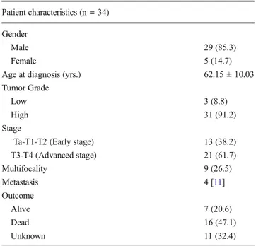
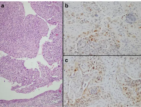
Mean age was 62.15 ± 10.03 (range 46–79) years, and 85.3 % of the patients were male. Patients’ characteristics are shown in Table1. Histologically, 6 out of 34 tumors (17.6 %) had nodular pattern, 16 tumors (47.1 %) had papillary pattern and 12 tumors (35.3 %) had invasive pattern (Fig.1/A). Three cases (8.8 %) had low grade tumors, whereas 31 cases (91.2 %) had high grade tumors.
p21 and p27 Expression
The median p21 and p27 expression scores were 6.97 % ± 10.03 (range 0–36) and 25.62 % ± 25.92 (range 0–84), respectively. The results of p21 and p27 nuclear immunoreactivity are shown in Fig. 1. p21 and p27 expression was observed in 52.9 % (n = 18) and 88.2 % (n = 30) of the cases respectively. A total of 21 tumors (61.7 %) showed either total loss of p21 expression (n = 16, 47 %) or showed lower p21 expression (n = 5, 14.7 %). But no correlation was found between p21 expression and the tested clinicopathological variables. However, cases showing loss of p27 expression (n = 4, 11.7 %) and cases with lower p27 expression (n = 19, 55.8 %) constituted 67.6 % (n = 23) of the cases totally (Table2).
Associations between Variables
The immunostaining in relation to the tumor characteristics of the patients are summarized in Table 2. There was no significant association between p21expression and tumor grade, stage, multifocality, architecture, development of subsequent bladder tumor and p27 expression (Table2). On the other hand, presence of p27 expression correlated with tumor architecture, none of the noninvasive tumors (papillary and nodular tumors) showed total loss of p27 (p = 0.016, Table 2). However, grade, stage, multifocality and development of subsequent bladder tumor did not correlate with p27 expression either. Six of the 34 (17.6 %) patients treated for UUTUC had subsequent bladder carcinoma development, at a mean interval of 66 (range 24–120 months) months. Of these, 16,7 % cases had low-grade and 83.3 % had high-grade tumors. Noninvasive (papillary and nodular) tumor architecture of the UUTUC correlated with subsequent bladder cancer development (p = 0.032) and invasive tumor architecture correlated with late stage (p = 0.003).
Outcome
The follow-up period comprised the time between the diagno-sis and the time of death or last control. The mean follow-up period in this series was 59.13 ± 52.0 (range, 2–180) months. The mean survival period was 87.35 ± 14.55 months (95%CI of 59–116 months). In the overall group, 1, 5 and 10 year-survival rates were 86 %, 49 % and 29.4 %, respectively.
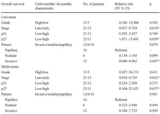
Patients with lower levels of p27 showed a shorter overall survival period (p = 0.023) as 63 months (95 % CI 30–95). For overall survival, p27 was found to be a significant prognostic marker in univariate and multivariate analysis (p = 0.039 and p = 0.037, respectively, Table 3). On the other hand, p21 expression did not correlate with survival (p > 0.05) and the mean survival of cases with low level p21 expression was 71 months (95 % CI 46–96).
Stage was also found to be significantly associated with prognosis in both univariate and multivariate analysis
Table 1 Patient characteristics
Patient characteristics (n = 34) Gender
Male 29 (85.3)
Female 5 (14.7)
Age at diagnosis (yrs.) 62.15 ± 10.03
Tumor Grade
Low 3 (8.8)
High 31 (91.2)
Stage
Ta-T1-T2 (Early stage) 13 (38.2)
T3-T4 (Advanced stage) 21 (61.7) Multifocality 9 (26.5) Metastasis 4 [11] Outcome Alive 7 (20.6) Dead 16 (47.1) Unknown 11 (32.4)
(p = 0.014 and p = 0.022, respectively, Table3). However, only invasive architecture was found to be significant in uni-variate analysis (p = 0.047, Table3).
Discussion
Different factors determine the prognosis and clinical outcome in patients with UUTUC. Tumor grade, stage, lymph node
involvement and type of surgical procedure are widely con-sidered as significant prognostic factors [2–4]. In the present study, stage was found to be an independent predictor of over-all survival.
The role of p21 and p27 in the pathogenesis of different malignancies have been investigated in several previous stud-ies [5–16]. We showed that higher p21 and lower p27 expres-sion was correlated with adverse prognostic factors in prostate carcinoma in a previously published study [21]. Similarly,
Fig. 1 Papillary pattern and high grade tumor (A: Hematoxylin-eosin ×10) Immunohistochemical staining in tumor cells (B: p21 immunoexpression, ×20; C: p27 immunoexpression (×20)
Table 2 Comparison of p21 and p27 staining with clinicopathological features
Total number of cases p21 total loss n (%) p Low p21 n (%) High p21 n (%) p p27 total loss n (%) p Low p27 n (%) High p27 n (%) p Total number of cases 34 16 (47) 5 (14.7) 13 (38.2) 4 (11.7) 19 (55.8) 11 (32.3) Architecture 0.14 0.38 0.016* 0.101 Nodular 6 5 (83.3) 0 1 (16.6) 0 2 (33.3) 4 (66.6) Papillary 16 6 (37.5) 4 [20] 6 (37.5) 0 11 (68.7) 5 (31.2) Invasive 12 5 (41.6) 1 (8.3) 6 (50) 4 (33.3) 6 (50) 2 (16.6) Multifocality 0.55 0.72 0.25 0.44 Present 9 5 (55.5) 1 (11.1) 3 (33.3) 2 (22.2) 5 (55.5) 2 (22.2) Absent 25 11 (44) 4 [16] 10 (40) 2 [8] 14 (56) 9 (36) Stage 0.53 0.71 0.12 0.54 Early 13 7 (53.8) 2 (15.3) 4 (30.7) 0 8 (61.5) 5 (38.4) Advanced 21 9 (42.8) 3 (14.2) 9 (42.8) 4 [19] 11 (52.3) 6 (28.5) Grade 0.47 0.27 0.50 1.00 Low 3 2 (66.6) 1 (33.3) 0 0 2 (66.6) 1 (33.3) High 31 14 (45.1) 4 (12.9) 13 (41.9) 4 (12.9) 17 (54.8) 10 (32.2) Subsequent bladder carcinoma development 0.28 0.78 1.00 0.07 Present 6 4 (66.6) 0 2 (33.3) 0 2 (33.3) 4 (66.6) Absent 28 12 (42.8) 5 (17.8) 11 (39.2) 4 (14.2) 17 (60.7) 7 [20]
Romics et al. have reported that grade progression in prostate carcinoma was accompanied with down-regulation of p27kip1 protein [22]. Down-regulation of p21 was found to be associated with HPV-18 positive cervical carcinomas, sug-gesting that loss of p21 molecule may contribute to the ma-lignant transformation of HPV-18 positive carcinomas [23].
There are discrepancies in previous reports regarding the association between low p27 expression and high tumor stage and grade for UUTUC. Although Masuda et al. [13] did not find any association between these parameters, Kamai et al. [14] reported that low p27 expression correlated with higher tumor stage. In the present study, we did not find any associ-ation between p27 expression and tumor stage or grade; how-ever, p27 expression was associated with overall survival. This finding is consistent with Kamai et al’s [14] report sug-gesting that p27 may play a role in the progression of urothelial carcinoma arising from pelvis renalis and ureter. However, in a more recent study of 62 patients, using tissue microarray technology, Fromont et al. reported that p27 had no prognostic value in UUTUC [15].
Tumor architecture has been demonstrated to predict clin-ical outcomes in international multicenter studies [20,24,25]. Sessile pattern was shown to be significantly associated with metastasis and an independent predictor of cancer specific mortality [24]. In a multi-institutional study of 1363 patients, Remzi et al. found that in 40 % of patients with papillary tumors and in 77 % of those with sessile tumors disease re-curred five years after radical nephroureterectomy [20]. Similarly, all invasive tumors were advanced-stage tumors in
our study group and invasive architecture was found to be a significant prognostic factor in univariate analysis.
Previous studies have been conducted on the association between tumor architecture and p21 and p27 expression in bladder tumors. Korkolopoulou et al. reported that no signif-icant relationship was detected between p21 expression and noninvasive papillary carcinoma of bladder [10]. On the con-trary, Franke et al. reported that the highest p27 expression was observed in papillary tumors of bladder [26]. However, the prognostic significance of tumor architecture and p21 or p27 expression has not been conclusively investigated in UUTUC. In the present study, we observed p27 negativity in 33.3 % of cases with invasive pattern whereas p27 expression was present in all cases with noninvasive UUTUC, similar to papillary bladder carcinomas. Similar to loss of p27 expres-sion, cases with lower p27 expression was also more frequent among cases with invasive pattern of growth pattern. Of note; in this study Blow expression^ for both p21 and p27 was defined as expressions lower than median expression ratios. Therefore, the cut-off levels presented herein should be eval-uated with caution as there is no pioneering study on their expression in UUTUC and as there is a lack of agreed cut-off levels in the literature for other tumors either.
Few studies to date have focused on the potential risk fac-tors for the development of subsequent bladder tumors after managing their UUTUC. It has been reported that 15–50 % of patients who underwent radical nephroureterectomy for UUTUC have subsequent cancer development in the bladder [27–29]. Tumor multiplicity, stage, grade, size, and ureteric
Table 3 Cox regression analysis for various potential prognostic factors in overall survival
Overall survival Unfavourable/ favourable
characteristic
No. of patients Relative risk
(95 % CI) p Univariate Grade High/low 31/3 0.246–14.406 0.542 Stage Late/early 21/13 0.057–0.724 0.014* p21 Low/high 21/13 0.292–2.457 0.760 p27 Low/high 23/11 1.071–13.491 0.039* Pattern Invasive/nodular/papillary 12/6/16 0.079 Papillary 16 Referent -Nodular 6 0.139–1.185 0.099 Invasive 12 0.040–0.982 0.047* Multivariate Grade High/low 31/3 0.027–36.313 0.431 Stage Late/early 21/13 0.018–0.729 0.022* p21 Low/high 21/13 0.224–2.289 0.573 p27 Low/high 23/11 0.104–25.421 0.037* Pattern Invasive/nodular/papillary 12/6/16 0.961 Papillary 16 Referent -Nodular 6 0.323–3.948 0.849 Invasive 12 0.106–7.732 0.929 *p < 0.05
location, as well as patient gender and surgical method are the reported risk factors [27–29]. In our study, we analyzed whether p21 and p27 expression of the UUTUC is associated with the development subsequent urinary bladder cancer but no significant relationship was found, possibly because of the low number of cases. Our study showed that noninvasive (papillary and nodular) tumor architecture correlate with de-velopment of subsequent bladder tumor. This finding may be the result of longer survival in patients with noninvasive tumors.
p21 immunopositivity occurred in 52.9 % of our cases and 61.7 % of those expressed low levels of p21 (Table2). The frequency of p21 expression in bladder cancer was observed between 30 and 64 % in the literature [10,30–32]. No signif-icant relationship was detected between p21 expression and clinicopathological features consistent with previous bladder cancer studies. It has been proposed that p21 activation may be an early event in urothelial carcinogenesis [30,32]. Also, although p21 and p27 are the members of the same protein family, p21 may be less effective on tumor suppression, as suggested by Philipp-Staheli et al. recently [33]. Observation on p21 is more limited and further investigation is needed to explore its role in urothelial tumorigenesis.
We appreciate that our study has limitations, especially related with smaller number of cases due to lower prevalence of UUTUC. However, we believe that the observations pre-sented above can help to enlighten the prognosis of upper tract urothelial carcinoma. Further studies are needed to confirm the role of p21 and p27 in upper tract urothelial carcinoma.
Compliance with Ethical Standards
Conflicts of Interest None declared.
References
1. Jemal A, Siegel R, Ward E, Hao Y, Xu J, Thun MJ (2009) Cancer
statistics, 2009. CA Cancer J Clin 59(4):225–249
2. Novara G, De Marco V, Gottardo F, Dalpiaz O, Bouygues V,
Galfano A, et al. (2007) Independent predictors of cancer-specific survival in transitional cell carcinoma of the upper urinary tract: multiinstitutional dataset from 3 European centers. Cancer 110(8):
1715–1722
3. Chromecki TF, Cha EK, Fajkovic H, Margulis V, Novara G, Scherr
DS, et al. (2012) The impact of tumor multifocality on outcomes in patients treated with radical nephroureterectomy. Eur Urol 61(2): 245–253
4. Verhoest G, Shariat SF, Chromecki TM, Raman JD, Margulis V,
Novara G, et al. (2011) Predictive factors of recurrence and survival
of upper tract urothelial carcinomas. World J Urol 29(4):495–501
5. Catzavelos C, Bhattacharya N, Ung YC, Wilson JA, Roncari L,
Sandhu C, et al. (1997) Decreased levels of the cell-cycle inhibitor p27Kip1 protein; prognostic implications in primary breast cancer.
Nat Med 3(2):227–230
6. Cote RJ, Susan Y, Groshen S, Alexander GM, Gatti LA, Firpo EJ,
et al. (1998) Association of p27Kip1 levels with recurrence and survival in patients with stage C prostate carcinoma. J Natl
Cancer Inst 90(12):916–920
7. Loda M, Cukor B, Tam SW, Lavin P, Fiorentino M, Draetta GF,
et al. (1997) Increased proteasomedependent degradation of the cyclin-dependent kinase inhibitor p27 in aggressive colorectal
car-cinomas. Nat Med 3(2):231–234
8. Shariat SF, Zlotta AR, Ashfaq R, Sagalowsky AI, Lotan Y (2007)
Cooperative effect of cell-cycle regulators expression on bladder cancer development and biologic aggressiveness. Mod Pathol 20(4):445–459
9. Lloyd RV, Erickson LA, Jin L, Kulig E, Qian X, Cheville JC, et al.
(1999) p27kip1: a multifunctional cyclin-dependent kinase inhibi-tor with prognostic significance in human cancers. Am J Pathol 154(2):313–323
10. Korkolopoulou P, Konstantinidou AE, Thomas-Tsagli E,
Christodoulou P, Kapralos P, Davaris P (2000) WAF1/p21 protein expression is an independent prognostic indicator in superficial and invasive bladder cancer. Appl Immunohistochem Mol Morphol 8(4):285–292
11. Shariat SF, Karakiewicz PI, Ashfaq R, Lerner SP, Palapattu GS,
Cote RJ, et al. (2008) Multiple biomarkers improve prediction of bladder cancer recurrence and mortality in patients undergoing cystectomy. Cancer 112(2):315–325
12. Matsushima H, Sasaki T, Goto T, Hosaka Y, Homma Y, Kitamura T,
et al. (1998) Immunohistochemical study of p21WAF1 and p53 proteins in prostatic cancer and their prognostic significance. Hum
Pathol 29(8):778–783
13. Masuda M, Takano Y, Iki M, Makiyama K, Ikeda I, Noguchi S,
et al. (2000) Cyclin-dependent kinase inhibitor p27kip1 expression in transitional cell carcinoma of renal pelvis and ureter. Cancer Lett
150(2):183–189
14. Kamai T, Takagi K, Asami H, Ito Y, Arai K, Yoshida KI (2000)
Prognostic significance of p27kip1 and Ki-67 expression in
carci-noma of the renal pelvis and ureter. BJU Int 86(1):14–19
15. Fromont G, Rouprêt M, Amira N, Sibony M, Vallancien G, Validire
P, et al. (2005) Tissue microarray analysis of the prognostic value of E-cadherin, Ki67, p53, p27, survivin and MSH2 expression in
up-per urinary tract transitional cell carcinoma. Eur Urol 48(5):764–
770
16. Gakis G, Schwentner C, Todenhöfer T, Stenzl A (2012) Current
status of molecular markers for prognostication and outcome in
invasive bladder cancer. BJU Int 110(2):233–237
17. Simsir A, Sarsik B, Cureklibatir I, Sen S, Gunaydin G, Cal C (2011)
Prognostic factors for upper urinary tract urothelial carcinomas:
stage, grade, and smoking status. Int Urol Nephrol 43(4):1039–
1045
18. Sobin LH, Gospodarowicz MK, Wittekind C, eds. TNM
classifica-tion of malignant tumors, ed. 7. UICC Internaclassifica-tional Union Against
Cancer. Hoboken, NJ: Wiley-Blackwell; 2010. p. 258–265.
19. Eble JN, Sauter G, Epstein JI, et al. World Health Organization
classification of tumours. Pathology and genetics of tumours of the urinary system and male genital organs. Lyon, France: IARC Press; 2004, p.90.
20. Remzi M, Haitel A, Margulis V, Karakiewicz P, Montorsi F,
Kikuchi E, et al. (2009) Tumour architecture is an independent predictor of outcomes after nephroureterectomy: a
multi-institutional analysis of 1363 patients. BJU Int 103(3):307–311
21. Doganavsargil B, Simsir A, Boyacioglu H, Cal C, Hekimgil M
(2006) A comparison of p21 and p27 immunoexpression in benign glands, prostatic intraepithelial neoplasia and prostate
adenocarci-noma. BJU Int 97:644–648
22. Romics I, Bánfi G, Székely E, Krenács T, Szende B (2008)
receptor in low and high Gleason score prostate cancer. Pathol
Oncol Res 14(3):307–311
23. Huang LW, Seow KM, Lee CC, Lin YH, Pan HS, Chen HJ (2010)
Decreased p21 expression in HPV-18 positive cervical carcinomas.
Pathol Oncol Res 16(1):81–86
24. Fritsche HM, Novara G, Burger M, Gupta A, Matsumoto K,
Kassouf W, et al. (2012) Macroscopic sessile tumor architecture is a pathologic feature of biologically aggressive upper tract
urothelial carcinoma. Urol Oncol 30(5):666–672
25. Margulis V, Youssef RF, Karakiewicz PI, Lotan Y, Wood CG,
Zigeuner R, et al. (2010) Preoperative multivariable prognostic model for prediction of nonorgan confined urothelial carcinoma
of the upper urinary tract. J Urol 184(2):453–458
26. Franke KH, Miklosi M, Goebell P, Clasen S, Steinhoff C,
Anastasiadis AG, et al. (2000) Cyclin-dependent kinase inhibitor P27 (KIP1) is expressed preferentially in early stages of urothelial carcinoma. Urology 56(4):689–695
27. Hisataki T, Miyao N, Masumori N, Takahashi A, Sasai M, Yanase
M, et al. (2000) Risk factors for the development of bladder cancer after upper tract urothelial cancer. Urology 55(5):663–667
28. Koga F, Nagamatsu H, Ishimaru H, Mizuo T, Yoshida K (2001)
Risk factors for the development of bladder transitional cell
carcinoma following surgery for transitional cell carcinoma of the
upper urinary tract. Urol Int 67(2):135–141
29. Raman JD, Ng CK, Scherr DS, Margulis V, Lotan Y, Bensalah K,
et al. (2010) Impact of tumor location on prognosis for patients with upper t ract urothelial c arci noma managed by radical
nephroureterectomy. Eur Urol 57(6):1072–1079
30. Clasen S, Schulz WA, Gerharz CD, Grimm MO, Christoph F,
Schmitz-Dräger BJ (1998) Frequent and heterogeneous expression of cyclin-dependent kinase inhibitor WAF1/p21 protein and mRNA
in urothelial carcinoma. Br J Cancer 77(4):515–521
31. Zlotta AR, Noel JC, Fayt I, Drowart A, Van Vooren JP, Huygen K,
et al. (1999) Correlation and prognostic significance of p53, p21 WAF1/CIP1 and Ki67 expression in patients with superficial blad-der tumors treated with bacillus Calmette-Guerin intravesical ther-apy. J Urol 161(3):792–798
32. Stein JP, Ginsberg DA, Grossfeld GD, Chatterjee SJ, Esrig D,
Dickinson MG, et al. (1998) Effect of p21 WAF1/CIP1 expression on tumor progression in bladder cancer. J Natl Cancer Inst 90(14): 1072–1079
33. Philipp-Staheli J, Kim KH, Liggitt D, Gurley KE, Longton G,
Kemp CJ (2004) Distinct roles for p53, 27Kip1, and p21Cip1 dur-ing tumor development. Oncogene 23(4):905–913