Research Article
Synthesis and Antimicrobial Assessment of Fe
3+
Inclusion
Complex of p-tert-Butylcalix[4]arene Diamide Derivative
Anwar Ali Chandio,
1Ayaz Ali Memon,
1Shahabuddin Memon,
1Fakhar N. Memon,
2Qadeer Khan Panhwar ,
3Fatih Durmaz,
4Shafi Muhammad Nizamani,
1and Nazir Ahmed Brohi
51National Centre of Excellence in Analytical Chemistry, University of Sindh, Jamshoro, Pakistan
2Department of Chemistry, University of Karachi, Karachi 75270, Pakistan
3Dr. M. A. Kazi, Institute of Chemistry, University of Sindh, Jamshoro, Pakistan
4Department of Chemistry, Selçuk University, 42031 Konya, Turkey
5Department of Microbiology, University of Sindh, Jamshoro, Pakistan
Correspondence should be addressed to Qadeer Khan Panhwar; [email protected] Received 29 November 2018; Accepted 18 February 2019; Published 28 March 2019
Academic Editor: Muhammad J. Habib
Copyright © 2019 Anwar Ali Chandio et al. This is an open access article distributed under the Creative Commons Attribution License, which permits unrestricted use, distribution, and reproduction in any medium, provided the original work is properly cited.
Present study deals with the synthesis of the p-tert-butylcalix[4]arene diamide derivative as ligand (L) and its Fe3+complex, followed by its characterization using TLC and FT-IR, while UV-Vis and Job’s plot study were performed for complex formation. Antimicrobial activity of the derivative (L) and its metal complex was carried out by the disc diffusion method against bacteria (Escherichia coli and Staphylococcus albus) and fungi (R. stolonifer). Different concentrations of the derivative (L) (6, 3, 1.5, 0.75, and 0.37 μg/mL) and its Fe3+complex were prepared, and Mueller–Hinton agar was used as the medium for the growth of microorganisms. Six successive dilutions of the derivative (L) and Fe3+complex were used against microorganisms. Two successive dilutions (6 and 3 μg/mL) of the derivative (L) showed antibacterial action against both positive and Gram-negative bacteria. In addition, three successive dilutions (6, 3, and 1.5 μg/mL) of the derivative (L) showed antifungal activity. However, all of six dilutions of the Fe3+complex showed antimicrobial activity. Derivative (L) showed 3 and 1.5 μg/mL minimum inhibitory concentrations (MIC) against bacteria and fungi, respectively. On the contrary, its Fe3+complex showed 0.37 μg/mL value of MIC against bacteria and fungi. Hence, Fe3+complex of the derivative (L) was found to be a more effective antimicrobial agent against selected bacteria and fungi than the diamide derivative (L).
1. Introduction
During the past thirty years, calixarenes have been success-fully merged into different fields because of their remarkable architectural distinctiveness. Sufficient literature has been published by researchers in which various applications of calixarenes have been explored like in catalysis, sensor technology, HPLC as the stationary phase, membrane tech-nology, enzyme mimetic, chiral separation, nonlinear optics, and ion selective study and also as antimicrobial agents [1–6]. On the basis of their low mammalian toxicity, calixar-enes were reported to have widespread applications in the
field of pharmaceutical and biological sciences [7–9]. A group of researchers first time published research on anti-bacterial action of calixarenes. In their research, calixarene-based compounds were used against tuberculosis and mycobacteriosis models [10]. Another group of researchers showed the antibacterial activity of vancomycin-modified calix[4]arenes. They explored their synthesized compounds for the antimicrobial activity against bacteria, i.e., Bacillus
cereus, Staphylococcus epidermidis, and Staphylococcus au-reus [11]. In 2006, researchers designed novel antifungal
agents hybrids of calix[4]arene and amphotericin B [12]. They observed that the antifungal action of both compounds
Volume 2019, Article ID 2534072, 8 pages https://doi.org/10.1155/2019/2534072
was either superior or equivalent to amphotericin B. The calixarene-based derivatives also proved to be excellent antifungal agents against selected fungi [13]. Recently, in 2012, the bioactivity assessment of water-soluble calix[4] arene derivatives was performed [6]. The compound showed good antimicrobial activity against selected microorganisms. Recently, some calix[4]arene-based diamide compounds as well as the Cu2+ complex were explored for antimicrobial activity [14].
The current study addresses the synthesis and charac-terization of the diamide derivative of p-tert-butylcalix[4] arene (L) and its Fe3+complex and the assessment of relative antimicrobial studies of ligand and its complex.
2. Materials and Methods
Gallenkamp (UK) model was used to check the melting points on a M.F.B. 595.01 M, using conserved capillary tubes. FT-IR spectrophotometer (Thermo Nicollet AVATAR 5700) was also used (in the range 4000 to 400 cm−1) by applying the KBr pellet method. CHNS instrument model Flash EA 1112 elemental analyzer (20090. Rodano, Milan, Italy) was used for elemental analysis. PerkinElmer (Shelton, CT06484, USA) Lambda-35 double-beam spectrophotometer was used for UV-Vis analysis, and standard 1.00 cm quartz cells were used.
Analytical grade chemicals were used without further purification. They were obtained from different sources: Merck (Darmstadt, Germany), Alfa Aesar (Germany), and Sigma (St. Louis, MO, USA). Plates (obtained from Merck), precoated with silica gel, were used for thin layer chro-matography analysis. Glass apparatus was carefully cleaned and washed with 5 M nitric acid and subsequently with deionized water prior to use. Millipore Milli-Q Plus water purification system (ELGA Model CLASSIC UVF, UK) was used to obtain deionized water for the preparation of aqueous solutions. For the antimicrobial activity, culture medium was obtained from Oxoid Ltd, Basingstoke, Hampshire, England (Batch no. CM0337).
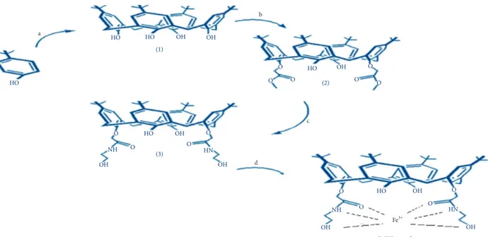
2.1. Experimental Scheme. The sequential scheme for the
present study is given in Figure 1.
2.2. Synthesis. Compounds 1 and 2 were synthesized by the
reported methods [15, 16]. Derivative (L) and its metal complex were synthesized according to the procedure depicted below.
Synthesis of the derivative (L) by the modified method was carried out by adding 1 mmol of compound (2) in 15 mL of chloroform and stirred for 5–10 minutes at room temperature. 50 mL solution of ethanolamine and meth-anol (1 : 3 v : v) was added dropwise to a flask containing compound (2), and the reaction was kept on stirring at room temperature. After 48 hours, white gummy material appeared, and the solvent was removed via the rotary evaporator. The remaining residue was dissolved in methanol, and distilled water was added into it; after vigorous shaking, white precipitates appeared which were
filtered and dried. Recrystallization in mixture of solvents (CH2Cl2 and CH3OH) gave white crystals, and the yield
was 69.89% [17]. Derivative (L) was characterized with FT-IR and elemental.
2.3. UV-Spectroscopic Measurements. Complexation study
was carried out by using appropriate concentrations of the derivative (L) and metal cations. Stock solution of the de-rivative (L) (2.6 × 10⁻³M) was prepared in 10 mL ethanol, which was further diluted to 100 mL (2.58 × 10⁻⁵M) in ethanol. This method was used for spectroscopic analysis of the derivative (L). Using absorption cell of 1 cm, 10 equivalents of the essential metals were measured. 2 mL of the derivative (L) (2.6 × 10⁻⁵M) and 2 mL of metal salts (2.6 × 10⁻⁴M) were mixed together in 5 mL glass test tubes separately. Spectral response of the derivative (L) before and after the mixing of metals was recorded.
2.4. Determination of Complex Stoichiometry. Stoichiometric
ratio of the derivative (L) and metal (Fe3+) was determined using Job’s plot method. For this method, equimolar so-lutions (2.6 × 10⁻⁵M) of both host and guest were added to each other in 1 : 9 to 9 : 1 varying ratios [18, 19].
2.5. Synthesis of Fe3+Complex. Iron complex was prepared
according to Job’s plot. The saturated solution of ligand was separately prepared followed by addition of stoichiometric amount of metal ions into the ligand solution. Subsequently, the solution was stirred at normal temperature for 24 hours. Finally, the solvent was evaporated, and the bright crystals obtained were dried in a vacuum oven. Complex was characterized with FT-IR and elemental analysis.
2.6. Microbial Study. The modified disc diffusion method of
Kirby–Bauer [20] was used to check the in vitro antimi-crobial activity of the derivative (L) and its Fe3+complex. With the help of American Type Culture Collection (ATCC), antimicrobial activity was studied against different micro-organisms. The growth of microorganism species including bacteria and a fungus was carried out using the Mueller–
Hinton agar (MHA) medium. Under the instructions of
manufacturer, the culture medium was prepared and used. To check the antimicrobial activity of the derivative (L) and its Fe3+complex, five successive dilutions 6, 3, 1.5, 0.75, and 0.37 (μg/mL) were prepared in absolute ethanol. Discs used were of Whatman No. 3 (Filter paper discs) with a diameter size of 6 mm. They were soaked in each prepared dilutions for a short period of time, and then under sterile circum-stances, discs were dried (to get rid of residual solvent) at room temperature. After that, the discs were placed on microbial agar plates (made in streaking fashion) and in-cubated at 37°C for 24 hours (for bacteria) and 48 hours (for
fungi). At the end of incubation period, the antimicrobial activity of compounds against each microbial species was recorded by measuring the zone of the inhibition diameter in millimeter (mm), and MIC levels were calculated. The tests were carried out in triplicate as suggested [21].
3. Results and Discussion
3.1. UV-Vis Study. UV-Vis spectroscopic technique was
used to explore the complexation behavior of the derivative (L) against various essential metal ions, i.e., K+, Na+, Mg2+,
Fe3+, Co2+, Zn2+, and Ni2+. 10 eq. of various metal ions was
used against the derivative (L) (2.6 ×10⁻³M) in absolute
ethanol to check the complexation ability. Figure 2 shows the UV-Vis spectra of the derivative (L) after the addition of metal ions.
The result showed the prominent variations in peaks during complexation. The obtained spectra showed that the derivative (L) possesses good attraction towards the essential metal ions (showing the difference in region 220–230 nm), but it showed noticeable interaction to Fe3+.
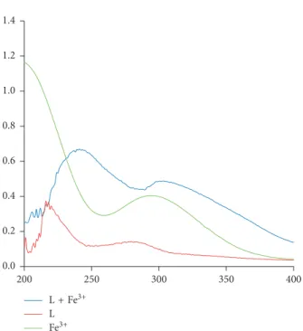
The electronic spectra of the derivative (L), Fe3+, and
Fe3+complex were recorded in ethanol, as given in Figure 3,
and values of wavelength maximum are given in Table 1. The reported complex is novel having brownish color. Actually, the transition metals having incomplete d orbital absorb radiation in the UV-Vis region and show electronic tran-sitions between different energy levels, whose separation is largely controlled by ligand species. The spectrum observed in Figure 3 for the derivative (L) indicates that 215 and 280 nm peaks are produced by intraligand transitions. In case of the Fe3+complex, red shifts were observed. In the
complex, peaks observed at 250 and 300 nm are assigned to characteristic metal d-d transitions. It indicates that metal and ligand molecules are well incorporated to give observed spectral changes. Likewise, when metal and ligand are co-ordinated, ligand causes splitting within d orbitals of metal shifting it to longer wavelength. Spectrum of ligand has shown a bathochromic shift after coordination.
Iron gives octahedral geometry here because it produces six coordination environments. Since Fe3+ gives the d5
system, whereas Fe2+gives d6, the high charge density and
small size of Fe3+make it a hard base, where O and N donor
ligands appear to be small sized, and relatively non-polarisable ligands are hard bases; hence, this type of hard-hard type acid-base combination favors significantly stable Fe3+ derivative (L) complex, whereas Fe2+ is classified as
borderline acid which will probably give less stable complex. The complex has faint brownish color mostly due to the d-d transition that gives weak colors since it is a Laporte bidden transition. However, transitions after complex for-mation may involve electronic rearrangements of valence electrons. Moreover, complex color is mostly dependent on metal, oxidation state of metal, and number of d electrons in metal, as Fe2+gives green color, while Fe3+produces brown
color. Contrary to this, charge transfer is less probable here
a HO HO OH OH OH d b c HO NH HN HN NH HO HO HO Fe(III) complex Fe3+ OH (1) (3) OH OH OH OH OH O O O O O O O O O O (2) O O O O
Figure1: Experimental scheme for the synthesis of 1–3 and Fe3+complex of 3 (derivative L). The conditions specified for each step are (a) HCHO/NaOH, (b) KCO3/C4H7BrO2, (c) C2H7NO/CH3OH, and (d) FeCl3·6H2O, respectively.
L L + Co2+ L + Fe3+ L + K1+ L + Mg2+ L + Na1+ L + Ni2+ L + Zn2+ 0.0 0.1 0.2 0.3 0.4 0.5 0.6 0.7 Ab so rba nce 265 315 365 415 215 Wavelength (nm)
Figure 2: Absorption spectra of the derivative L (ligand) (2.6 ×10⁻⁵M) with different essential metals ions (2.6 ×10⁻⁴M).
because internally it involves the oxidation-reduction pro-cess. LMCT and MLCT cause the redox changes and may give very strong/sharp peaks. Such change is not observed here, but instead weak peaks are observed here evidencing the peaks to be produced from d-d transitions. However, it is believed that variation in absorption peaks is associated with stable chelate complex formation in case of iron, which further supported the complexation. Furthermore, the study was carried out on the Fe3+complex.
3.2. Concentration Study. In order to evaluate the
com-plexation efficiency of the derivative (L), the concentration of Fe3+ was steady increased from 1.0 to 10 equivalents. The
results shown in Figure 4 indicate that adsorption intensity of the derivative (L) was increased linearly with the increasing dose of Fe3+ ions. These results suggest that derivative (L)
possesses appreciable binding efficiency with Fe3+.
3.3. Job’s Plot for Fe3+ Complex. Technique of constant
variation was performed for Job’s plot using absorbance versus mole fraction (X) to know the stoichiometric ratio between the derivative (L) and Fe3+metal ion. In case of
Job’s plot study for the Fe3+ complex, it is revealed that
derivative (L) shows good complexation at 0.5 mole fraction (Figure 5), which reflects that derivative (L)and Fe3+
pos-sesses good relation in 1 :1. Therefore, the reaction was set
between derivative (L) and Fe3+in absolute ethanol using
indicated appropriate ratios for complex synthesis.
3.4. Characterization of Fe3+Complex. FT-IR study helps to
confirm the complex formation by showing disappearance and appearance or shifting of some peaks present in the derivative (L) spectrum after complexation. Figure 6 shows the comparable spectra of the derivative (L) and its Fe3+
complex in which reasonable changes are observed. A band 1666.49 cm−1for C O is shifted to 1659.76 cm−1in the Fe3+
complex spectrum. Also a band 1549.27 cm−1 for NH
bending is shifted to 1541.39 cm−1after complexation. Two
peaks at 1110.4 cm−1and 1059.34 cm−1for C-O stretch of
hydroxyl of the ethanolamine group are shifted to 1104.2 cm−1 and 1051.6 cm−1 after complexation. The OH stretch peak at 3382.99 cm−1is also shifted to 3378.9 cm−1on
complexation with Fe3+. These remarkable changes suggest
that Fe3+coordinates with nitrogen, oxygen of the carbonyl
group, and oxygen of the hydroxyl group. In addition, these changes indicate the complex formation.
Elemental analysis found (%): C, 73.36; H, 8.20; N, 3.35. Anal. Calcd for compound 3 (%): C, 73.41: H, 8.24: N, 3.39. Similarly elemental analysis found (%): C, 68.91; H, 7.7; N, 3.06. Anal. Calcd for Fe complex (%): C, 68.95; H, 7.7; N: 3.09.
3.5. Antimicrobial Study. Derivative (L) and its Fe3+complex
are good antimicrobial agents, as shown in Tables 2 and 3.
L + Fe3+ L Fe3+ 0.0 0.2 0.4 0.6 0.8 1.0 1.2 1.4 250 300 350 400 200
Figure3: UV-Vis spectra of ligand (L), Fe3+, and Fe3+complex.
Table1: λmaxof various transitions in the derivative (L) and iron complex. Compounds Transitions (λ in nm) Derivative (L) 215 280 L-Fe3+complex 250 300 10 0 0.0 0.1 0.2 0.3 0.4 0.5 0.6 0.7 Ab so rba nce 290 340 390 440 490 240 Wavelength
Figure4: UV-Vis titration spectra of derivative (L) (2.6 ×10⁻⁵M) by adding various equivalents (0–10 eq.) of Fe3+.
0.000 0.050 0.100 0.150 0.200 0.250 0.300 Ab so rba nce 0.1 0.2 0.3 0.4 0.5 0.6 0.7 0.8 0.9 1 0 Mole fraction
Figure5: Job’s plot produced by mixing various equivalents of the derivative (L) (2.6 ×10⁻⁵) and Fe3+(2.6 ×10⁻⁵).
They showed antimicrobial activity against selected micro-organisms (S. albus, E. coli, and R. stolonifer). However, it is important to note that the Fe3+ complex showed higher
antimicrobial activity as compared to the derivative (L). In-crease in the activity of complex can be explained on the basis of Tweedy’s chelating theory [22] and overtone concept [23, 24]. A preliminary evaluation of the antimicrobial activity of the derivative (L) and Fe3+complex against S. albus and E. coli was performed and gauged according to the following
four stage criteria:
(+++) High antimicrobial activity; zone of inhibition is 12–17 mm
(++) Relatively high antimicrobial activity; zone of inhibition is 8–11 mm
(+) Weak antimicrobial activity; zone of inhibition is 1–7 mm
(−) No antimicrobial activity
3.6. Antibacterial Activity. From Figures 7 and 8, it is clear
that the Fe3+complex showed higher antimicrobial activity
(+++) at 6 μg/mL against S. albus, and for E. coli, it showed higher antimicrobial activity (+++) at 6, 3, and 1.5 μg/mL, while it showed relatively high antimicrobial activity (++) at 3 μg/mL against S. albus and at 0.75 μg/mL against E. coli. It showed weak antimicrobial activity (+) below 3 μg/mL against S. albus and at 0.37 μg/mL against E. coli. How-ever, derivative (L) showed relatively high antimicrobial activity (++) at 6 μg/mL and weak antimicrobial activity (+) at 3 μg/mL against E. coli. It showed weak antimicrobial activity (+) at 6 and 3 μg/mL against S. albus. No antimi-crobial study (−) was observed below 3 μg/mL for both bacterial strains.
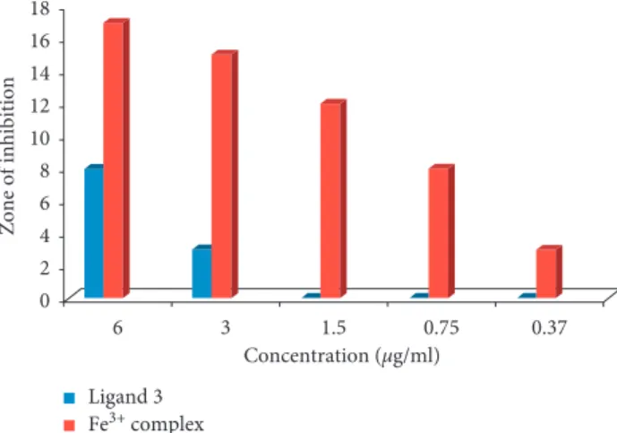
3.7. Antifungal Activity. Antifungal activity of the derivative
(L) and its Fe3+complex was carried out against R. stolonifer.
Figure 9 shows that the Fe3+complex showed higher
an-871.85 1040.28 1059.34 1110.4 1195.55 1300.2 9 1361.84 1484.59 1549.27 1666.49 2867.90 2960.10 3382.99 590.56 872.58 1044.84 1051.6 1104.2 1203.84 1299.43 1362.45 1393.06 1483.91 1542.39 1659.84 2867.79 2959.51 3378.9 Compound 3 Fe complex 3500 3000 2500 2000 1500 1000 500 4000 Wavenumber (cm−1) –100 –80 –60 –40 –20 0 20 40 60 80 100 % T
Figure6: FT-IR spectra of compound 3 (L) and its complex with Fe3+. Table2: Zones of inhibition for antimicrobial activities of the tested derivative (L).
Microorganism strains Derivative (L) MIC value (μg/mL) (concentration in μg/mL) 6 3 1.5 0.75 0.37 (zones of inhibition in mm) S. albus 6 2 — — — 3 E. coli 8 3 — — — 3 R. stolonifer 9 4 1 — — 1.5
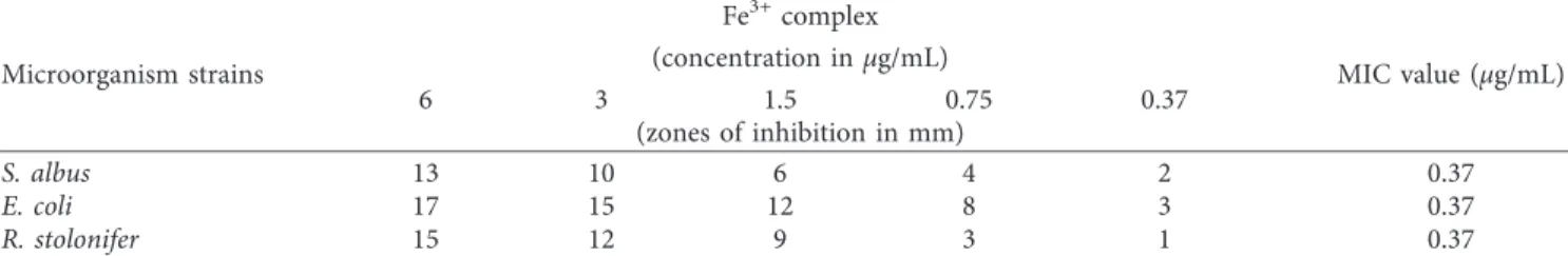
Table3: Zones of inhibition for antimicrobial activities of the tested Fe3+complex of the derivative (L).
Microorganism strains Fe3+complex MIC value (μg/mL) (concentration in μg/mL) 6 3 1.5 0.75 0.37 (zones of inhibition in mm) S. albus 13 10 6 4 2 0.37 E. coli 17 15 12 8 3 0.37 R. stolonifer 15 12 9 3 1 0.37
timicrobial activity (+++) at 6 and 3 μg/mL, relatively high antimicrobial activity (++) at 1.5 μg/mL, and weak antimi-crobial activity (+) at 0.75 and 0.37 μg/mL. However, de-rivative (L) showed no higher antimicrobial activity but relatively high antimicrobial activity (++) at 6 μg/mL and weak antimicrobial activity (+) at 3 and 1.5 μg/mL against R.
stolonifer.
3.8. Minimum Inhibitory Concentration (MIC). The
mini-mum concentration at which the growth of microorganisms (bacteria and fungi) is inhibited is usually known as in vitro minimum inhibitory concentration (MIC) [25]. Different concentration level solutions (6, 3, 1.5, 0.75, and 0.37 μg mL−1) of the derivative (L) and its metal complex were prepared against bacteria and fungi (Tables 2 and 3). Figures 10 and 11 show that derivative (L) had the same MIC values (3 μg/mL) for both selected bacterial strains, which means it is less effective against bacteria because at lower concentrations, its effect was not observed. However, the Fe3+ complex showed high activity against E. coli and S. albus with an MIC value of 0.37 μg/mL for both bacteria,
because it showed antimicrobial action even at lower con-centrations. Thus, the Fe complex of the derivative (L)
synthesized in this study is the most potent antimicrobial agent than calix[4]arene derivative reported so far such as calix[4]arene-based vancomycin mimics [11], antibacterial
tetra-para-guanidinoethyl-calix[4]arenes [26], water-soluble nalidixic acid/calix [4]arene ester adducts [27], and water-soluble calix[4]arenes derivatives [6].
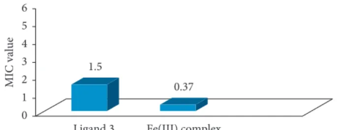
In case of antifungal action, Figure 12 shows that de-rivative (L) had an MIC value of 1.5 μg/mL, which means it is more effective against R. stolonifer as compared to selected bacteria for which it showed an MIC value of 3 μg/mL. However, the Fe3+complex was equally effective against the
fungi and two bacterial species tested with an MIC value of 0.37 μg/mL.
4. Conclusions
The derivative (L) was synthesized and explored for the complexation study with essential metals. It was revealed that derivative (L) showed selective behavior toward the Fe3+
ion. Job’s plot study evaluated that derivative (L) and Fe3+
6 3 1.5 0.75 0.37 0 2 4 6 8 10 12 14 Zone of inhibition Concentration (µg/ml) Ligand 3 Fe3+ complex
Figure 7: Antimicrobial activity of ligand 3 (L) and its Fe3+ complex against S. albus (G +ve).
6 3 1.5 0.75 0.37 Concentration (µg/ml) Ligand 3 Fe3+ complex 0 2 4 6 8 10 12 14 16 18 Zone of inhibition
Figure8: Antimicrobial activity of the derivative (L) and its Fe3+ complex against E. coli (G−ve).
6 3 1.5 0.75 0.37 Concentration (µg/ml) Ligand 3 Fe3+ complex 0 2 4 6 8 10 12 14 16 Zone of inhibition
Figure9: Antifungal activity of ligand 3 (L) and its Fe3+complex against R. stolonifer (fungus) concentration (μg/mL).
Ligand 3 Fe(III) complex
0.37 3 0 1 2 3 4 5 6 MIC value
Figure10: MIC values of ligand 3 (L) and its metal complex against
S. albus (G +ve).
Ligand 3 Fe(III) complex
3 0.37 0 1 2 3 4 5 6 MIC value
Figure11: MIC values of ligand 3 (L) and its metal complex against
metal ion form good complex in 1 :1 ratio. Finally, derivative (L) and its Fe3+complex were bioassayed for antimicrobial
activity. On the basis of MIC values, it is concluded that the Fe3+complex of the derivative (L) is more effective against
selected bacteria and fungi, as compared to the derivative (L) because the Fe3+ complex showed higher antimicrobial
sensitivity towards selected bacteria and fungi by showing an MIC value of 0.37 μg/mL. Thus, the Fe3+ complex of the
derivative (L) may prove to be a good candidate for further studies as a pharmaceutical drug.
Data Availability
All the data relevant to the study are present in this article.
Disclosure
This work was performed as part of employment at Uni-versity of Sindh, Jamshoro.
Conflicts of Interest
The authors declare that there are no conflicts of interest.
Acknowledgments
We acknowledge the National Center of Excellence in Analytical Chemistry and Dr. M. A. Kazi Institute of Chemistry, University of Sindh, Jamshoro, Pakistan, for providing all facilities for this work.
References
[1] C. D. Gutsche and L.-G. Lin, “Calixarenes 12,” Tetrahedron, vol. 42, no. 6, pp. 1633–1640, 1986.
[2] M. Yilmaz and S. Sayin, “Calixarenes in organo and bio-mimetic catalysis,” in Calixarenes and Beyond, pp. 719–742, Springer, Cham, Switzerland, 2016.
[3] J. S. Kim and D. T. Quang, “Calixarene-derived fluorescent probes,” Chemical Reviews, vol. 107, no. 9, pp. 3780–3799, 2007.
[4] C. Redshaw, “Coordination chemistry of the larger calixar-enes,” Coordination Chemistry Reviews, vol. 244, no. 1-2, pp. 45–70, 2003.
[5] M. Yigiter Bayrakcı, S. Ertul, and M. Yilmaz, “Synthesis of novel silica gel immobilized-calix4arene amide ionophores and their anion binding abilities toward phosphate and chromate anions,” Journal of Applied Polymer Science, vol. 124, no. 5, pp. 3831–3839, 2012.
[6] A. M. Soomro, R. K. Oad, S. Memon, and I. Qureshi, “Pak. Bioactivity assessment of water soluble calix[4]arene de-rivative,” Pakistan Journal of Analytical and Environmental
Chemistry, vol. 13, pp. 36–39, 2012.
[7] J. S. Millership, “A preliminary investigation of the solution complexation of 4-sulphonic calix[n]arenes with testoster-one,” Journal of Inclusion Phenomena and Macrocyclic
Chemistry, vol. 39, no. 3/4, pp. 327–331, 2001.
[8] E. D. Silva, P. Shahgaldian, and A. W. Coleman, “Haemolytic properties of some water-soluble para-sulphonato-calix-[n]-arenes,” International Journal of Pharmaceutics, vol. 273, pp. 57–62, 2004.
[9] J. Gualbert, P. Shahgaldian, and A. W. Coleman, “Interactions of amphiphilic calix[4]arene-based Solid Lipid Nanoparticles with bovine serum albumin,” International Journal of
Phar-maceutics, vol. 257, no. 1-2, pp. 69–73, 2003.
[10] J. W. Conforth, P. D. Hart, G. A. Nicholls, R. J. W. Rees, and J. A. Stock, “Antituberculous effects of certain surface-active polyoxyethylene ethers,” British Journal of Pharmacology and
Chemotherapy, vol. 10, no. 1, pp. 73–86, 1955.
[11] A. Casnati, M. Fabbi, N. Pelizzi et al., “Synthesis, antimi-crobial activity and binding properties of calix[4]arene based vancomycin mimics,” Bioorganic and Medicinal Chemistry
Letters, vol. 6, no. 22, pp. 2699–2704, 1996.
[12] V. Paquet, A. Zumbuehl, and E. M. Carreira, “Biologically active amphotericin B-Calix[4]arene conjugates,”
Bio-conjugate Chemistry, vol. 17, no. 6, pp. 1460–1463, 2006.
[13] R. Lamartine, M. Tsukada, D. Wilson, and A. Shirata, “An-timicrobial activity of calixarenes,” Comptes Rendus Chimie, vol. 5, no. 3, pp. 163–169, 2002.
[14] S¸. Ç. ¨Ozkan, A. Yilmaz, E. Arslan, L. Açık, ¨U. Mutlu, and E. G. Elif, “Novel copper(II) complexes ofp-tert-butylcalix[4] arene diamide derivatives: synthesis, antimicrobial and DNA cleavage activities,” Supramolecular Chemistry, vol. 27, no. 4, pp. 255–267, 2015.
[15] C. D. Gutsche, M. Iqbal, and D. Stewart, “Calixarenes. 19. Syntheses procedures for p-tert-butylcalix[4]arene,” Journal
of Organic Chemistry, vol. 51, no. 5, pp. 742–745, 1986.
[16] D. Maity, A. Chakraborty, R. Gunupuru, and P. Paul, “Calix [4]arene based molecular sensors with pyrene as fluoregenic unit: effect of solvent in ion selectivity and colorimetric de-tection of fluoride,” Inorganica Chimica Acta, vol. 372, no. 1, pp. 126–135, 2011.
[17] S. Inese and D. B. Smithrud, “Condensation of calix[4]arenes with unprotected hydroxyamines and their resulting water solubilities,” Tetrahedron, vol. 57, no. 47, pp. 9555–9561, 2001. [18] D. C. Harris, Quantitative Chemical Analysis, Vol. 4, W.H.
Freeman and Company, New York, NY, USA, 1995. [19] Q. K. Panhwar, S. Memon, and M. I. Bhanger, “Synthesis,
characterization, spectroscopic and antioxidation studies of Cu(II)-morin complex,” Journal of Molecular Structure, vol. 967, no. 1–3, pp. 47–53, 2010.
[20] A. W. Bauer, W. M. A. Kirby, J. C. Sherris, and M. Truck, “Antibiotic susceptibility testing by a standardized single disk method,” American Journal of Clinical Pathology, vol. 45, pp. 493–496, 1996.
[21] D. A. V. Berghe and A. J. Vlietinck, “Screening methods for antibacterial and antiviral agents from higher plants,” in
Meth. Plant Biochem., P. M. Dey and J. D. Harbone and,
Eds., p. 47, Academic Press, London, UK, Academic Press, 1991.
[22] J. Joseph, K. Nagashri, and G. A. B. Rani, “Synthesis, char-acterization and antimicrobial activities of copper complexes
Ligand 3 Fe(III) complex
0.37 1.5 0 1 2 3 4 5 6 MIC value
Figure12: MIC values of ligand 3 (L) and its metal complex against
derived from 4-aminoantipyrine derivatives,” Journal of Saudi
Chemical Society, vol. 17, no. 3, pp. 285–294, 2013.
[23] S. A. Sadeek, W. H. El–Shwiniy, W. A. Zordok, and A. M. El-Didamony, “Synthesis, spectroscopic, thermal and biological activity investigation of new Y(III) and Pd(II) Norfloxacin complexes,” Journal of the Argentine Chemical Society, vol. 97, pp. 128–148, 2009.
[24] Q. K. Panhwar and S. Memon, “Synthesis, characterization and microbial evaluation of metal complexes of molybdenum with ofloxacin (levo (S-form) and dextro (R-form)) isomers,”
Journal of Modern Medicinal Chemistry, vol. 2, pp. 1–9, 2014.
[25] F. Mojab, M. Poursaeed, Mehrgan, and S. Pakdaman, “An-tibacterial activity of Thymus daenensis methanolic extract,”
Pakistan Journal of Pharmaceutical Sciences, vol. 21,
pp. 210–213, 2008.
[26] M. Mourer, R. E. Duval, C. Finance, and J.-B. Regnouf-de-Vains, “Functional organisation and gain of activity: the case of the antibacterial tetra-para-guanidinoethyl-calix[4]arene,”
Bioorganic and Medicinal Chemistry Letters, vol. 16, no. 11,
pp. 2960–2963, 2006.
[27] H. M. Dibama, I. Clarot, S. Fontanay et al., “Towards calixarene-based prodrugs: drug release and antibacterial behaviour of a water-soluble nalidixic acid/calix[4]arene ester adduct,” Bioorganic and Medicinal Chemistry Letters, vol. 19, no. 10, pp. 2679–2682, 2009.
Tribology
Advances in Hindawi www.hindawi.com Volume 2018 Hindawi www.hindawi.com Volume 2018International Journal ofInternational Journal of
Photoenergy
Hindawi www.hindawi.com Volume 2018 Journal ofChemistry
Hindawi www.hindawi.com Volume 2018 Advances inPhysical Chemistry
Hindawi www.hindawi.com Analytical Methods in Chemistry Journal of Volume 2018 Bioinorganic Chemistry and Applications Hindawi www.hindawi.com Volume 2018Spectroscopy
International Journal of Hindawiwww.hindawi.com Volume 2018 Hindawi Publishing Corporation
http://www.hindawi.com Volume 2013 Hindawi www.hindawi.com
The Scientific
World Journal
Volume 2018Medicinal ChemistryInternational Journal of
Hindawi www.hindawi.com Volume 2018
Nanotechnology
Hindawi www.hindawi.com Volume 2018 Journal ofApplied Chemistry
Journal of Hindawi www.hindawi.com Volume 2018 Hindawi www.hindawi.com Volume 2018 Biochemistry Research International Hindawi www.hindawi.com Volume 2018Enzyme
Research
Hindawi www.hindawi.com Volume 2018 Journal ofSpectroscopy
Analytical Chemistry International Journal of Hindawi www.hindawi.com Volume 2018Materials
Journal of Hindawi www.hindawi.com Volume 2018 Hindawi www.hindawi.com Volume 2018 BioMedResearch International
Electrochemistry
International Journal of Hindawi www.hindawi.com Volume 2018