Introduction
The 26S proteasome, an ATP-dependent proteolytic system, is engaged in the degradation of short-lived proteins under normal metabolic conditions, as well as bulk degradation of long-lived proteins, partial digestion/processing of some proteins (e.g., NF-κB), and antigen presentation. Among the key regulatory proteins degraded by the 26S proteasome are the transcription factor c-Fos, M-, S-, and G1-phase specific cyclins, cyclin-dependent kinase inhibitors, p53, ornithine decarboxylase (ODC), and a variety of oncoproteins (1-3). The 26S proteasome has a molecular mass of approximately 2.5 MDa and is formed by the assembly of one 20S proteasome complex (the core particle) with 2 molecules of the 19S complex, the regulatory complex. The 19S regulatory complexes are attached at both ends of the 20S proteasome (4).
The 20S proteasome complexes (~700 kDa) of prokaryotes and eukaryotes both contain 28 subunits but differ in complexity. The archaebacterium Thermoplasma acidophilum consists of 14 copies of 2 distinct but related polypeptides (αand β), whereas eukaryotic proteasomes are made up of 2 copies each of 7 distinct αand 7 distinct β-type subunits (5). The αsubunits make up the 2 outer layers, and the β subunits the 2 inner rings of the structure [i.e. α7β7β7α7(prokaryotes); (α1-α7)(β1-β7)(β1
-β7)(α1-α7) (eukaryotes)]. The hallmark of this protein
fold is a 4 layer cylindrical structure (6-8). Studies have indicated that proteasomes are weakly susceptible to the inhibitors of serine and cysteine proteases. A combination of genetic, structural and inhibitor studies have revealed the amino terminal threonine (Thr) at the βsubunit as a nucleophile catalyst in the 20S proteasome-mediated proteolysis (9). The classification of the 20S proteasome
Cellular Functions of the 26S Proteasome
Azmi YERL‹KAYA
Department of Biology, Faculty of Art & Science, Dumlup›nar University, 43100, Kütahya - TURKEY
Received: 26.08.2003
Abstract: The 26S proteasome plays a critical role in many cellular processes by degrading a wide range of both short-lived and
long-lived proteins. In this review, proteasome structure and the mechanisms of substrates targeting to the 26S proteasome are described. In addition, this review focuses on the cellular functions that are affected after inhibition of the proteasome, including immune-response, cell-cycle, and transcription. The proteasome is involved in these cellular processes because it degrades numerous short-lived proteins and regulatory proteins that control these cellular functions. Even though it is known that inhibition of the 26S proteasome results in the induction of the synthesis of heat-shock proteins (e.g., Hsp 70), several groups indicated that 26S proteasome inhibition also causes a significant loss of global protein synthesis activity in a very short time.
Key Words: 26S proteasome; proteasome inhibitors; transcription; translation.
26S Protezomun Hücresel Görevleri
Özet: 26S protezom, bir çok uzun-ömürlü ve k›sa-ömürlü proteinleri y›karak çok say›da hücresel fonksiyonda önemli roller
oynamaktad›r. Bu derlemede, protezomun yap›s› ve substratlar›n 26S protezoma gönderilme mekanizmalar› aç›klanm›flt›r. Ayr›ca, bu derleme ba¤›fl›kl›k-sistemi, hücre-döngüsü ve transkripsiyon gibi protezomun inhibisyonundan etkilenen hücresel fonksiyonlar üzerinde özenle durmufltur. Protezom, bu hücresel ifllemlere onlar›n fonksiyonlar›n› kontrol eden bir çok k›sa-ömürlü ve regülatör proteinlerin y›k›l›m›n› sa¤layarak kat›lmaktad›r. 26S protezomun inhibisyonun heat-shock proteinlerin (örne¤in, Hsp 70 ) sentezini uyard›¤› bilinmesine ra¤men, baz› gruplar 26S protezomun inhibisyonun k›sa bir sürede önemli derecede global protein sentez aktivitesinin de düflmesine neden oldu¤unu göstermifllerdir.
as a threonine protease was supported by the covalent labeling of a Thr residue at an active β subunit in mammalian proteasomes by the specific proteasome inhibitor lactacystin, a fungal metabolite (10). In complex eukaryotic proteasomes, at least 3 of 14 βsubunits have functional active sites (11). The 20S proteasome exhibits at least 3 distinct proteolytic activities, e.g., the trypsin-like, the chymotrypsin-trypsin-like, and the post-glutamyl peptidyl hydrolytic (PGPH) activities. The active sites are generated by adjacent pairs of identical β-type subunits residing in different β rings (12,13). As chymotrypsin, trypsin and elastase are synthesized as inactive proenzymes, the catalytic β-type subunits of the 20S proteasome are also synthesized as inactive proproteins and processed to their mature forms upon assembly into the 20S proteasome core (14,15). The active sites face a chamber in the 20S proteasome in which access to the central chamber is blocked in the free 20S proteasome. Assembly of the 19S regulatory complex at both ends of the 20S proteasome was shown to result in the formation of pores at the ends of the 20S proteasome complex through which the substrates can be fed into active sites (16). The 19S complex is composed of a lid and a base. A portion of the lid recognizes the ubiquitinated substrates, while the base, which contains 6 ATPases and caps the end of the 20S proteasome core, unfolds protein substrates and threads them into the catalytic chamber of the 20S particle in an ATP-dependent manner (17). Unlike other proteases, the chamber arrangement of the 20S proteasome ensures that virtually all peptide bonds within a protein substrate are susceptible to cleavage, since the multiple proteolytic activities (i.e. chymotrypsin and trypsin-like activities) in one catalytic chamber can efficiently trap the proteins until they are degraded to peptides with an average length of 7 to 9 residues (18,19). While the 20S proteasome in vitro will only degrade small peptides and certain denatured or oxidized proteins (without the need for ATP), the 26S proteasome degrade ubiquitinated proteins in an ATP-dependent reaction (20-22).
Targeting of Substrates to the 26S Proteasome
One of the first steps in the degradation of a substrate by the 26S proteasome involves the conjugation of ubiquitin (a 76 amino acid protein) to protein substrates. This is a multistep process. The first step is catalyzed by an ubiquitin-activating enzyme (E1). In the presence of
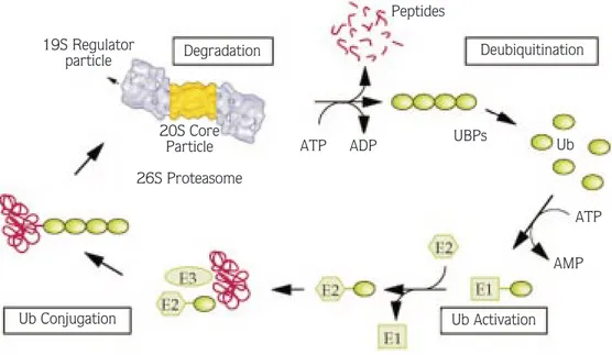
ATP, E1 forms a high-energy thiol ester bond with the C-terminus of ubiquitin. Activated ubiquitin is then transferred from an E1 enzyme to the active site cysteine residue of one of several ubiquitin-conjugating enzymes (also known as E2 enzymes). These E2 enzymes can then either donate ubiquitin directly to a protein substrate, often with the aid of one of numerous E3 enzymes (called ubiquitin-recognition or ubiquitin ligase enzymes, which can help to direct the E2 enzymes to a specific protein substrate), or in some cases can act as an intermediate carrier of the activated ubiquitin in a process in which the E2 enzyme transfers the activated ubiquitin to the E3 enzyme, which then performs the final transfer of ubiquitin to the target protein substrate. In this final reaction, whether accomplished by an E2 or an E3 enzyme, the C-terminal glycine residue of ubiquitin is linked by an isopeptide bond to a specific internal lysine residue of a target protein (23,24). After the first attachment of a single ubiquitin molecule to a substrate, the mono-ubiquitinated substrate can be further ubiquitinated by the linkage of additional ubiquitins to lysine 48 of the previously attached ubiquitin, known as polyubiquitination (25). Ubiquitin protein conjugates are then recognized by the 19S regulatory complex of the 26S proteasome, unfolded by the same complex and fed into the interior of the 20S complex, where they are degraded into oligopeptides (Figure) (5). Before this reaction occurs, ubiquitin chains are selectively removed from the target protein by the action of deubiquitinating enzymes, and used repeatedly in the degradation pathway (24). Although it is not known exactly how ubiquitination machinery is targeted to certain lysine residues, phosphorylation of certain residues has been shown to serve as an essential part of a specific recognition site for subsequent ubiquitination in certain target proteins, e.g., IκBα, cyclin D1, and cell cycle inhibitor p27 (26,27).
Although the process outlined above probably accounts for the great majority of protein degradation performed by the 26S proteasome, a lysine residue is not always required for ubiquitination in a target protein. For example, it has been shown that the NH2groups of the
N-terminal residues of MyoD, a tissue-specific transcriptional activator, can serve as an essential and sufficient conjugation site for subsequent degradation by the 26S proteasome (28). In addition, it has been reported that polyamine biosynthetic enzyme ornithine decarboxylase (ODC) is degraded only in an
ATP-dependent, but ubiquitin-independent manner by the 26S proteasome (29). This important finding indicated that a substrate can be degraded by the 26S proteasome without ubiquitination (4).
Cellular Functions of the 26S Proteasome
A variety of cell-permeable inhibitors of the 26S proteasome have proved very valuable tools for clarifying the proteasome’s in vivo functions. Probably the most widely used inhibitors are peptide aldehydes (e.g., MG-132), which are reversible transition state analogs. As mentioned above, lactacystin, a natural product, targets the 26S proteasome by an irreversible modification of the amino terminal threonine of β-subunits (10,30). Studies with these inhibitors have shown that the bulk of cellular proteins (80-90%) are degraded by the 26S proteasome (31). In recent years, it was shown that misfolded proteins in the endoplasmic reticulum (ER) undergo a retrograde transport from the ER to the cytosol, and are subsequently degraded by the 26S proteasome (32,33). The 26S proteasome was also shown to be involved in the regulation of metabolic adaptation, cell differentiation,
cell-cycle control, the stress response and immune system, retrovirus assembly and budding, and regulation of transcription and translation.
Cell-Cycle Control
The eukaryotic cell-cycle is controlled by periodic synthesis and degradation of positive regulators (i.e. cyclins and cyclin-dependent kinases or Cdks) and negative regulators (i.e. cyclin-dependent kinase inhibitors). The 26S proteasome is involved in the cell-cycle control by degrading different cyclins that are specific for G1-, S-, or M-phases. These cyclins accumulate and activate their cognate Cdks at appropriate times during the cell cycle and then are degraded, causing Cdks inactivation (8). G1 cyclins, mitotic B-type cyclins, and S-phase cyclins such as cyclin A are ubiquitinated before proteolysis, suggesting that the proteasome is responsible for their degradation (34). Genetic studies have also provided important information regarding the role of the 26S proteasome in cell-cycle control. For example, the yeast temperature-sensitive nin1-1 mutant, which contains a mutation in a non-ATPase regulatory
19S Regulator particle Degradation Peptides Deubiquitination Ub UBPs ATP ADP 20S Core Particle 26S Proteasome Ub Conjugation ATP AMP Ub Activation
Figure . Schematic representation of degradation of a target protein by the proteasome.
The target protein is first conjugated to ubiquitin chains involving the ubiquitin activating enzyme E1, the ubiquitin conjugating enzyme E2, and the ubiquitin ligase E3. The polyubiquitinated target protein is recognized by the 26S proteasome. After or during degradation by the 26S proteasome, the ubiquitin chain is released from the target protein remnant, and the free ubiquitin chain is disassembled by deubiquitinating enzymes (e.g., UBP, ubiquitin-specific processing protease). The figure is from Reference 5, modified slightly with the permission of Dr. W. Baumeister.
subunit of the 26S proteasome, is blocked in both G1-S transition and G2-M transition (35). In mammalian cells, the activities of CDK can be controlled by Cdk inhibitors p21, p27 and p57. The p27 inhibitor is abundant in quiescent and G1 cells, whereas it is down regulated in proliferating and S-phase cells. Studies have indicated that p27 is targeted for ubiquitination and degradation by the 26S proteasome. The degradation of p27 by the proteasome is required for the progression through the G1 to S-phase of the cell-cycle (36,37). Although the degradation of cyclins causes inactivation of Cdks and a cell-cycle arrest, the degradation of a Cdk inhibitor by the 26S proteasome results in activation of the Cdk and restart of DNA replication (38).
Stress Response and Immune System
Heat stress and other harsh conditions increase the level of heat-shock proteins (e.g., Hsp72, Hsp60 and Hsp32). These heat-shock proteins protect the cells against stressors like oxidative stress, heat, ischemia and other insults. They mainly function as molecular chaperones involved in the protein transport, folding, assembly, and degradation of misfolded or damaged proteins. Thus, they appear to prevent the accumulation of damaged proteins in stress-affected cells. Recently, various proteasome inhibitors (e.g., MG-132, MG-115 and lactacystin) were shown to cause rapid increases in the levels of mRNAs encoding cytosolic Hsps (e.g., Hsp70) and ER chaperones (Bip, Grp94 and ERp72) and to protect the cells from subsequent lethal hyperthermic injury (39). In cardiomyocytes MG-132 induced Hsp72 and Hsp32 in a p38 MAP kinase-dependent manner and protected these cells against oxidative stress (40).
Use of proteasome inhibitors has also clearly demonstrated that the 26S proteasome is involved in the generation of antigenic peptides from cytosolic proteins for presentation on MHC class I molecules, which initiate the immune response (41). After stimulation of human cells with the cytokine interferon-γ(INF-γ) 3 proteasomal β-subunits (X, Y and Z) are exchanged by LMP7, LMP2 and MECL1, respectively (42). These new subunits allow a more efficient generation of peptides that are recognized by MHC class I molecules (30). NF-κB is a transcriptional factor involved in immune and inflammatory responses. It is a heterodimer composed of p50 and p65 (RelA) (43). NF-κB is held in cytoplasm by
association of the inhibitory protein IκB to the p50-p65 complex. Upon phosphorylation at Ser32 and Ser36, IκB is polyubiquitinated and targeted for degradation by the 26S proteasome, resulting in activation and nuclear translocation of NF-κB (44,45). The 26S proteasome is also involved in the processing of the 105 kDa NF-κB precursor to the active p50 form through limited proteolysis (46). This indicates that the proteasomes are not only involved in complete degradation of proteins, but also in activation of proteins through limited proteolysis of inactive precursor proteins (47).
Retrovirus Assembly and Budding
The 26S proteasome pathway is implicated in retrovirus assembly through the finding that retroviruses contain a high concentration of ubiquitin, a tag for targeting substrate proteins to the 26S proteasome for degradation as mentioned above (48). Moreover, it was found that up to 50% of Gag proteins (the viral protein responsible for assembly and budding) were conjugated to ubiquitin (49). Recently, it was discovered that the budding and assembly of human immunodeficiency virus type 1 (HIV-1) are reduced when infected cells were treated with proteasome-specific inhibitors, e.g., lactacystin or epoxomicin (50). Similarly, it was shown that treatment of infected cells with proteasome inhibitors reduced the release of Rous Sarcoma virus (an avian virus) and that this effect can be suppressed by over-expressing ubiquitin and also by directly fusing ubiquitin to the C-terminus of Gag protein (51). These results suggested that the block to viral budding was due to a rapid reduction in the free ubiquitin level as a consequence of massive conjugation of ubiquitin to cellular proteins in the presence of proteasome inhibitors. Further efforts are required to determine additional factors that are involved in the mechanisms causing abrogation of retroviruses budding after inhibition of the 26S proteasome.
Regulation of Transcription and Translation (Protein Synthesis)
Actively transcribing genes have a higher ubiquitinated histone content, and when transcription is inhibited the level of nucleosomal ubiquitinated H2B specifically decreases (52,53). Recently, it was demonstrated that
proteasome inhibitors caused a rapid loss of ubiquitinated histones H2A and H2B, and under these conditions DNA replication and RNA transcription was strongly inhibited (54). Ubiquitination of histones H2A and H2B is thought to facilitate the action of polymerases and transcription factors by weakening the interaction of the histone tails with either DNA or accessory nonhistone proteins (55,56). As is the case in viral budding, under the conditions of blocking proteasome function, massive conjugation of ubiquitin to cellular proteins eventually consumes the free ubiquitin pool (since Ub recycling is blocked by proteasome inhibition) and results in the redistribution of ubiquitin away from nucleosomal H2A and H2B (54). Since the proteasome is involved in the degradation of a number of transcriptional factors (e.g., c-Jun, p53, and NF-κB) DNA replication and transcription can also be abolished indirectly as consequence of inhibition of the normal cellular function of these transcription factors. Nonetheless, specific transcription of a subset of genes is induced under the conditions of blocking the 26S proteasome (e.g., Hsp25 and Hsp75). The 26S proteasome inhibitors MG-132 and lactacystin induced hyperphosphorylation and the DNA binding activity of heat-shock transcription factor 1 (HSF1), which is the key factor in the regulation of heat-shock protein expression (e.g., Hsp 70 and Hsp 27) (57). With the exception of specific induction of heat-shock protein expression, inhibition of the 26S proteasome seems to inhibit general protein synthesis, as several reports have indicated that proteasome inhibition results in a global decrease in protein synthesis (54,58), although no mechanism by which proteasome inhibition causes this decrease in protein synthesis rates has been proposed. In contrast to these studies, Bush et al. reported that the proteasome inhibitor MG-132 did not affect the total protein synthesis even after 18 h of treatment in a different cell line: Madin-Darby canine kidney cells (39). Whether the result reported by Bush et al. is an experimental artifact or whether it is a cell-specific response to the 26S proteasome inhibitors remains to be determined. If 26S proteasome inhibition leads to a global decrease in protein synthesis activity, it will be intriguing to examine how the synthesis of the heat-shock proteins is induced under the conditions of the down-regulation of global protein synthesis activity caused by blocking 26S proteasome function.
Conclusions
The proteasome is engaged in the selective degradation of short-lived proteins under normal metabolic conditions, bulk degradation of long-lived proteins, partial digestion/processing of some proteins (e.g., NF-κB) as well as regulation of proteins that control the cell-cycle. Therefore, the proteasome has become an attractive target for the treatment of a broad range of human diseases. Indeed, proteasome inhibition has shown impressive antitumor activity by inducing apoptosis in numerous tumor types (59,60). Apoptosis is probably induced by proteasome inhibitors as a consequence of the accumulation of some crucial proteins (e.g., p53 and p27) that are normally degraded by the proteasome. Furthermore, the proteasome inhibitors might be active against tumor growth by inhibiting global protein synthesis and causing a significant increase in S-adenosylmethionie (SAM) levels (61). In addition to being a precursor in polyamine biosynthesis, SAM is also the major methyl donor in DNA methylation reactions (62). It is thus possible that this increased SAM level following inhibition of the proteasome causes transcriptional inactivation of cancer related genes by an increase in global hypermethylation of DNA. Further experiments are warranted to determine whether proteasome inhibition causes hypermethylation of DNA. Our observations indicate that down-regulation of protein synthesis by 26S proteasome inhibition is probably due to an increase in eIF2α phosphorylation (Yerlikaya, A., Stanley, B.A., manuscript in preparation). It is well known that a dominant mechanism of control of global protein synthesis is phosphorylation/dephosphorylation of translational components, especially that of eIF2α (63,64). Existence of a feedback mechanism between protein synthesis and degradation is quite logical to prevent accumulation of excess levels of proteins in cells when protein degradation is compromised. It is thought that eIF2αis an intermediate in this feedback mechanism. The factors that lead to an increase in eIF2α phosphorylation following inhibition of the 26S proteasome remain to be determined.
In conclusion, the 26S proteasome plays diverse roles in numerous cellular functions by controlling the fine balance of a plethora of proteins implicated in proliferation, cell-cycle, transcription, immune-response, and apoptosis. As discussed, inhibition of the 26S proteasome not only causes accumulation of proteins in
cells but also results in activation/inactivation of cellular proteins through either limited proteolysis (i.e. activation of NFκB) or phosphorylation (i.e. activation of HSF1 or inactivation of eIF2α). Further investigations will greatly increase our understanding of the interaction of the 26S proteasome with other intracellular components. New information obtained will be crucial for designing better therapeutic approaches for cancer and neurodegenerative diseases (e.g., Alzheimer’s disease), since a growing body of evidence indicates the involvement of the 26S proteasome in these diseases.
Acknowledgments
I am grateful to Dr. Bruce A. Stanley for his guidance throughout my academic carreer and for his critical review of this manuscript. I thank Dr. W. Baumeister for permission to use the Figure, published previously in Reference 5. Many contributions to the protein degradation field have been omitted because of space limitations.
References
1. Etlinger JD, Goldberg AL. A soluble ATP-dependent proteolytic system responsible for the degradation of abnormal proteins in reticulocytes. Proc Natl Acad Sci USA. 74: 54-58, 1977. 2. He H, Qi XM, Grossmann J et al. c-Fos degradation by the
proteasome. An early, Bcl-2-regulated step in apoptosis. J Biol Chem. 273: 25015-25019, 1998.
3. Hilt W, Wolf DH. Proteasomes: destruction as a programme. Trends Biochem Sci. 21: 96-102, 1996.
4. Pickart CM. Targeting of substrates to the 26S proteasome. FASEB J. 11: 1055-1066, 1997.
5. Voges D, Zwickl P, Baumeister W. The 26S proteasome: a molecular machine designed for controlled proteolysis. Ann Rev Biochem. 68: 1015-1068, 1999.
6. Puhler G, Weinkauf fi, Bachmann L et al. Subunit stiochiometry and three-dimensional arrangement in proteasomes from Thermoplasma acidophilum. EMBO J. 11: 1607-1616, 1992. 7. Kopp F, Kristensen P, Hendil KB et al. The human proteasome
subunit HsN3 is located in the inner rings of the complex dimer. J Mol Biol. 248: 264-272, 1995.
8. Hershko A, Ciechanover A. The ubiquitin system. Ann Rev Biochem. 67: 425-479, 1998.
9. Seemüller E, Lupas A, Zuhl F et al. The proteasome from Thermoplasma acidophilum is neither a cysteine nor a serine protease. FEBS Lett. 359: 173-179, 1995.
10. Fenteany G, Staendaert RF, Lane WS et al. Inhibition of proteasome activities and subunit-specific amino-terminal threonine modification by lactacystin. Science. 268: 726-731, 1995.
11. Orlowski M. The multicatalytic proteinase complex, a major extralysosomal proteolytic system. Biochemistry. 29: 10289-10297, 1990.
12. Arendt CS, Hochstrasser M. Identification of the yeast 20S proteasome catalytic centers and subunit interactions required for active-site formation. Proc Natl Acad Sci USA. 94: 7156-7161, 1997.
13. Kopp F, Hendil KB, Dahlmann B et al. Subunit arrangement in the human 20S proteasome. Proc Natl Acad Sci USA. 94: 2939-2944, 1997.
14. Yang Y, Fruh K, Ahn K et al. In vivo assembly of the proteasomal complex, implications for antigen processing. J Biol Chem. 270: 27687-27694, 1995.
15. Coux O, Tanaka K, Goldberg AL. Structure and function of the 20S and 26S proteasomes. Ann Rev Biochem. 65: 801-847, 1996.
16. Groll M, Bajorek M, Kohler A et al. A gated channel into the proteasome core particle. Nat Struct Biol. 7: 1062-1067, 2000. 17. Braun BC, Glickman M, Kraft R et al. The base of the proteasome regulatory particle exhibits chaperone-like activity. Nat Cell Biol. 1: 221-226, 1999.
18. Nussbaum AK, Dick TP, Keilholdz W et al. Cleavage motifs of the yeast 20S proteasome beta subunits deduced from digests of enolase 1. Proc Natl Acad Sci USA. 95: 12504-12509, 1998. 19. Kisselev AF, Akopian TN, Goldberg AL. Range of sizes of peptide
products generated during degradation of different proteins by archaeal proteasomes. J Biol Chem. 273: 1982-1989, 1998. 20. Peters JM, Cejka Z, Harris JR et al. Structural features of the 26S
proteasome complex. J Mol Biol. 234: 932-937, 1993. 21. Peters JM. Proteasomes: protein degradation machines of the
cell. Trends Biochem Sci. 19: 377-382, 1994.
22. Akopian TN, Kisselev AF, Goldberg AL. Processive degradation of proteins and other catalytic properties of the proteasome from Thermoplasma acidophilum. J Biol Chem. 272: 1791-1798, 1997.
23. Hershko A, Ciechanover A, Heller H et al. Proposed role of ATP in protein breakdown: conjugation of protein with multiple chains of the polypeptide of ATP-dependent proteolysis. Proc Natl Acad Sci USA. 77: 1783-1786, 1980.
24. Jentsch S. The ubiquitin-conjugation system. Ann Rev Genet. 26: 179-207, 1992.
25. Chau V, Tobias JW, Bachmair A et al. A multiubiquitin chain is confined to specific lysine in a targeted short-lived protein. Science. 243: 1576-1583, 1989.
26. Pagano M. Cell cycle regulation by the ubiquitin pathway. FASEB J. 11: 1067-1075, 1997.
27. Jesenberger V, Jentsch S. Deadly encounter: ubiquitin meets apoptosis. Nat Rev Mol Cell Biol. 3: 112-121, 2002.
28. Breitschopf K, Bengal E, Ziv T et al. A novel site for ubiquitination: the N-terminal residue, and not internal lysines of MyoD, is essential for conjugation and degradation of the protein. EMBO J. 17: 5964-5973, 1998.
29. Murakami Y, Matsufuji S, Kameji T et al. Ornithine decarboxylase is degraded by the 26S proteasome without ubiquitination. Nature. 360: 597-599, 1992.
30. Myung J, Kim KB, Crews CM. The ubiquitin-proteasome pathway and proteasome inhibitors. Med Res Rev. 21: 245-273, 2001. 31. Rock KL, Gramm C, Rothstein L et al. Inhibitors of the
proteasome block the degradation of most cell proteins and the generation of peptides presented on MHC class I molecules. Cell. 78: 761-771, 1994.
32. Plemper RK, Bohmler S, Bordallo J et al. Mutant analysis links the translocon and BiP to retrograde protein transport for ER degradation. Nature. 388: 891-895, 1997.
33. Pilon M, Schekman R, Römisch K. Sec61p mediates export of a misfolded secretory protein from the endoplasmic reticulum to the cytosol for degradation. EMBO J. 16: 4540-4548, 1997. 34. Sudakin V, Ganoth D, Dahan A, et al. The cyclosome, a large
complex containing cyclin-selective ubiquitin ligase activity, targets cyclins for destruction at the end of mitosis. Mol Biol Cell. 6: 185-198, 1995.
35. Ghislain M, Udvardy A, Mann C.S. cerevisiae 26S protease mutants arrest cell division in G2/metaphase. Nature. 366: 358-362, 1993.
36. Loda M, Cukor B, Tam, SW et al. Increased proteasome-dependent degradation of the cyclin-proteasome-dependent kinase inhibitor p27 in aggressive colorectal carcinomas. Nature Medicine. 3: 231-234, 1997.
37. Sutterluty H, Chatelain E, Marti A et al. p45SKP2 promotes p27Kip1 degradation and induces S phase in quiescent cells. Nat Cell Biol. 1: 207-214, 1999.
38. Schwob E, Bohm T, Mendenhall MD et al. The B-type cyclin kinase inhibitor P40SICI controls the G1 to S transition in S. cerevisiae. Cell. 79: 233-244, 1994.
39. Bush KT, Goldberg AL, Nigami SK. Proteasome inhibition leads to a heat-shock response, induction of endoplasmic reticulum chaperons, and thermotolerance. J Biol Chem. 272: 9086-9092, 1997.
40. Lüss H, Schmitz W, Neumann J. A proteasome inhibitor confers cardioprotection. Cardiovascular Research. 54: 140-151, 2002.
41. Rock KL, Goldberg AL. Degradation of cell proteins and the generation of MHC class I-presented peptides. Ann Rev Immunol. 17: 739-779, 1999.
42. Gaczynska M, Rock KL, Goldberg AL. Gamma-interferon and expression of MHC genes regulate peptide hydrolysis by proteasomes. Nature. 365: 264-267, 1993.
43. Baldwin AS. The NF-kappa B and I kappa B proteins: new discoveries and insights. Ann Rev Immunol. 14: 649-683, 1996. 44. Alkalay I, Yaron A, Hatzubai A et al. Stimulation-dependent I kappa B alpha phosphorylation marks the NF-kappa B inhibitor for degradation via the ubiquitin-proteasome pathway. Proc Natl Acad Sci USA, 92: 10599-10603, 1995.
45. Maniatis T. Catalysis by a multiprotein Ikappa B kinase complex. Science. 278: 818-819, 1997.
46. Palombella VJ, Rando OJ, Goldberg AL et al. The ubiquitin-proteasome pathway is required for processing of NF-kappa B1 precursor. Cell. 78: 773-785, 1994.
47. Hilt W, Wolf DH. Proteasomes: destruction a programme. Trends Biochem Sci. 21: 96-102, 1996.
48. Ott DE, Coren LV, Copeland TD et al. Ubiquitin is covalently attached to the p6Gag proteins of human immunodeficiency virus type 1 and simian immunodeficiency virus and to the p12Gag protein of Moloney murine leukemia virus. J Virol. 72: 2962-2968, 1998.
49. Strack B, Calistri A, Accola MA et al. A role for ubiquitin ligase recruitment in retrovirus release. Proc Natl Acad Sci USA. 97: 13063-13068, 2000.
50. Schubert U, Ott D, Chertova EN et al. Proteasome inhibition interferes with gag polyprotein processing, release, and maturation of HIV-1 and HIV-2. Proc Natl Acad Sci USA. 97: 13057-13062, 2000.
51. Patnaik A, Chau V, Wills JW. Ubiquitin is part of the retrovirus budding machinery. Proc Natl Acad Sci USA. 97: 13069-13074, 2000.
52. Levinger L, Varshavsky A. Selective arrangement of ubiquitinated and D1 protein-containing nucleosomes within the Drosophila genome. Cell. 28: 375-385, 1982.
53. Davie JR, Murphy LC. Level of ubiquitinated H2B in chromatin is coupled to ongoing transcription. Biochemistry. 29: 4752-4757, 1990.
54. Mimnaugh EG, Chen HY, Davie JR et al. Rapid deubiquitination of nucleosomal histones in human tumor cells caused by proteasome inhibitors and stress response inducers: effects on replication, transcription, translation, and the cellular stress response. Biochemistry. 36: 14418-14429, 1997.
55. Cook J, Chock PB. Ubiquitin: a review on a ubiquitous biofactor in eukaryotic cells. Biofactors. 1: 133-146, 1988.
56. Van Holde KE, Lohr DE, Robert C. What happens to nucleosomes during transcription. J Biol Chem. 267: 2837-2840, 1992.
57. Kim D, Kim S-H, Li GC. Proteasome inhibitors MG-132 and lactacystin hyperphosphorylate HSF1 and induce hsp70 and hsp27 expression. Biochem Biophy Res Com. 254: 264-268, 1999.
58. Schubert U, Anton LC, Gibbs J et al. Rapid degradation of a large fraction of newly synthesized proteins by proteasomes. Nature. 404: 770-774, 2000.
59. Nawrocki ST, Bruns CJ, Harbison MT et al. Effects of the proteasome inhibitor PS-341 on apoptosis and angiogenesis in orthotopic human pancreatic tumor xenografts. Mol Cancer Ther. 1: 1243-1253, 2002.
60. Ling YH, Liebes L, Jiang JD et al. Mechanism of proteasome inhibitor PS-341 induced G(2)-M-Phase arrest and apoptosis in human non-small cell lung cancer cell lines. Clin Cancer. 9: 1145-1154, 2003.
61. Yerlikaya A, Stanley BA. S-adenosylmethionine decarboxylase degradation by the 26S proteasome is accelerated by substrate-mediated transamination. J Biol Chemistry. 279: 12469-12478, 2004.
62. Lu SC. S-Adenosylmethionine. Int J Biochem Cell Biol. 32: 391-395, 2000.
63. Hershey JW. Translational control in mammalian cells. Ann Rev Biochem. 60: 717-755, 1991.
64. Kimball SR. Eukaryotic initiation factor eIF2. Int J Biochem Cell Biol. 31: 25-29, 1999.