Reduction and arthrodesis with sublaminar
spiral silk in atlantoaxial joint instability
Correspondence: Kadir Kotil, MD, Prof. İstanbul Arel Üniv. Sağlık Bilimleri Yüksekokulu,
Fizyoterapi ve Rehabilitasyon Bölümü, Tepekent 34537 Büyükçekmece, Istanbul, Turkey. Tel: +90 212 – 867 25 00 e-mail: [email protected]
Submitted: January 06, 2013 Accepted: February 28, 2014 ©2014 Turkish Association of Orthopaedics and Traumatology
Available online at www.aott.org.tr doi: 10.3944/AOTT.2014.3156 QR (Quick Response) Code
doi: 10.3944/AOTT.2014.3156
Kadir KOTIL1, Mustafa Gökhan BILGILI2, Selim KAYACI3
1Department of Physiotherapy and Rehabilitation, School of Health Sciences, Istanbul Arel University, Istanbul, Turkey; 2Department of Orthopedics and Traumatology, Bakırköy Dr. Sadi Konuk Training and Research Hospital, Istanbul, Turkey;
3Department of Neurosurgery, Faculty of Medicine, Recep Tayyip Erdoğan University, Rize, Turkey
Objective: The aim of this study was to evaluate the clinical and radiologic results of the use of thick spiral silk knotting instead of sublaminar wiring for C1-C2 arthrodesis in patients with atlantoaxial instability.
Methods: We retrospectively evaluated 16 patients (10 females, 6 males; mean age: 43.4 years; mean follow-up: 34 months) with atlantoaxial instability who underwent C1-C2 fusion by reduction and sublaminar spiral silk knotting. All patients underwent open reduction, bounding both laminae with thick spiral silk instead of wiring and arthrodesis with autografting. Reduction rates, screw position and fusion rates were evaluated using computed tomography.
Results: Preoperative mean atlantodental interval (ADI) was 8 (range: 6 to 11) mm and postoperative ADI was 2.1 (range: 0.5 to 2.5) mm. There was no dural or spinal cord injury. Complete reduction was observed in all cases. Fusion was unsuccessful in 1 case (6.25%). Postoperative mean flexion ADI was 10 mm and mean extension ADI was 1 mm. Graft separation between C1-C2 was observed in slice tomographic examination in one patient. Malposition was observed in 2 screws (4%).
Conclusion: The sublaminar silk knotting technique appears to provide safe anatomical reduction. As this method is cheap and simple and does not require extra implantation, loosen, create neurologic compromise or cause radiologic crowding, it can be considered an alternative surgical technique to sublaminar wiring.
Key words: Atlantoaxial stabilization; cervical spine; interlaminar fusion. Atlantoaxial joint (AAJ) instability may occur due to
degenerative joint disease, rheumatologic diseases, in-fectious reasons, os odontoideum, tumors or trauma. [1] Regardless of etiology, reduction can be provided by several different techniques in case of disrupted integ-rity of the atlas and axis. Posterior open reduction is the preferred method except in cases that are irreducible or that require circumferential decompression. If posterior
decompression and reduction is not successful, anterior transoral surgery should be performed.[1] The C1-C2 transarticular screwing stabilization technique is used in AAJ instability cases which cannot be treated by an-terior odontoid screw.[2,3] The C1-C2 dislocation must be reduced and anatomic alignment should be provided before application of the transarticular screw (Magerl) technique to avoid neurovascular complications.[4-6]
There are many different techniques for reduction and fusion, without standardization. Surgeons who be-lieve the advantages of sublaminar wiring and autograft-ing prefer to use the Sonntag technique to increase fu-sion.[6-8] Fusion with sublaminar wiring technique was defined for the first time by Gallie in 1939.[9] Sublaminar wiring provides reduction and settlement of the fusion material. However, many publications have reported loosening of wires in time with this technique, eventu-ally leading to neurological damages.[5,10-13] Different modifications of this technique were defined by Brooks, Sonntag, Magerl and Harms.[3] Wires can wear the lam-ina over time, particularly when performed in children. [14] Although soft and titanium wires appear to solve this problem, their application is more difficult and expen-sive. In order to prevent neurological damage, some sur-geons prefer unrestricted graft application or tightening of the graft material between the C1-C2 lamina after decortication.[4] However, the main disadvantage of this technique is pseudarthrosis.[2]
The aim of this study was to report the results of a sublaminar spiral silk technique to eliminate the wire-related complications.
Patients and methods
We retrospectively evaluated 16 patients (10 females, 6 males; mean age: 43.4 years, range: 10 to 61 years) treated with C1-C2 fixation for AAJ instability between 2006 and 2012 by a single surgeon (KK). Neurological conditions, radiological examination, operation notes and follow-up period were noted. Mean follow-up time was 34 (range: 27 to 45) months. Third plan, thin slice tomographic analysis was performed preoperatively to determine the dominant vertebral artery and cases with a highly corrugated vertebral artery. A string was formed by winding three no. 2 silks to each other. Silk was
pre-ferred due to its endurance to tension and better knot-ting power when compared to other suture materials. The neurological status of the patients was evaluated us-ing the Nurick scale. A Philadelphia collar was used for 6 weeks postoperatively. Patients were assessed clinically and radiologically in the 1st week and at the 3rd, 6th, 12th, 24th and 60th months.
All patients were evaluated by preoperative and post-operative plain radiographs, open mouth dens views, dynamic X-ray views, computed tomography (CT) and magnetic resonance image (MRI). Atlantodental inter-val (ADI) of 5 mm or higher was accepted as instabil-ity. Preoperative ADI values were measured by a blinded radiologist and compared with the postoperative values. Retro-odontoid tumor-like pannus formation was ob-served in 2 cases. There was no contrast enhancement in these two cases. Because of poor bone quality, aug-mentation was achieved with pedicle screws reaching to the middle of the cervical region. Patients were evaluated using CT in the early postoperative period and at the 6th month follow-up. Reduction values of the odontoid migrations in basilar invagination were assessed post-operatively. Values were in the physiological limits in all patients. Iliac crest autograft was used in all patients and fusion occurred in all but one (93.8%). Inappropriate positioning was seen in 2 (4%) screws but revision was not required.
Operations were performed using the dorsal ap-proach with the patient in the military posture in a prone position. The C1-C2 complex was exposed. The head was placed into a Mayfield helmet with spinous processes positioned longitudinally on the same mid-line. In cases with a deeply seated C2, thoracic kyphosis was corrected and brought to the neutral position be-cause of angulation problems in screw director and in-sufficient anterior tubercle fixation. A skin incision was
Fig. 1. (a) Compression of the spinal cord with scissoring effect at the dislocation site. (b) Radiography of AAD showing widened interspinous interval of C1-C2. [Color figure can be viewed in the online issue, which is available at www.aott.org.tr]
made between the inion and C3 and muscles were dis-sected subperiosteally. Dissection was continued to the transverse foramen of the atlas and inferior part of the C3 without disrupting the C2-C3 facet joint capsule. Monopolar cautery was preferred since high tempera-ture might damage bones and soft tissues. The C2 was dissected to feel the isthmus and arcus depth and the di-rection of the screw was properly assessed. Dislocation between C1 and C2 was reduced by extension of the lower cervical spine and the flexion of the inion. Surgi-cal challenges associated with the dislocation were the deep location of C1’s lamina, the compression of spinal cord by C1’s lamina and the highly-localized C2. The spinal cord was compressed due to the shearing effect of dislocation (Fig. 1). The C2 lamina was pressed and the joint reduced. The surrounding soft tissue around the C1 and C2 lamina was dissected using a 360-degree-angled dissector. The silk string was placed in the sub-laminar region (Fig. 2a) and knotted over both laminas to provide anatomic reduction by passing through the C1 and C2 laminas (Figs. 2b and c). The posterior facet of both laminas was decorticated before knotting. Re-duction was checked using fluoroscopy (Fig. 3a). The transarticular screw was positioned and controlled us-ing fluoroscopy; an iliac crest autograft was placed on the prepared site and the second knot was tied on the graft (Fig. 3b). Autograft bone chips were used in order to increase fusion.
Results
The most frequent symptom in our series was neck pain, which occurred in 15 cases (93.8%). Spastic quadripa-resis occurred in 4 (25%) cases. These four cases were ambulatory using walking aids. Clinical findings and postoperative assessment results are summarized in Ta-ble 1. Neck pain at different levels and restriction in neck movements were observed in all patients. In rheumatoid arthritis patients, there was generalized cervical muscle spasm. None of our cases had neurological deterioration during their follow-ups. There was os odontoideum in 6 cases (37.5%), Type 2 dens fracture in 5 (31.3%), rheu-matoid arthritis in 4 (25%) and previous Type 2 dens fracture in one (6.3%).
Complications were grouped in two; early and late. There was one early non-dominant vertebral artery mi-nor injury in which bleeding was easily tamponed and stopped using hemostatic agents. Late complications in-cluded delayed fusion (in the 15th month) in two cases with rheumatoid arthritis and insufficiency of the C1-C2 bone bridge in these two cases, and pseudarthrosis in one rheumatoid arthritis patient. None of these patients were smokers.
Reduction was complete in all cases. Mean preop-erative and postoppreop-erative ADI values were 8 (range: 6 to 11) mm and 2.1 (range: 0.5 to 2.5) mm, respectively. No dural or spinal cord injury complications were observed. Mean flexion ADI value was 10 mm and mean extension
Fig. 2. (a) Passing of spiral silk through the C1 lamina. (b) Passing of spiral silk through the C2 lam-ina. (c) Knotted spiral silk after passing through both laminas and reduction. (d) Same pro-cedure followed on the op-posite side. (e) Osteosynthesis and placed transarticular screw with knotting. [Color figure can be viewed in the online issue, which is available at www.aott. org.tr] (a) (d) (b) (e) (c)
ADI value was 1 mm. Malposition was observed in two screws (4%).
Posterior transodontoid screwing technique for C1-C2 fixation was used instead of transarticular screwing in two trauma cases.[2] Postoperative Nurick scale and neck pain visual analog scale (VAS) values of the pa-tients are defined in Table 1. Neck pain continued in 2 patients. One patient had the same VAS value postop-eratively and one patient had a one point higher VAS value.
In the long-term follow-up, there was no restriction movement of the neck. No complaints were noted in the
level of disturbance of the patients in terms of neck com-fort.
Discussion
Dislocation of the AAJ can occur in the axial, coronal or sagittal planes. In some cases, osteosynthesis can be pro-vided by anterior odontoid screwing. However, it is also possible to treat with C1-C2 arthrodesis. Post reduction fusion should be provided in all cases of anatomic dislo-cation. If perioperative reduction is possible, the fixation of the C1-C2 complex after open reduction is provided by a suitable technique.[3]
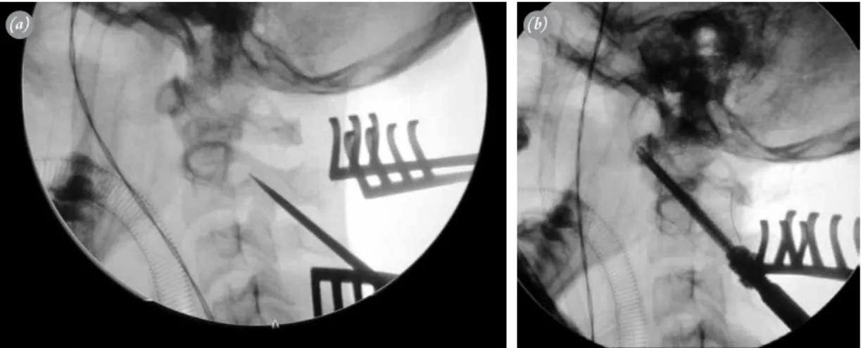
Fig. 3. (a) Perioperative fluoroscopy image; after knotting of laminas, the dislocation is reduced and screwing is securely made. (b) Fluoroscopy image after reduction and osteosynthesis.
(a) (b)
Table 1. Demographic distribution of the patients and assessment results.
No Age Gender Diagnosis VAS Pre-op Post-op Pre-op Nurick Post-op Nurick Fusion at VAS score
ADD ADD score score 12th of the
month neck 1 10 F RA 7 6 1 2 1 Complete 2 2 56 F OsO 7 7 1 2 1 Complete 2 3 45 M OsO 6 7 1 1 1 Complete 1 4 34 F RA 9 6 2 3 1 Complete at 15th month 3 5 61 M RA 5 8 1 3 1 Pseudarthrosis 5 6 60 F DF 8 9 1 2 1 Complete 2 7 30 M DF 8 11 1 1 2 Complete 1 8 38 F DF 7 12 2 1 2 Complete 2 9 55 F DF 9 9 1 1 1 Complete 1 10 48 F RA 7 7 1 1 1 Complete 2 11 43 M OsO 5 8 1 1 1 Complete 1 12 58 F PF 6 9 1 1 1 Complete 3 13 51 M OsO 5 11 2 1 1 Complete 3 14 50 F OsO 6 12 2 1 1 Complete 7 15 23 F DF 7 7 1 1 1 Complete 1 16 33 M RA 7 8 1 2 1 Complete at 15th month 3
C1-C2 fixation techniques include posterior insulat-ed C1 side mass and C2 pars screwing, pinsulat-edicular screw-ing, transarticular screwscrew-ing,[7] laminar screwing or hook usage techniques.[8] There are specific advantages and disadvantages of each C1-C2 fixation techniques defined in the literature.[3,5,15-18] However, regardless of which is used, reduction maneuver and fusion with autograft is essential. In C1-C2 luxation, fixation with screws only is not sufficient. The Sonntag sublaminar wire reconstruc-tion technique, with binding of the iliac crest autograft to the surgical area, is the most preferred and frequently used with fusion rates of 82.6 to 100%.[19] Bendable wires or soft titanium wires can be used for bone-wire con-struction. However, titanium wires may cause neural or dural injury due to loosening and are expensive.[10,14,20,21] Another disadvantage of titanium cables or wires is that they may cause radiologic crowding and misinterpreta-tion of fusion degree during follow-up. Conversely, there is no such disadvantage to the use of silk.
Controversy exists on the stability and fusion follow-ing fixation of the autograft with sublaminar wires. Mat-sumoto et al. reported successful fusion in all patients, with loosening of wires in 18 of 52 cases, causing spinal cord problems.[19] Ito et al. reported wire loosening but successful fusion in all patients.[22,23] Unfortunately, the wiring technique is difficult and time-consuming and may cause severe complications, especially in cases of canal stenosis.[24] For these reasons, some authors rec-ommend only bone lamella placement technique after decortication.[19] Freely placed grafts without knotting to the lamina has resorption and displacement risks. If the interlaminar distance is wide, the free graft may compress the spinal cord. Nonunion might also occur if allograft bone chips are scattered and placed around the surgical field.
The main goals of our technique are reduction and arthrodesis. Reduction primarily corrects instability, which causes scissoring and compression of the spinal cord. After reduction, secure fixation is obtained. Ar-throdesis creates an interlaminar connection or bridge with the tricortical iliac crest. There are several advan-tages of using sublaminar spiral silk. Primarily, knotting by twine shaped silk decreases the interspinous distance. Two-side application of this process makes it more cure. After reduction, transarticular screwing can be se-curely applied in an anatomical position. Additionally, by placing the iliac crest graft on the silk knots, the graft is tightly exposed to laminar contact, before a second graft knot is tied. This technique has many advantages for se-cure screwing and to create standard fusion between the C1 and C2.
Their high price and lack of consistent availability in the surgical room are the main disadvantages of titanium sublaminar wires. However, silk can be found easily in the surgical room and is a very cheap material. In addi-tion, it does not create radiological visual pollution.
Fusion in particular is highly important in rheu-matoid arthritis cases due to the high incidence rate of pseudarthrosis. Additionally, it is essential to provide contact between the bone decortication face and auto-graft cancellous chips in order to increase fusion strength in its revisions. A strong contact between the graft and the laminar decortication field should be provided. Al-though pseudarthrosis was present in one of our cases, implant insufficiency did not develop even in the 4th year. Transarticular screws provide 3-dimensional fixing and prevent implant failure.
In conclusion, sublaminar spiral silk for C1-C2 ar-throdesis is an alternative technique to sublaminar wir-ing. The method provides secure anatomic reduction, is cheap and easily applied and does not require extra implant or cause loosening or neurological damage. In addition, it is free of radiological visual pollution. Yet, for more precise results, further comparative studies with higher proof values are required.
Conflicts of Interest: No conflicts declared.
References
1. Kotil K, Kalayci M, Bilge T. Management of cervicome-dullary compression in patients with congenital and ac-quired osseous-ligamentous pathologies. J Clin Neurosci 2007;14:540-9.
2. Kotil K, Köksal NS, Kayacı S. Posterior transodontoid fixation: A new fixation (Kotil) technique. J Craniovertebr Junction Spine 2011;2:41-5.
3. Dickman CA, Sonntag VK. Posterior C1-C2 transarticu-lar screw fixation for atlantoaxial arthrodesis. Neurosur-gery 1998;43:275-81.
4. Gluf WM, Brockmeyer DL. Atlantoaxial transarticular screw fixation: a review of surgical indications, fusion rate, complications, and lessons learned in 67 pediatric patients. J Neurosurg Spine 2005;2:164-9.
5. Gluf WM, Schmidt MH, Apfelbaum RI. Atlantoaxial transarticular screw fixation: a review of surgical indica-tions, fusion rate, complicaindica-tions, and lessons learned in 191 adult patients. J Neurosurg Spine 2005;2:155-63. 6. Paramore CG, Dickman CA, Sonntag VK. The
anatomi-cal suitability of the C1-2 complex for transarticular screw fixation. J Neurosurg 1996;85:221-4.
7. Madawi AA, Casey AT, Solanki GA, Tuite G, Veres R, Crockard HA. Radiological and anatomical evaluation of the atlantoaxial transarticular screw fixation technique. J
Neurosurg 1997;86:961-8.
8. Melcher RP, Puttlitz CM, Kleinstueck FS, Lotz JC, Harms J, Bradford DS. Biomechanical testing of posterior atlantoaxial fixation techniques. Spine 2002;27:2435-40. 9. Gallie WE. Fracture and dislocations of the cervical spine.
Am J Surg 1939;46:495-9.
10. Fraser AB, Sen C, Casden AM, Catalano PJ, Post KD. Cervical transdural intramedullary migration of a sub-laminar wire. A complication of cervical fixation. Spine 1994;19:456-9.
11. Geremia GK, Kim KS, Cerullo L, Calenoff L. Complica-tions of sublaminar wiring. Surg Neurol 1985;23:629-35. 12. Lundy DW, Murray HH. Neurological deterioration after
posterior wiring of the cervical spine. J Bone Joint Surg Br 1997;79:948-51.
13. Schrader WC, Bethem D, Scerbin V. The chronic lo-cal effects of sublaminar wires. An animal model. Spine 1988;13:499-502.
14. Kakarla UK, Valdivia JV, Sonntag VK, Bambakidis NC. Intracranial hemorrhage and spinal cord injury from a fractured C1-C2 sublaminar cable: case report. Neurosur-gery 2010;66:E1203-4.
15. Grob D, Crisco JJ 3rd, Panjabi MM, Wang P, Dvorak J. Biomechanical evaluation of four different posterior atlan-toaxial fixation techniques. Spine 1992;17:480-90. 16. Harms J, Melcher RP. Posterior C1-C2 fusion with
poly-axial screw and rod fixation. Spine 2001;26:2467-71. 17. Henriques T, Cunningham BW, Olerud C, Shimamoto
N, Lee GA, Larsson S, et al. Biomechanical comparison
of five different atlantoaxial posterior fixation techniques. Spine 2000;25:2877-83.
18. Lapsiwala SB, Anderson PA, Oza A, Resnick DK. Biome-chanical comparison of four C1 to C2 rigid fixative tech-niques: anterior transarticular, posterior transarticular, C1 to C2 pedicle, and C1 to C2 intralaminar screws. Neuro-surgery 2006;58:516-21.
19. Matsumoto M, Chiba K, Nakamura M, Ogawa Y, Toyama Y, Ogawa J. Impact of interlaminar graft materials on the fusion status in atlantoaxial transarticular screw fixation. J Neurosurg Spine 2005;2:23-6.
20. Garcia R Jr, Gorin S. Failure of posterior titanium atlanto-axial cable fixation. Spine J 2003;3:166-70.
21. Bahadur R, Goyal T, Dhatt SS, Tripathy SK. Transar-ticular screw fixation for atlantoaxial instability - modi-fied Magerl’s technique in 38 patients. J Orthop Surg Res 2010;5:87.
22. Ito T, Hayashi M, Takei H. Loosening of supplemental cable in transarticular screw fixation and bone grafting. J Orthop Surg 1998;6:71-4.
23. Ito H, Neo M, Fujibayashi S, Miyata M, Yoshitomi H, Nakamura T. Atlantoaxial transarticular screw fixation with posterior wiring using polyethylene cable: facet fu-sion despite posterior graft resorption in rheumatoid pa-tients. Spine 2008;33:1655-61.
24. Stillerman CB, Wilson JA. Atlanto-axial stabilization with posterior transarticular screw fixation: technical descrip-tion and report of 22 cases. Neurosurgery 1993;32:948-55.