ONLINE FIRST This is a provisional PDF only. Copyedited and fully formatted version will be made available soon.
ISSN: 0015-5659 e-ISSN: 1644-3284
The determination of the pituitary gland, optic chiasm, and
intercavernous distance measurements in healthy subjects
according to age and gender
Authors: Sema Özandaç Polat, Fatma Yasemin Öksüzler, Mahmut Öksüzler, Ayse
Gül Uygur, Ahmet Hilmi Yücel
DOI: 10.5603/FM.a2019.0058 Article type: ORIGINAL ARTICLES Submitted: 2019-03-11
Accepted: 2019-04-24
Published online: 2019-05-10
This article has been peer reviewed and published immediately upon acceptance.
It is an open access article, which means that it can be downloaded, printed, and distributed freely, provided the work is properly cited.
Articles in "Folia Morphologica" are listed in PubMed.
The determination of the pituitary gland, optic chiasm, and intercavernous distance measurements in healthy subjects according to age and gender
Running Title: The morphometry of the pituitary gland, optic chiasm, and
intercavernous distance
Sema Özandaç Polat1, Fatma Yasemin Öksüzler2, Mahmut Öksüzler3, Ayşe Gül Uygur1, Ahmet Hilmi Yücel1
1Cukurova University Faculty of Medicine, Department of Anatomy, Adana, Turkey 2Adana City Research and Training Hospital University of Health Sciences, Department
of Radiology, Adana, Turkey
3Adana Medline Hospital, Department of Radiology, Adana, Turkey
Address for correspondence: Dr. Sema Polat, Cukurova University Faculty of
Medicine, Department of Anatomy, Adana, Turkey, fax: +90 3223387147, e -mail: [email protected]
Abstract
Background: This paper was undertaken to determine the morphometry of pituitary
gland diameter, pituitary gland height, intercavernous distance, optic chiasm diameter and optic chiasm height in skulls of Turkish population aged between 18-60 years.
Materials and Methods: It was a retrospective study in which 292 subjects were
included 187 females and 105 males, ranging from 18 up to 60 years. Subjects having brain MRI in the Radiology Department. Statistical analysis were evaluated with SPSS 21.00 programme. ANOVA Test, Chi-Square Test, and Pearson Correlation analysis were used to determine the relation and significance between measurements and age group. The p<0.05 value was considered as significant.
Results: The groups were divided into five groups according to age. The overall means
mm; pituitary gland height, 4.91±1.10 mm; intercavernous distance, 15.93±3.05 mm; optic chiasm width, 12.82±1.27 mm; and optic chiasm height, 2.80±0.49 mm in
females, respectively whereas, the same measurements were 12.96±1.74 mm; 4.79±0.95 mm; 16.08±3.11 mm; 13.13±1.37 mm; 2.86±0.70 mm in males, respectively. Height of the pituitary gland reached a maximum in the 18 to 20 years of age group in both females and males, after there was a decrease in the pituitary gland height in the subsequent age groups.
Conclusions: Knowledge of the variation in the size of pituitary gland, intercavernous
distance and optic chiasm is more important to evaluate the dimensions of this structures for clinical and pathological processes.
Key words: pituitary gland, optic chiasm, intercavernous distance, morphometry
Introduction
Sella turcica which is found in the middle cranial fossa, is comprised of three section named as the tuberculum sella, the pituitary gland and the hypophyseal fossa [1-3]. Hypophyseal fossa was located in the deepest of the sella turcica [4]. The important structures surround the pituitary gland; sphenoidal sinus from anteriorly, dorsum sellae, basilar artery and pons from posteriorly, optic chiasm from anterior superior side, hypothalamus from inferiorly, and carotis artery, and cavernous sinus from lateral side, and also from lateral and superior side by the dura thin layer [5-10, 11]. Sella turcica is a critical reference landmark related with pathologies of pituitary gland, optic chiasm, and craniofacial region. Therefore, the knowledge of this region’s normal anatomy, variations or morphometry may provide to determine of the growth in a subject and evaluate orthodontic treatment results and help to neurologists and neurosurgeons in preventing damage during surgery [2-4].
In the studies of the pituitary gland, optic chiasm, and intercavernous distance’s anatomy, the differences between females and males and age related changes were observed [11-18]; whereas, in some researches there were no found significant
difference in dimensions of pituitary gland, optic chiasm, and intercavernous distance. Also, some diferent results have been reported about both pituitary gland and optic chiasm measurements in decades[11, 12, 14, 15, 18, 19]. There were very few studies
about morphometry of the hypohysis cerebri, intercavernous distance or optic chiasm in Turkish population in literature [18, 20, 21]. Knowing the normal dimensions of
pituitary gland and other structures in this critical area of the brain is very important for clinical and pathological evaluations. The purpose of this study was to reveal the normative data related to the pituitary gland, optic chiasm, intercavernous distance dependent on age groups and to determine measurements with age and sex in Turkish population.
Materials and Methods
This study was carried out from the 292 healthy adult subjects (187 females; 105 males) aged 18-60 years. The study period extended from January 2015 and 2018. This study was approved by the Institutional Review Ethics Committee at Cukurova
University. This study was a retrospective observational study which done in
Department of Radiology at Medline Hospital in Turkey. Brain MRI protocol including coronal T2-weighted turbo spin echo (TR:3600, TE:87 ms; slice thickness 5 mm; gap 1.5 mm) and sagittal T2-weighted spin echo (TR:3600, TE: 87 ms; slice thickness 5 mm; gap 1.5 mm) was used. The measurements of the pituitary gland width, pituitary gland height, the intercavernous distance, optic chiasm width, and optic chiasm height were evaluated in this study. The measurements were performed from digital MRI images on a hospital using caliper function with x2 magnification. MRI was performed using a 1.5 T MRI system (Siemens; Essenza, Erlangen, Germany).
Healthy adult subjects were selected by criteria of optimal health. The main exclusion criteria are:
a) Adult subjects who were history of endocrine disturbance, pregnancy, breast-feeding.
b) Those were undergone hormonal treatment such as thyroid, postmenopausal estrogen, progesterone, steroid therapy.
c) Subjects, who was on a medication (like phenothiazine, reserpine i.e). The data were divided into both two groups according to gender: healthy adult female and male subjects, and age groups. Estimations were expressed as millimeter.
The SPSS 21.0 program was used for statistical analysis of the measurement results. From these measurements, means, standard deviations (SD), minimum and maximum values were calculated; In all statistical analyses; p value under 0.05 was considered statistically significant. ANOVA Test, Chi-Square Test, and Pearson Correlation analysis were also used.
Results
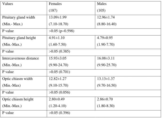
The means, associated standard deviations, and range of values for the various measurements from sella turcica region were presented in Table 1-4. No significant difference (p>0.05) was observed in the corresponding mean values of gender,
consequently the means and standard deviations of the measurements from sella turcica were calculated. The overall means and standard deviations of the measurements were: the pituitary gland width, 13.09±1.99 mm; the pituitary gland height, 4.91±1.10 mm; the intercavernous distance, 15.93±3.05 mm; the optic chiasm width, 12.82±1.27 mm; the optic chiasm height, 2.80±0.49 mm in females, respectively whereas, the same
measurements were 12.96±1.74 mm; 4.79±0.95 mm; 16.08±3.11 mm; 13.13±1.37 mm; 2.86±0.70 mm in males, respectively (Table 1).
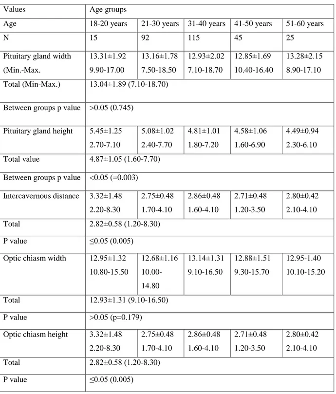
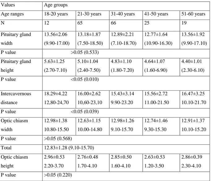
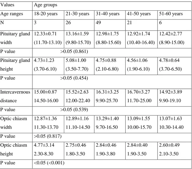
The mean of the pituitary gland height in the age group 18-20 years was the highest value (5.45±1.25 mm). The same measurement was 5.08±1.02 mm in the age group 21-30 years; 4.81±1.01 mm in the age group 31-40 years; 4.58±1.06 mm in the age group 41-50 years and, 4.49±0.94 mm in the age group 51-60 years. The same value was observed to be 4.87±1.05 mm in both subjects (Table 1). P value for the pituitary gland height and intercavernous distance measurements were found to be 0.010 and 0.039 in females between second decade and sixth decade which were statistically significant, however p value for optic chiasm height was calculated as 0.001 in between the age groups of males which was statistically significant (p<0.05) (Table 3 and 4).
The values of pituitary gland height in different age group and in both sexes were shown in the Table 1, Table 3 and Table 4. The means of the pituitary gland height of female subjects in all age groups under 51 years were higher than in male subjects in all age groups under 51 years. Whereas, in female subjects above 50 years of age, the means of the pituitary gland height was lower than in male subjects the same age groups. Moreover, the means of these height was found as the highest in 18-20 years
and after, 21-30 years old age groups (Table 1). In males the corresponding value showed the highest value in 21-30 years old (third decade), while in females the same measurement was highest between 18-20 years (second decade). Additionally, the height of pituitary gland in females decreased gradually with increasing age, whereas there were no such gradually decrease in males (Table 3 and 4).
Significant differences were no found in pituitary gland height and width, intercavernous distance, optic chiasm width and height according to gender (>0.05). While pituitary gland width was showed positive high degree correlation with
intercavernous distance (r=0.712), positive weak correlation with optic chiasm width (r=0.279). Intercavernous distance was showed weak positive correlation with optic chiasm width (r=0.337). There were obtained negative moderate degree correlation in pituitary gland height and age.
Discussion
Magnetic resonance imaging (MRI) is an important, useful and accurate
diagnostic technique using in examinations of pituitary gland. Many pathologies such as physiological hypertrophy, neoplasia (microadenoma or macroadenomas), inflammatory disease, empty sella, embryonic abnormalities and increased lobulated margin are determined with this method [1, 12-15]. Moreover, the MRI can be alternative to computed tomograpy (CT) measurements due to provide greater facility to the patient and with having potentially lesser risk [22]. Also, preoperative imaging of the tumors provides surgical approach to be practical (20). Additionally, MRI is reliable than CT and it does not use any harmful ionizing radiations. It allows detailed visualization of the pituitary gland, optic chiasm etc (11).
The pituitary gland plays an important role in neuroendocrine regulation (11). The height of the pituitary has a critical significance in both determine of intrasellar mass and evaluation of pituitary tumour diagnosis and prognosis [11, 22]. It also allows to be used as a guideline of response to medical therapy [22]. Argyropoulou et al reported that pituitary height is an important measurement marker, because of a sign of the pituitary gland growth [23]. Suzuki et al. stated that if the pituitary gland height exceed 9.00 mm in females and 8 mm in males, that means pituitary gland is abnormal [15]. According to Elster rules which was used as a guideline for height of the pituitary,
this dimension was declared as 6 mm in child; 8 mm in males or post menopause period; 10 mm in fertility period; and in 12 mm pospartum/lactation period [20, 21]. Tsunoda et al reported that pituitary height could show physiological neuroendocrine variations between both young and old subjects, and females and males [12]. The height of the pituitary gland increased in during puberty, third and fifth decades. The reason for this increase in puberty period could be with luteinizing hormone hypersecretion.
Furthermore, the height increased in the third and the fifth decades was due to more active as endocrinolologically and both fertility in females and increase in basal cerum concentrations of gonadotrofic hormones such as luteinizing hormone or follicle stimulating hormone [12, 14, 16, 23]. The decrease in pituitary height with age could show the endocrinology of aging and physiological pituitary atropy [12, 23]. Moreover, the one reason of decreasing in pituitary height was reduction in basal cerum
concentrations of gonadotrofic hormones after puberty until to the ages of 50 years [11-13]. Additionally, an another reason of decreasing in pituitary gland height was
regression of pituitary gland tissue causing loss of function, and progressive compression of the pituitary gland from cerebrospinal fluid pressure and ischemic changes in the anterior lobe [15].
In studies, there were different reports about the period of peak height of the pituitary. Some authors stated the peak height of the pituitary in females was observed in second decade [15, 20, 24, 25], in the ages of 16-25 years [14], and in third decade [11, 12], whereas in males the pituitary peak height was seen in the age of 11-20 years [25], in the age of 10-29 years [15], in the ages of 16-25 years [14], in the age of 21-30 years [11, 24], in the age of 51-60 years [20] and in the age of 20-29 years [12]. In this study the peak height of pituitary was found in second decade (the ages of 18-20 years) in females and in third decade (the ages of 21-30) in males, respectively. We also found differences in females the peak values of pituitary height of above studies except the populations of Nigerian, Turkish, Japanese and Pakistan. According to this data, our results of males were similar to studies of Japanese and Pakistan populations. On the other hand, in a studying consisting of German subjects with idiopathic intracranial hypertension (IICH) and healthy subjects, the same measurements were found as 3.23 mm and 5.55 mm, respectively [26]. In other study, Wiener et al. stated that pituitary height of patients with pituitary tumour was found as 15.5 mm (range 5 – 35 mm) in CT
and 14.5 mm (range 5-32 mm) in MRI [22]. These results are important for knowing normal values of the pituitary height to research whether a tumor is present or not [20]. In literature, pituitary height means were found as 5.52 mm and 5.66 mm in Scottish male and female subjects [14]; 4.7 mm and 4.9 mm and 5 mm and 5.2 mm in Japanese males and females [15, 16]; 5.60 mm and 5.80 mm in Pakistan male and female population [11]; 6.45 mm and 6.46 mm in Nigerian male and female population [25], and 5.37 mm and 6.27 mm in Indians, respectively [27, 28]. When we analyzed our data including pituitary height was found to be 4.79 mm and 4.91 mm in males and females, respectively. According to this data, our values were lower than literature except Suzuki et al’s study. We consider that these differences could be a result of such factors like race, genetic variables, demographic variables (age, gender), the ratio of females in study, mean age of participants, hormonal status, stress and menstrual cycle. Moreover, pituitary gland diseases cause to excessive secretion or growth of the pituitary gland such as acromegaly, prolactinoma, gigantism, hyperthyroidism or diseases including diabetes insipidus, hypothyroidism, Addison’s or Graves diseases cause to less secretion of gland, and the growth deficiency in genital system, or cavernous sinus infection, presence of tumour as glioma, meningioma, lymphoma etc. play a role in differences of pituitary gland dimensions [6-10, 12]. Furthermore, the immaturity of the hormonal feed back system between hypothalamo-hypophyseal tract and target organs is responsible for decrease in pituitary height during the first year of age. Therefore in newborn children, there are high hypophyseal concentrations and blood level of the pituitay hormones [23].
The pituitary dimensions vary accross age groups. It is a sign of changes in pituitary gland’s complicated hormonal milieu [25]. The means of the pituitary width measured as 8 mm [27]. These measurements were significantly different between male and female. It was known that females had larger glands [25]. Peak value of pituitary width were seen in the ages of 25-35 years, whereas the lowest value of pituitary width was obtained between 55-65 years [14]. In this study, there were no correlation with age in pituitary width measurements, which is in agreement with the literature [25]. In researches, the means of the pituitary width were between 11.22 mm and 13 mm in Indians [4, 27, 28];11.57 mm and 11.91 mm in Scottish males and females [14]; 12.4 mm and 13.5 mm in Japanese males and females [16], 9.08 mm and 9.21 mm in
Nigerian males and females, respectively [25]. However, mean value of the pituitary width were found as 12.32 mm both subjects with IICH and healthy subjects [26]. In this study, the pituitary width of females (13.09 mm) were higher than males’results (12.96 mm) in accordance with the literatüre [14, 16, 25, 28]. We also found differences in the mean values of pituitary width of above studies except Indians with our
population. The Scottish, Nigerian and Japanese males having lower, and Japanese females having greater values than ours. On the other hand, in this study, the maximum values of the pituitary width were recorded in the second and sixth decades whereas, the lowest value of pituitary width was obtained in fifth decade in females. Conversely, in males the maximum values were reached in third decade whereas the lowest value was recorded in second decade. According to above studies’s data, we thought that the pituitary width was exposed to many variations at different periods of life. It can vary from individuals, depending on age, sex, menstrual period, race, and presence tumors or diseases.
Many critical structures are present close to optic chiasm. The optic chiasm forms with combining optic nerves anteriorly and leaving optic tracts posteriorly. Optic chiasm gets in touch with cerebrospinal fluid anteriorly, within the subarachnoid space and posteriorly within the third ventricle. It is an important parameter for comment MRI examinations of brain [17, 29]. A small optic chiasm could be a sign of many disorders or a large optic chiasm could be an indication of the glioma, meningioma, lymphoma etc [29]. The mean of the optic chiasm width was found as 14.0 mm (range from 10.3 mm to 18.3 mm) in Americans aged between 18 to 82 years [29]. Bilal et al. stated the means of optic chiasm width and height were 13.08 mm and 2.49 mm in females having abnormal brain MR respectively, whereas the corresponding values were found as 13.58 mm and 2.58 mm in males, respectively. In that study, researchers reported that the optic chiasm width ranged between 11.11 mm and 15.09 mm, whereas the optic chiasm height ranged from 2.11 mm to 2.89 mm. Furthermore, the maximum value were seen in six decade, whereas the lowest value were measured in fifth decade [17]. Schmitz et al measured the the same dimension as 10.3 mm and 12.9 mm in subjects with albinism and healthy group [30]. In this study, the means of the optic chiasm height and width values were found as 3.5 mm and 15.0 mm in healthy subjects, while the corresponding values were 3.5 mm and 15.00 mm in subjects with optic athophy [31]. We found the
means of the optic chiasm height and width higher in males than females with similar to literature [17].
There were very few studies about intercavernous distance measurement (18, 32, 33). Intercavernous distance defined the distance between the two medial walls of the cavernous sinus. It was stated that the medial wall of the cavernous sinus was weakest and thinner part of the pituitary gland’s cover thereforeallowing lateral tumoral growth into the cavernous sinus [18, 34, 35].
The mean of this dimension was measured as 14.9 mm (range 10.1 mm and 18.2 mm) by Romano et al [32]. In Turkish female and male population, the intercavernous distance was found as 14.1 mm and 13.0 mm, respectively. There were no significant differences in between genders. Additionally, the intercavernous distances showed negative correlation with age. In the studies, it has been reported that the intracavernous
distance decreases with increasing age [18]. The intercavernous distance was 12.7 mm
in healthy Americans, whereas the corresponding value was 15.9 mm in subjects with sella based lesions [33]. In this study, this dimensions was measured as 15.93 mm and 16.08 mm in females and males. There were no significant differences between genders. However, in evaluation of intercavernous distance with age, there were significant differences in females. But there were no significant differences in intercavernous distance of males. Intercavernous distance decreased from second decade to fifth decade, with increasing age. Beside these findings, in fifth decade this value was increased whereas intercavernous distance was decreased in sixth decade in females.
As a result, according to this study data, some important differences between the Turkish population and other nations were shown. At the same time, some measurement values were different in also Turkish population with age and sex. The fact that data differences are well known in normal individuals is of great importance in evaluating the structures of organs in clinical and pathological processes.
Acknowledgement
This study was supported by a grant from Cukurova University Scientific Research Projects Coordination Unit (Project number: TSA-2018-11285).
References
1. Venieratos D, Anagnostopoulou S, Garidou A. A new morphometric method fort he sella turcica and the hypophyseal fossa and its clinical relevance. Folia Morphol. 2005;4(64):240-7.
2. Chaukan P, Kalra S, Mongia SM, Ali S, Anurag A. Morphometric analysis of sella turcica in North Indian population: a radiological study. International Journal of Research in Medical Sciences. 2014;2(2):521-6.
3. Sakran AMEA, Khan MA, Altaf FMN, Faragalla HEH, Mustafa AYAE, Hijazi MM, Niyazi RA, Tawakul AJ, Malebari AZ, Salem AA. Int J Anat Res.
2015;3(1):927-34.
4. Singh AK, Thenmozhi MS. Morphology and morphometric study of hypophyseal fossa. Drug invention today. 2018;10(12):2342-4.
5. Ju KS, Bae HG, Park HK, Chang JC, Choi SK, Sim KB. Morphometric study of the Korean adult pituitary glands and the diaphragma sellae. J Korean Neurosurg Soc. 2010;47:42-7.
6. Waugh A, Grant A. Ross AND. Wilson anatomy and physiology in health and illness. 11th Ed. Churchill Livingstone Elsevier, Toronto, 2010.
7. Pansky B, Gest TR, Tüccar E. Lippincott Açıklamalı İnsan Anatomi Atlası: Baş ve Boyun. Cilt 3. Güneş Tıp Kitabevleri, Ankara, 2015.
8. Ergun M, Hayran M, Demiyürek D, Bayramoğlu A. Anatomi. MN Medikal &Nobel Tıp Kitapevi. Ankara, 2014.
9. Snell RS. Yıldırım M, Topografik Klinik Anatomi. 9.Baskı. Palme Yayıncılık. Ankara, 2015.
10. Kidd D. The optic chiasm. Clinical anatomy. 2014;27:1149-58.
11. Bughio S, Ali M, Mughal AM. Estimation of pituitary gland volume by magnetic resonance imaging and its correlation with sex and age. Pakistan Journal of Radiology. 2017;27(4):304-8.
12. Tsunoda A, Okuda O, Sato K. MR Height of the Pituitary Gland as a Function of Age and Sex: Especially Physiological Hypertrophy in Adolescence and in Climacterium. AJNR Am J Neuroradiol 1997;18:551-4.
13. Yadav P, Singhal S, Chauhan S, Harit S. MRI Evaluation of size and shape of nomal pituitary gland: Age and sex related changes. Journal of Clinical and diagnostic Research. 2017:11(12):1-4.
14. Sinclair J, Kanodia AK, Schembri N, Sudarshan T, Guntur P. MRI Measurement of Normal Pituitary Size Using Volumetric Imaging in Scottish Patients. Curr Trends Clin Med Imaging. 2017;1(3): 001-5.
15. Suzuki M, Takashima T, Kadoya M, Konishi H, Kameyama T, Yoshikawa J,
Gabata T, Arai K, Tamura S, Yamamoto T, Kawahara K. Height of normal pituitary gland on MR imaging: age and sex differentiation. Journal of Computer Assisted Tomography. 1990;14(1):36-9.
16. Kato K, Saeki N, Yamaura A. Morphological changes on MR imaging of the normal pituitary gland related to age and sex: main emphasis on pubescent females. Journal of clinical neuroscience. 2002;9(1):53-6.
17. Bilal D, Yousef M, Abukonna A, Bushara L, Salih M. Assessment of Optic Chiasm Measurements in Abnormal MRI Brain. IOSR Journal of Dental and Medical Sciences. 2018;17(7):50-6.
18. Farımaz M. Çelik HH, Ergun KM, Akgöz A, Urfalı B. The morphometry of the cavernous part of the internal carotid artery. Folia Morphologica. 2018;1-15. 19. Yamashita S, Resende LA, Trindade AP, Zanini MA. A radiologic morphometric
study of sellar, infrassellar and parasellar regions by magnetic resonance in adults. Springer Plus. 2014;3:291-7.
20. Denk CC, Önderoğlu S, İlgi S, Gürcan F. Height of normal pituitary gland on MRI: Differences between age groups and sexes. Okajimas Folia Anatomica
Japonica.1999;76(2-3):81-8.
21. Ünlü E, Turamanlar O, Acay MB, Yıldız Y, Acay A, Kaçar E, Balçık Ç, Horata E. Assessment of the effect of age and gender on pituitary gland volume by magnetic resonance imaging. Journal of Clinical and Analytical Medicine. 2015;6 (Suppl 4): 434-7.
22. Wiener SN, Rzeszotarski MS, Droege RT, Pearlstein AE, Shafron M. Measurement of Pituitary Gland Height with MR Imaging. American Roentgen Ray Society. 1985;6:717-22.
23. Argyropoulou M, Perignon E, Brunelle F, Brauner R, Rappaport R. Height of normal pituitary gland as a function of age evaluated by magnetic resonance imaging in children. Pediatric Radiology. 1991;21:247-9.
24. Ikram MF, Sajjad Z, Shokh I, Omair A. Pituitary height on magnetic resonance imaging observation of age and sex related changes. Journal of the Pakistan Medical Association. 2008;58 (5):261-5.
25. Ibinaiye PO, Akorede SO, Kajogbola O, Bakari AG. Magnetic resonance imaging determination of normal pituitary gland dimensions in Zaria, northwest Nigerian population. Journal of Clinical Imaging Science. 2015;2(5):1-6.
26. Hoffmann J, Huppertz HJ, Schmidt C, Kunte H, Harms L, Klingebiel R, Wiener E. Morphometric and volumetric MRI changes in idiopathic intracranial hypertension. International Headache Society.2013;33(13):1075-84.
27. Sanjay SC, Subbaramaiah M, Jagannatha SR. Variation in size and shape of a normal adult female pituitary gland: A radiological study. Journal of Evolution of Medical and Dental Sciences. 2014;18(3):4934-39.
28. Singh AKC, Kandasamy D, Garg A, Jyotsna VP, Khadgawat R. Study of pituitary morphometry using MRI in Indian subjects. Indian Journal of Endocrinology and Metabolism. 2018;5(22):605-9.
29. Wagner AL, Murtagh FR, Hazlett KS, Arringhton JA. Measurement of the normal optic chiasm on Coronal MR Images. AJNR Am J Neuroradiol. 1997;18:723-6. 30. Schmitz B, Krick C, Kellner BK. Morphologie des Chiasm opticum bei Albinismus.
The neurosurgical anatomy of the sphenoid sinus and sellar floor in endoscopic transsphenoidal surgery. Ophthalmologe. 2007; 104:662-5.
31. Parravano JG, Toledo A, Kucharczyk W.Dimensions of the optic nerves, chiasm, and tracts: MR quantitative comparison between patients with optic atrophy and normals. J Comput Assist Tomogr. 1993;17(5):688-90. Abstract.
32. Romano A, Zuccarello M, Van loveren HR, Keller JT. Expanding the Boundaries of the Transsphenoidal Approach: A Microanatomic Study. Clinical Anatomy.
2001;14:1-9.
33. Zada G, Agarwalla PK, Mukundan S, Dunn L, Golby AJ, Laws ER. The neurosurgical anatomy of the sphenoid sinus and sellar floor in endoscopic transsphenoidal surgery. J Neurosurg. 2011; 114(5): 1319-30.
34. Campero A, Campero AA, Martins C, Yasuda A, Rhoton Jr AL. Surgical anatomy of the dural walls of the cavernous sinus. Journal of Clinical Neuroscience.
2010;17:746-50.
35. Farımaz M. Arteria carotis interna’nın pars cavernosa’sının morfometrik olarak değerlendirilmesi. Hacettepe Universitesi Sağlık Bilimleri Enstitüsü. Yüksek Lisans Tezi. Ankara, 2016.
LEGENDS OF TABLES
Table 1. The means, standard deviations, and ranges of the MRI measurements (mm) taken in the sellar region of Turkish population.
Table 2. Changes age related of sellar region measurements according to gender. Table 3. The MRI measurements according to age groups in females.
Table 1. The age related changes of the pituitary gland, optic chiasm, and
intercavernous distance MRI measurements
Values Age groups
Age 18-20 years 21-30 years 31-40 years 41-50 years 51-60 years
N 15 92 115 45 25
Pituitary gland width (Min.-Max. 13.31±1.92 9.90-17.00 13.16±1.78 7.50-18.50 12.93±2.02 7.10-18.70 12.85±1.69 10.40-16.40 13.28±2.15 8.90-17.10 Total (Min-Max.) 13.04±1.89 (7.10-18.70)
Between groups p value >0.05 (0.745) Pituitary gland height 5.45±1.25
2.70-7.10 5.08±1.02 2.40-7.70 4.81±1.01 1.80-7.20 4.58±1.06 1.60-6.90 4.49±0.94 2.30-6.10 Total value 4.87±1.05 (1.60-7.70)
Between groups p value <0.05 (=0.003) Intercavernous distance 3.32±1.48 2.20-8.30 2.75±0.48 1.70-4.10 2.86±0.48 1.60-4.10 2.71±0.48 1.20-3.50 2.80±0.42 2.10-4.10 Total 2.82±0.58 (1.20-8.30) P value ≤0.05 (0.005) Optic chiasm width 12.95±1.32
10.80-15.50 12.68±1.16 10.00-14.80 13.14±1.31 9.10-16.50 12.88±1.51 9.30-15.70 12.95-1.40 10.10-15.20 Total 12.93±1.31 (9.10-16.50) P value >0.05 (p=0.179)
Optic chiasm height 3.32±1.48 2.20-8.30 2.75±0.48 1.70-4.10 2.86±0.48 1.60-4.10 2.71±0.48 1.20-3.50 2.80±0.42 2.10-4.10 Total 2.82±0.58 (1.20-8.30) P value ≤0.05 (0.005)
Table 2. Sex related changes of the pituitary gland, optic chiasm, and intercavernous distance MRI measurements
Values Females
(187)
Males (105) Pituitary gland width
(Min.- Max.) 13.09±1.99 (7.10-18.70) 12.96±1.74 (8.80-16.40) P value >0.05 (p=0.598)
Pituitary gland height (Min.-Max.) 4.91±1.10 (1.60-7.50) 4.79±0.95 (1.90-7.70) P value >0.05 (0.385) Intercavernous distance (Min.-Max.) 15.93±3.05 (9.90-24.70) 16.08±3.11 (9.90-25.70) P value >0.05 (0.701)
Optic chiasm width (Min.-Max) 12.82±1.27 (9.10-15.70) 13.13±1.37 (9.70-16.50) P value >0.05 (0.056)
Optic chiasm height (Min.-Max.) 2.80±0.49 (1.20-4.10) 2.86±0.70 (1.80-8.30) P value >0.05 (0.396)
Table 3. Age related changes of the pituitary gland, optic chiasm, and intercavernous distance MRI measurements in females
Values Age groups
Age ranges 18-20 years 21-30 years 31-40 years 41-50 years 51-60 years
N 12 65 66 25 19 Pituitary gland width 13.56±2.06 (9.90-17.00) 13.18±1.87 (7.50-18.50) 12.89±2.21 (7.10-18.70) 12.77±1.64 (10.90-16.30) 13.56±1.92 (9.90-17.10) P value >0.05 (0.533) Pituitary gland height 5.63±1.25 (2.70-7.10) 5.10±1.04 (2.40-7.50) 4.83±1.10 (1.80-7.20) 4.64±1.07 (1.60-6.90) 4.40±1.01 (2.30-6.10) P value <0.05 (0.010) Intercavernous distance 18.29±4.22 12,80-24,70 16.00±2.62 10,60-23,10 15.43±3.14 9.90-23.20 15.56±2.72 11.00-21.50 16.47±3.25 10.10-21.70 P value <0.05 (0.039) Optic chiasm width 12.98±1.38 10.80-15.50 12.63±1.15 10.00-14.80 12.98±1.26 9.10-15.70 12.74±1.46 9.30-15.30 12.91±1.37 10.10-15.20 P value >0.05 (0.568) Total 12.83±1.28 (9.10-15.70) Optic chiasm height 2.96±0.53 2.20-3.70 2.76±0.48 1.70-4.10 2.85±0.50 1.60-4.10 2.63±0.53 1.20-3.50 2.86±0.39 2.30-4.10 P value >0.05 (0.220)
Table 4. Age related changes of the pituitary gland, optic chiasm, and intercavernous distance MRI measurements in males
Values Age groups
Age ranges 18-20 years 21-30 years 31-40 years 41-50 years 51-60 years
N 3 26 49 21 6 Pituitary gland width 12.33±0.71 (11.70-13.10) 13.16±1.59 (9.80-15.70) 12.98±1.75 (8.80-15.60) 12.92±1.74 (10.40-16.40) 12.42±2.77 (8.90-15.00) P value >0.05 (0.861) Pituitary gland height 4.73±1.23 (3.70-6.10) 5.08±1.00 (3.50-7.70) 4.75±0.88 (2.10-6.80) 4.56±1.06 (1.90-6.10) 4.78±0.64 (3.70-6.50) P value >0.05 (0.454) Intercavernous distance 15.00±0.87 14.50-16.00 15.52±2.63 12.00-22.40 16.31±3.25 9.90-25.70 16.70±3.27 11.70-25.00 14.92±3.89 9.90-19.10 P value >0.05 (0.539) Optic chiasm width 12.87±1.36 11.30-13.70 12.89±1.16 11.10-14.50 13.29±1.40 9.70-16.50 13.09±1.55 10.00-15.70 13.07±1.63 10.30-14.40 P value >0.05 (0.817) Optic chiasm height 4.77±3.14 2.30-8.30 2.75±0.46 1.80-3.50 2.84±0.46 1.90-3.80 2.84±0.40 1.90-3.50 2.60±0.49 2.10-3.50 P value <0.05 (<0.001) LEGENDS OF FIGURE
Figure 1. The measurements of pituitary gland, optic chiasm, and intercavernous