i
SMALL FUNCTIONAL GROUPS PRESENTED ON PEPTIDE NANOFIBERS FOR DETERMINING FATE OF RAT MESENCHYMAL STEM CELLS
A THESIS
SUBMITTED TO THE MATERIALS SCIENCE AND NANOTECHNOLOGY PROGRAM OF THE GRADUATE SCHOOL OF ENGINEERING AND SCIENCE
OF BILKENT UNIVERSITY
IN PARTIAL FULFILLMENT OF THE REQUIREMENTS FOR THE DEGREE OF
MASTER OF SCIENCE
By Öncay Yaşa December, 2014
ii
SMALL FUNCTIONAL GROUPS PRESENTED ON PEPTIDE NANOFIBERS FOR DETERMINING FATE OF RAT MESENCHYMAL STEM CELLS
By Öncay Yaşa December, 2014
I certify that I have read this thesis and that in my opinion it is fully adequate, in scope and in quality, as a thesis of the degree of Master of Science.
...
Assist. Prof. Dr. Ayşe Begüm Tekinay (Advisor)
I certify that I have read this thesis and that in my opinion it is fully adequate, in scope and in quality, as a thesis of the degree of Master of Science.
... Assoc. Prof. Dr. Mustafa Özgür Güler
I certify that I have read this thesis and that in my opinion it is fully adequate, in scope and in quality, as a thesis of the degree of Master of Science.
... Assoc. Prof. Dr. Çağdaş Devrim Son
Approved for the Graduate School of Engineering and Science: ...
Prof. Dr. Levent Onural
iii
ABSTRACT
SMALL FUNCTIONAL GROUPS PRESENTED ON PEPTIDE NANOFIBERS FOR DETERMINING FATE OF RAT MESENCHYMAL STEM CELLS
Öncay Yaşa
M.S. in Materials Science and Nanotechnology Supervisor: Assist. Prof. Dr. Ayşe Begüm Tekinay
December, 2014
Glycosaminoglycans (GAGs) are negatively-charged, unbranched polysaccharides that play important roles in various biological processes and are vital for the regeneration of damaged tissues. Like other natural extracellular matrix components, glycosaminoglycans and proteoglycans show considerable variation in local concentration and chemical composition depending on tissue type. They are found in various connective tissues, including bone, cartilage and fat, and display strong water-binding capacity due to their negative charges. Mechanical characters of GAGs are heavily influenced by the degree and pattern of sulfation, which may greatly alter their viscoelasticity and physiological functions. Variations in GAG sulfation patterns are created principally through extracellular matrix modeling. Due to their extracellular matrix-organizing abilities, glycosaminoglycans are promising biomacromolecules for the design of new bioactive materials for tissue engineering and tissue reconstruction applications. In this study, we functionalized peptide amphiphile molecules with carboxylate and sulfonate groups to develop nanofibrous
iv
networks displaying a range of chemical patterns, and evaluated the effect of the chemical groups over the differentiation fate of rat mesenchymal stem cells. We demonstrate that higher sulfonate-to-glucose ratios are associated with adipogenesis, while higher carboxylate-to-glucose ratios resulted in chondrogenic and osteogenic differentiation of the rat mesenchymal stem cells.
Keywords: Peptide nanofibers, extracellular matrix, glycosaminoglycans, biomimetic, mesenchymal stem cells.
v
ÖZET
PEPTİT NANOFİBERLER ÜZERİNDE SUNULAN KÜÇÜK FONKSİYONEL GRUPLARIN RAT MEZENKİMAL KÖK HÜCRELERİ KADERİNİN
BELİRLENMESİ İÇİN KULLANILMASI Öncay Yaşa
Malzeme Bilimi ve Nanoteknoloji Programı, Yüksek Lisans Tez Yöneticisi: Yrd. Doç. Dr. Ayşe Begüm Tekinay
Aralık, 2014
Glikozaminoglikanlar çeşitli biyolojik olaylarda ve zarar görmüş dokuların yenilenmesinde önemli roller oynayan negatif yüklü, dallanmamış polisakkaritlerdir. Diğer doğal hücre dışı matris elemanları gibi, glikozaminoglikanlar ve proteoglikanlar da dokunun tipine göre yerel konsantrasyon ve kimyasal kompozisyon olarak dikkate değer değişiklikler göstermektedirler. Kemik, kıkırdak ve yağ olmak üzere çeşitli bağ dokularda yer alırlar ve negatif yüklerinden dolayı güçlü su tutma özellikleri vardır. Sülfatlanma dereceleri ve motifleri glikozaminoglikanların mekanik özelliklerini etkileyip, viskoelastik ve fizyolojik fonksiyonlarını önemli derecede değiştirmektedir. Glikozaminoglikanların sülfatlanma motiflerindeki farklılıklar birincil olarak hücre dışı matris modelleme yöntemlerinden kaynaklanmaktadır. Hücre dışı matrisi organize edebilme özelliklerinden dolayı, glikozaminoglikanlar doku mühendisliği ve doku yenilenmesi uygulamalarında kullanılabilecek yeni biyoaktif malzemelerin geliştirilmesinde umut
vi
vaat eden biyomakromoleküllerdir. Bu çalışmada, nanoipliksi yapıda ve farklı derecelerde kimyasal gruplar içeren ağlar geliştirebilmek adına peptit amfifil molekülleri karboksilat ve sülfonat gruplarıyla fonksiyonlandırılmış, ve elde edilen farklı derecelerde kimyasal gruplar içeren nanoipliksi iskelelerin rat mezenkimal kök hücrelerinde hücre farklılaşmasına olan etkileri araştırılmıştır. Yüksek sülfonat/ glikoz oranının yağ doku farklılaşmasıyla, yüksek karboksilat/glikoz oranının ise kıkırdak doku farklılaşmasıyla bağlantılı olduğu rat mezenşim kök hücrelerinde gösterilmiştir.
Anahtar Kelimeler: Peptit nanofiberler, hücrelerarası iskele, glikozaminoglikanlar, biyomimetik, mezenkimal kök hücreler
vii
ACKNOWLEDGEMENTS
I would like to express the deepest appreciation to my advisor Prof. Ayşe Begüm Tekinay for her guidance and support. She always supported my scientific career and pushed me to perform at my best throughout my two years as a Master’s student. She also contributed to my personal development and gave me a different perspective about social issues. I also would like to thank Prof. Mustafa Özgür Güler for his guidance and support to my research, without which this work could not have been completed.
I would like to indicate my greatest thanks and most sincere gratitude to my family. In my whole life, I have always felt the love and endless support of my family with me. They always show respect to my decisions and never turn their backs. I am proud of them and I feel very lucky to have such a family.
I would like to express my special thanks to İmmihan Ceren Garip. She was always with me either in good times or bad, and her supports has been invaluable. I can say that the times I spent with her until now were the greatest times of my life.
I would like to express my most sincere thanks to Yasin Tümtaş for his collaboration and friendship in different projects, which would not have been possible without his sincere efforts.
I learned many techniques from my colleagues. I am grateful for their kindness, as well as their contributions to my technical skills and view of science. All NBT and BML laboratory members were there for me when I needed help. It was great to meet, know and work with them. I would like to thank Elif Arslan, Nuray Gündüz, Didem Mumcuoğlu, Berna Şentürk, Melis Göktaş, Seher Yaylacı, Gülistan
viii
Tansık, Gözde Uzunallı, Melike Sever, Gülcihan Gülseren, Mevhibe Geçer, Elif Ergül, Büşra Mammadov, Ahmet Emin Tolap, Alper Devrim Özkan, Ömer Faruk Sarıoğlu, Nalan Oya San, Giray Bulut, Emre Evin, Duygu Akgün, Melis Şardan and Ashif Shaikh for their support and friendship. I also would like to express my special thanks to Melis Şardan for her contribution to my work. I was lucky to be in such a warm working environment, and to work with such a wonderful friends.
Finally, I would like to thank the National Nanotechnology Research Center (UNAM) and Bilkent University for providing pleasant facilities and latest equipments for my research.
ix
TABLE OF CONTENTS
CHAPTER 1 ... 1
INTRODUCTION ... 1
1.1 AN OVERVIEW OF REGENERATIVE MEDICINE ... 2
1.1.1 PROGENITOR CELL OR STEM CELL TRANSPLANTATION ... 3
1.1.2 ARTIFICIAL TISSUE IMPLANTATION ... 4
1.1.3 INDUCTION OF REGENERATION BY INTRODUCING CHEMICALS INTO THE SITE OF INJURY ... 4
1.2 MESENCHYMAL STEM CELLS IN REGENERATIVE MEDICINE ... 7
1.2.1 THE DISCOVERY OF MESENCHYMAL STEM CELLS ... 7
1.2.2 ISOLATION OF MESENCHYMAL STEM CELLS ... 9
1.2.3 DIFFERENTIATION OF MESENCHYMAL STEM CELLS ... 9
1.3 EXTRACELLULAR MATRIX AND ITS COMPONENTS ... 10
1.4 NATURAL GLYCOSAMINOGLYCANS AND THEIR ROLES IN TISSUE REGENERATION ... 11
1.5 PEPTIDE AMPHIPHILE MOLECULES AND THEIR USE IN REGENERATIVE MEDICINE ... 16
1.6 MOTIVATION AND GOALS ... 19
CHAPTER 2 ... 20
EXPERIMENTAL ... 20
2.1 CHEMICALS AND SOLUTIONS ... 21
x
2.3 CHARACTERIZATION OF THE SELF-ASSEMBLED PEPTIDE
NANOSTRUCTURES ... 23
2.3.1 LIQUID CHROMATOGRAPHY-MASS SPECTROSCOPY ... 24
2.3.2 CIRCULAR DICHROISM ... 25
2.3.3 SCANNING ELECTRON MICROSCOPY ... 25
2.3.4 TRANSMISSION ELECTRON MICROSCOPY ... 26
2.4 PEPTIDE AMPHIPHILE NANOFIBER FORMATION ... 27
2.5 CELL CULTURE ... 27
2.5.1 CELL VIABILITY AND PROLIFERATION... 28
2.5.2 ALIZARIN RED-S STAINING ... 29
2.5.3 SAFRANIN-O STAINING ... 30
2.5.4 OIL RED-O STAINING ... 31
2.5.5 PROTEIN AND ALKALINE PHOSPHATASE ACTIVITY ASSAYS . 32 2.5.6 QUANTITATIVE REAL-TIME GENE EXPRESSION ANALYSES .... 33
2.6 STATISTICAL ANALYSIS ... 34
CHAPTER 3 ... 36
RESULTS AND DISCUSSIONS ... 36
3.1 SYNTHESIS AND CHARACTERIZATION OF PEPTIDE AMPHIPHILE MOLECULES ... 37
3.1.1 PURIFICATION OF THE PEPTIDE AMPHIPHILES WITH HIGH PRESSURE LIQUID CHROMATOGRAPHY ... 41
3.1.2 ANALYSES OF PURITY AND SIZE OF PEPTIDE AMPHIPHILES ... 41
xi
3.1.4 MORPHOLOGICAL ANALYSIS OF PEPTIDE AMPHIPHILES ... 45 3.2 EFFECTS OF PEPTIDE NANOFIBER NETWORKS ON RAT MESENCHYMAL STEM CELLS ... 48
3.2.1 THE DESIGN OF 2D CELL CULTURE EXPERIMENTS ... 48 3.2.2 BIOCOMPATIBILITY OF PEPTIDE NANOFIBER NETWORKS... 49 3.2.3 PROLIFERATION ANALYSIS OF RAT MESENCHYMAL STEM CELLS ON PEPTIDE NANOFIBER NETWORKS ... 50
3.3 INVESTIGATION OF THE DIFFERENTIATION OF RAT
MESENCHYMAL STEM CELLS ... 54 3.3.1 ANALYSIS OF CALCIUM DEPOSITION AND MINERALIZATION 54 3.3.2 ALKALINE PHOSPHATASE ACTIVITY ... 64 3.3.3 EXPRESSION LEVELS OF OSTEOGENIC DIFFERENTIATION MARKERS ... 69 3.3.4 NEUTRAL TRIGLYCERIDE AND LIPID PRODUCTION ANALYSES ... 72 3.3.5 EXPRESSION LEVELS OF ADIPOGENIC DIFFERENTIATION MARKERS ... 73 3.3.6 SULFATED GLYCOSAMINOGLYCAN PRODUCTION ANALYSES ... 78 3.3.7 EXPRESSION LEVELS OF CHONDROGENIC DIFFERENTIATION MARKERS ... 83 CHAPTER 4 ... 85 CONCLUSION AND FUTURE PERSPECTIVES ... 85
xii
xiii
LIST OF FIGURES
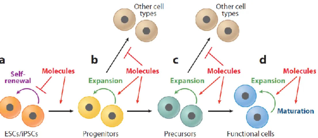
Figure 1.1 Chemical strategies for inducing differentiation. Differentiation in vitro is
typically carried out in a stepwise manner that recapitulates normal embryonic development. Several strategies have been developed to increase the efficiency of differentiation towards a specific lineage: (a) inhibiting stem cell self-renewal by small molecules to accelerate or enhance the differentiation process; (b) directing cells into specific cell types by a combination of small molecules that induce desired lineage-specific mechanisms and inhibit the activity of pathways and genes that lead to the induction of undesired cell types; (c) promoting progenitor cell expansion by small molecules to generate a large population of intermediate cells for various applications; and (d) maturing terminally differentiated cells by small molecules to generate desired functional cells. ESC, embryonic stem cell; iPSC, induced pluripotent stem cell 12. ... 6
Figure 1.2 Mesenchymal stem cells can give rise to osteocytes, hypertrophic
chondrocytes and adipocytes, or self-renew to produce identical cells that may later differentiate toward lineages that form the skeleton and bone marrow stroma. Trans-differentiation of MSCs into other non-mesodermal cell types has been also reported in vitro but remains controversial in vivo 25. ... 8
Figure 1.3 Chemical representations of the disaccharide units of glycosaminoglycans. (A) Chondroitin 4-sulfate, (B) Chondroitin 6-sulfate, (C) Dermatan sulfate, (D) Keratan sulfate, (E) Heparin, and (F) Hyaluronan. ... 12
xiv
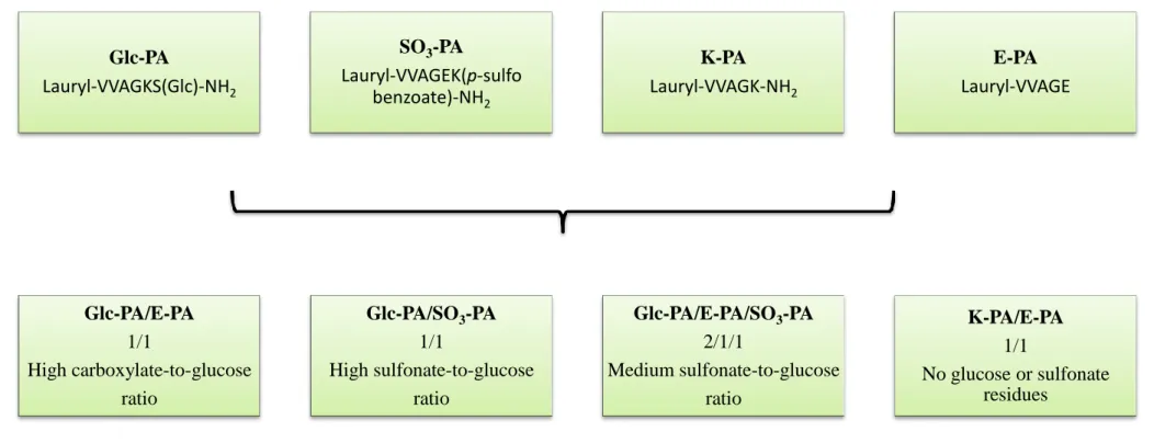
Figure 3.1 Chemical representations of peptide amphiphile molecules. (A) Glc-PA
[Lauryl-VVAGKS(Glc)-Am], (B) SO3-PA [Lauryl-VVAGEK(p-sulfo benzoate)-Am], (C) E-PA [Lauryl-VVAGE], and (D) K-PA [Lauryl-VVAGK-Am]. ... 39
Figure 3.2 Self-assembled nanofiber network systems used in this study. The peptide
nanofiber networks self-assemble when mixed with oppositely charged peptide amphiphiles and were modified to incorporate chemical groups found in natural glycosaminoglycans. ... 40
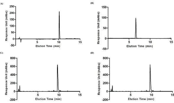
Figure 3.3 HPLC chromatogram of purified: (A) Glc-PA, (B) SO3-PA, (C) E-PA, and (D) K-PA K molecule at 220 nm... 43
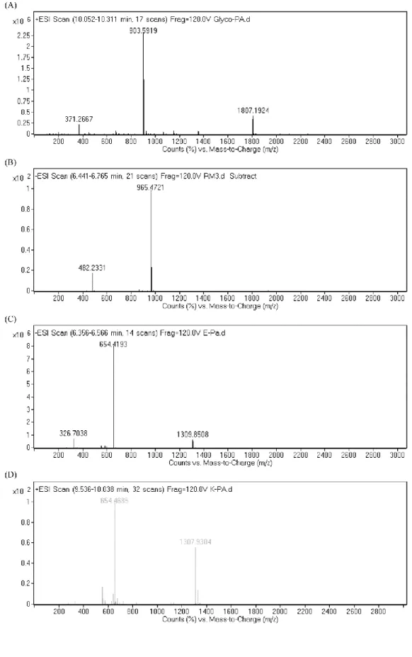
Figure 3.4 Electrospray ionization mass spectra of: (A) Glc-PA, (B) SO3-PA, (C)
E-PA, and (D) K-PA. ... 44
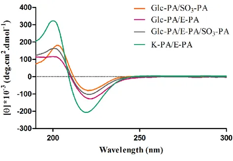
Figure 3.5 Circular dichroism (CD) spectra of peptide nanofibers used for the
characterization of the secondary structures. CD results indicate that peptide amphiphile nanofibers show a characteristic β-sheet structure at physiological pH. 45
Figure 3.6 Characterization of the peptide amphiphile nanofiber networks by using
scanning electron microscopy. (A) Glc-PA/E-PA, (B) Glc-PA/SO3-PA, (C) Glc-PA/SO3-PA/E-PA and (D) K-PA/E-PA. ... 46
Figure 3.7 Characterization of the peptide amphiphile nanofiber networks by using
transmission electron microscopy. (A) Glc-PA/E-PA, (B) Glc-PA/SO3-PA, (C) Glc-PA/SO3-PA/E-PA and (D) K-PA/E-PA. ... 47
Figure 3.8 Viability analyses of rat mesenchymal stem cells on different peptide
nanofiber networks and uncoated tissue culture plate. AlamarBlue® was used to quantitatively determine the viability of rat mesenchymal stem cells at 24 h. One-way ANOVA with Tukey post test (95% confidence interval) was applied for
xv
analyzing the results, and no significant difference was found between each groups. ... 52
Figure 3.9 Proliferation analyses of rat mesenchymal stem cells cultured with BrdU
on different peptide nanofiber networks and uncoated tissue culture plate at day 3. One-way ANOVA with Tukey post test (95% confidence interval) was applied for analyzing the results and significant differences were expressed as (* p<0.05, ** p<0.01, *** p < 0.001). ... 53
Figure 3.10 Alizarin Red-S calcium staining of cells cultured with growth medium at
day 7. (A) Glc-PA/E-PA, (B) Glc-PA/SO3-PA, (C) Glc-PA/SO3-PA/E-PA, (D) K-PA/E-PA, (E) TCP and (F) Quantitative analyses of Alizarin Red-S. One-way ANOVA with Tukey post test was applied to analyze the results and significant differences were expressed as (** p<0.01 and *** p < 0.001). (Scale bars = 200 μm) ... 56
Figure 3.11 Alizarin Red-S calcium staining of cells cultured with osteogenic
medium at day 7. (A) Glc-PA/E-PA, (B) Glc-PA/SO3-PA, (C) Glc-PA/SO3 -PA/E-PA, (D) K-PA/E--PA/E-PA, (E) TCP and (F) Quantitative analyses of Alizarin Red-S. One-way ANOVA with Tukey post test was applied to analyze the results and significant differences were expressed as (* p<0.05, ** p<0.01 and *** p < 0.001). (Scale bars = 200 μm) ... 57
Figure 3.12 Alizarin Red-S calcium staining of cells cultured with growth medium at
day 14. (A) Glc-PA/E-PA, (B) Glc-PA/SO3-PA, (C) Glc-PA/SO3-PA/E-PA, (D) K-PA/E-PA, (E) TCP and (F) Quantitative analyses of Alizarin Red-S. One-way ANOVA with Tukey post test was applied to analyze the results and significant differences (**) were expressed at p < 0.01. (Scale bars = 200 μm) ... 58
xvi
Figure 3.13 Alizarin Red-S calcium staining of cells cultured with osteogenic
medium at day 14. (A) Glc-PA/E-PA, (B) Glc-PA/SO3-PA, (C) Glc-PA/SO3 -PA/E-PA, (D) K-PA/E--PA/E-PA, (E) TCP and (F) Quantitative analyses of Alizarin Red-S. One-way ANOVA with Tukey post test was applied to analyze the results and significant differences were expressed as (* p<0.05 and ** p<0.01). (Scale bars = 200 μm) ... 59
Figure 3.14 Alizarin Red-S calcium staining of growth medium without cells at day
7, conducted as a control for non-specific calcium deposition. (A) Glc-PA/E-PA, (B) Glc-PA/SO3-PA, (C) Glc-PA/SO3-PA/E-PA, (D) K-PA/E-PA and (E) TCP. (Scale bars = 200 μm) ... 60
Figure 3.15 Alizarin Red-S calcium staining of osteogenic differentiation medium
without cells at day 7, conducted as a control for non-specific calcium deposition.
(A) Glc-PA/E-PA, (B) Glc-PA/SO3-PA, (C) Glc-PA/SO3-PA/E-PA, (D) K-PA/E-PA and (E) TCP. (Scale bars = 200 μm) ... 61
Figure 3.16 Alizarin Red-S calcium staining of growth medium without cells at day
14, conducted as a control for non-specific calcium deposition. (A) Glc-PA/E-PA,
(B) Glc-PA/SO3-PA, (C) Glc-PA/SO3-PA/E-PA, (D) K-PA/E-PA and (E) TCP. (Scale bars = 200 μm) ... 62
Figure 3.17 Alizarin Red-S calcium staining of osteogenic differentiation medium
without cells at day 14, conducted as a control for non-specific calcium deposition.
(A) Glc-PA/E-PA, (B) Glc-PA/SO3-PA, (C) Glc-PA/SO3-PA/E-PA, (D) K-PA/E-PA and (E) TCP. (Scale bars = 200 μm) ... 63
Figure 3.18 p-nitrophenol standard curve obtained by using known p-nitrophenol
standards. The standard curve was used to analyze the ALP activities of the rMSCs. The slope of the best line was 0.0723. ... 66
xvii
Figure 3.19 Alkaline phosphatase activity of rat mesenchymal stem cells at day 3.
Different nanofiber networks were used to investigate the changes in ALP activity according to the bio-functionality of the microenvironment. ALP activity of the rMSCs roughly correspond to the lipid accumulation characteristics of these cells. One-way ANOVA with Tukey post test was applied to analyze the results and significant differences were expressed as (* p<0.05, ** p<0.01 and *** p < 0.001). 67
Figure 3.20 Alkaline phosphatase activity of rat mesenchymal stem cells at day 7.
Different nanofiber networks were used to investigate the changes in ALP activity according to the bio-functionality of the microenvironment. ALP activity of the rMSCs roughly correspond to the lipid accumulation characteristics of these cells. One-way ANOVA with Tukey post test was applied to analyze the results and significant differences were expressed as (* p<0.05, ** p<0.01 and *** p < 0.001). 68
Figure 3.21 Gene expression analyses of rat mesenchymal stem cells cultured on
various nanofiber networks and tissue culture plate with growth medium at day 7.
(A) RUNX2 and (B) COL1. The expression level of each gene was normalized
against TCP and GAPDH, the latter of which was used as internal control. ... 70
Figure 3.22 Gene expression analyses of rat mesenchymal stem cells cultured on
various nanofiber networks and tissue culture plate with growth medium at day 14.
(A) RUNX2 and (B) COL1. The expression level of each gene was normalized
against TCP and GAPDH, the latter of which was used as internal control. ... 70
Figure 3.23 Gene expression analyses of rat mesenchymal stem cells cultured on
various nanofiber networks and tissue culture plate with osteogenic differentiation medium at day 7. (A) RUNX2 and (B) COL1. The expression level of each gene was normalized against TCP and GAPDH, the latter of which was used as internal
xviii
control. One-way ANOVA with Tukey post test was applied to analyze the results and significant differences were expressed as (** p<0.01 and *** p<0.001). ... 71
Figure 3.24 Gene expression analyses of rat mesenchymal stem cells cultured on
various nanofiber networks and tissue culture plate with osteogenic differentiation medium at day 14. (A) RUNX2 and (B) COL1. The expression level of each gene was normalized against TCP and GAPDH, the latter of which was used as internal control. One-way ANOVA with Tukey post test was applied to analyze the results and significant differences were expressed as (* p<0.05 and ** p<0.01). ... 71
Figure 3.25 Oil Red-O staining of cells in growth medium at day 7, showing the
extent of lipid deposition. (A) Glc-PA/E-PA, (B) Glc-PA/SO3-PA, (C) Glc-PA/SO3 -PA/E-PA, (D) K--PA/E-PA, (E) TCP and (F) Quantification of Oil Red O. One-way ANOVA with Tukey post test was applied to analyze the results and significant differences were expressed as (* p<0.05 and ** p<0.01). (Scale bars = 200 μm) ... 75
Figure 3.26 Oil Red-O staining of cells in growth medium at day 14, showing the
extent of lipid deposition. (A) Glc-PA/E-PA, (B) Glc-PA/SO3-PA, (C) Glc-PA/SO3 -PA/E-PA, (D) K--PA/E-PA, (E) TCP and (F) Quantification of Oil Red O. One-way ANOVA with Tukey post test was applied to analyze the results and significant differences were expressed as (** p<0.01 and *** p<0.001). (Scale bars = 200 μm) ... 76
Figure 3.27 Gene expression analyses of rat mesenchymal stem cells cultured on
various nanofiber networks and tissue culture plate with growth medium at day 7. (A) FABP4 and (B) ADIPOQ. The expression level of each gene was normalized against TCP and GAPDH, the latter of which was used as internal control. One-way
xix
ANOVA with Tukey post test was applied to analyze the results and significant differences were expressed as (* p<0.05 and ** p<0.01). ... 77
Figure 3.28 Gene expression analyses of rat mesenchymal stem cells cultured on
various nanofiber networks and tissue culture plate with growth medium at day 14.
(A) FABP4 and (B) ADIPOQ. The expression level of each gene was normalized
against TCP and GAPDH, the latter of which was used as internal control. ... 77
Figure 3.29 Safranin-O staining at day 7 for the analysis of sulfated
glycosaminoglycan incorporation. Cells were cultured with growth medium. (A) Glc-PA/E-PA, (B) Glc-PA/SO3-PA, (C) Glc-PA/SO3-PA/E-PA, (D) K-PA/E-PA and (E) TCP. (Scale bars = 200 μm) ... 81
Figure 3.30 Safranin-O staining at of cells in growth medium at day 7, showing the
extent of sulfated glycosaminoglycan incorporation. (A) PA/E-PA, (B) Glc-PA/SO3-PA, (C) Glc-PA/SO3-PA/E-PA, (D) K-PA/E-PA and (E) TCP. (Scale bars = 200 μm) ... 82
Figure 3.31 Gene expression analyses of rat mesenchymal stem cells cultured on
various nanofiber networks and tissue culture plate with growth medium at day 7.
(A) SOX9 and (B) COL2. The expression level of each gene was normalized against
TCP and GAPDH, the latter of which was used as internal control. ... 84
Figure 3.32 Gene expression analyses of rat mesenchymal stem cells cultured on
various nanofiber networks and tissue culture plate with growth medium at day 14.
(A) SOX9 and (B) COL2. The expression level of each gene was normalized against
TCP and GAPDH, the latter of which was used as internal control. One-way ANOVA with Tukey post test was applied to analyze the results and significant differences were expressed as (* p<0.05 and ** p<0.01). ... 84
xx
LIST OF TABLES
Table 1.1 The localization and primary functions of the natural glycosaminoglycans
in the body. ... 15
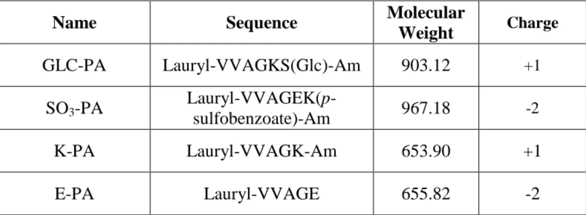
Table 2.1 Sequences, molecular weights, and charges of the synthesized peptide
amphiphile molecules... 23
Table 2.2 Primer sequences and annealing temperatures of runt-related transcription
factor 2, collagen 1, transcription factor SOX-9, collagen 2, adiponectin and fatty acid binding protein 4 for rat mesenchymal stem cells. ... 35
xxi
LIST OF ABBREVIATIONS
ECM : Extracellular Matrix
PA : Peptide Amphiphile
GAG : Glycosaminoglycan
DIEA : N,N-diisopropylethylamine
TFA : Trifluoroacetic Acid
TIS : Triisoproplysilane
DMF : Dimethylformamide
DCM : Dichloromethane
LC-MS : Liquid Chromatography Mass Spectroscopy
HPLC : High Pressure Liquid Chromatography
CD : Circular Dichroism
SEM : Scanning Electron Microscopy
TEM : Transmission Electron Microscopy
FBS : Fetal Bovine Serum
DMEM : Dulbecco’s Modified Eagle Medium
PS : Penicillin/Streptomycin
BrdU : Bromodeoxyuridine
ALP : Alkaline phosphatase
ORO : Oil Red O
TCP : Tissue Culture Plate
xxii
GAPDH : Glyceraldehyde 3-phosphate dehydrogenase
COL1 : Collagen 1
COL2 : Collagen 2
RUNX2 : Runt-related Transcription Factor 2
SOX9 : Transcription Factor SOX-9
ADIPOQ : Adiponectin
FABP4 : Fatty Acid Binding Protein 4
qRT-PCR : Quantitative Real-Time Polymerase Chain Reaction
GE : Glc-PA/E-PA
GS : Glc-PA/SO3-PA
GES : Glc-PA/SO3-PA/E-PA
1
CHAPTER 1
2
1.1 AN OVERVIEW OF REGENERATIVE MEDICINE
Regenerative medicine researches aim to assist the natural tissue regeneration process, and employ a variety of approaches to repair, renew or replace tissues or organs that have lost their functionality due to traumatic injury, disease, age or hereditary defects 1. The human body is a complex system, and its maintenance mechanisms include natural repair processes that operate at molecular, cellular and systemic levels to continuously regenerate lost tissue in healthy individuals. However, the body’s natural capacity for regeneration may not be enough in cases of severe injury or chronic disease, necessitating external assistance to fully restore the functions of the damaged tissue. Regenerative medicine studies deal with the organizational complexity and functional diversity of the cells and their surroundings to recover and regenerate tissues following traumatic injury. Its success, therefore, depends on our ability to understand the cellular and molecular mechanisms of regeneration, and to transfer this information to facilitate the functional regeneration of damaged tissues with limited capacity for renewal. Three main approaches are used in regenerative medicine studies in order to regenerate damaged tissues or organs. These approaches are: (1) differentiated cell (progenitor cell) or stem cell transplantation either alone or as part of a biomaterial 2-3, (2) artificial tissue implantation 4 and (3) induction of the regeneration by using chemicals at the sites of injuries 5. These methods can be employed alone or in combination to overcome the engineering limitations of the bionic devices and the donor shortage inherent to organ transplants 6.
3
The effectiveness of regenerative medicine hinges on its ability to mimic the microenvironment normally experienced by cells in their native tissues. This microenvironment typically contains at least one of these features:
1. The presence of the requisite bio-signals for cell attachment and migration; 2. The retention and presentation of biochemical factors;
3. A porous environment for the adequate diffusion of nutrients, and waste; and 4. Mechanical characteristics, such as rigidity or elasticity, that are naturally experienced by each cell type 7.
1.1.1 PROGENITOR CELL OR STEM CELL TRANSPLANTATION
Cell transplantation is an attractive alternative to whole organ transplantation because (1) it allows early intervention in diseases, (2) it reduces the immunogenecity of transplantation and the risk of organ rejection, and (3) it eliminates the risk of proper donor finding. Cells used in transplantation studies should be easy to access and propagate, be pluripotent and not subject to immuno-rejection. Progenitor cells and stem cells show these properties, and therefore can be used in cell transplantation studies either alone or part of a biomaterial. There are two main types of stem cells, embryonic stem cells and adult stem cells. Embryonic stem cells are totipotent cells, i.e. they can differentiate into all three embryonic germ layers. Adult stem cells are multipotent cells, and have more limited differentiation potentials compared to embryonic stem cells. They can differentiate into mesodermal and non-mesodermal cell types 8.
4
1.1.2 ARTIFICIAL TISSUE IMPLANTATION
Artificial tissue implantation involves the development of biomimetic devices or functionalized platforms that can be used to replace areas of damaged tissue and provide a suitable environment for the survival and regeneration of the remaining cells. Various artificial tissue transplants have been developed for the replacement of several tissue types, using methods such as hydrogels or 3D bio-printing. For example, it was shown that a mixture of human liver precursor cells and two other cell types can form 3D structures called “liver buds”, which were able to form functional connections with natural blood vessels and perform liver-specific functions in vivo 9. In another study, a complex 3D architecture with designed patterns was created by using pressure activated micro-syringe equipped with a fine-bore exit needle and used to constitute implantable bioartificial organs. It was demonstrated that cells remain viable and continue their biological functions in this platform because the microchannels present in the system provide an easy access to nutrients and facilitate the removal of waste materials 10.
1.1.3 INDUCTION OF REGENERATION BY INTRODUCING CHEMICALS INTO THE SITE OF INJURY
Small molecules can be used to induce regeneration directly at the site of injury. This method has several advantages compared to artificial tissue implantation and cell transplantation. First, these chemicals have physicochemically well-defined structures and can be synthesized in large quantities with little batch-to-batch
5
variation. In addition, most of these chemicals have the ability to penetrate the cells and modulate intracellular processes. Finally, being small molecules, they are relatively non-immunogenic compared to other regeneration strategies 11.
It is well-demonstrated that small chemicals with the ability to target specific signaling pathways and/or proteins can be used for the manipulation of cell fate, state and function 12. There are various strategies for efficient directed differentiation of the cells as shown in Figure 1.1. To illustrate; Noggin is a BMP4 inhibitor and SB431542 is a TGFβ receptor inhibitor, and the inhibition of BMP pathway and TGFβ pathway at the same time by using these molecules increased the efficiency of neural differentiation of a monolayer culture of human embryonic stem cells 13. Pumorphamine which is an adenine derivative is another example for the small molecules used in regenerative medicine applications. It is used as an inducer of osteoblast formation from multipotent mesenchymal progenitor cells and lineage-committed preosteoblasts 14.
The in vivo potential of small chemicals is also well-established. For instance, the 1,2-isoxazoles were shown to repair damaged heart tissue after infarction, activating a cardiac program in the Notch-activated epicardium-derived progenitor cells 15. Finally, in another in vivo study, a compound named as P7C3 was shown to improve the survival of newly formed neurons from apoptotic cell death and promote neurogenesis in the subgranular zone of the hippocampal dentate gyrus, but did not affect neural stem cell differentiation 16.
6
Figure 1.1 Chemical strategies for inducing differentiation. Differentiation in vitro is
typically carried out in a stepwise manner that recapitulates normal embryonic development. Several strategies have been developed to increase the efficiency of differentiation towards a specific lineage: (a) inhibiting stem cell self-renewal by small molecules to accelerate or enhance the differentiation process; (b) directing cells into specific cell types by a combination of small molecules that induce desired lineage-specific mechanisms and inhibit the activity of pathways and genes that lead to the induction of undesired cell types; (c) promoting progenitor cell expansion by small molecules to generate a large population of intermediate cells for various applications; and (d) maturing terminally differentiated cells by small molecules to generate desired functional cells. ESC, embryonic stem cell; iPSC, induced pluripotent stem cell 12.
7
1.2 MESENCHYMAL STEM CELLS IN REGENERATIVE MEDICINE
Mesenchymal stem cells (MSCs), also known as multipotent marrow stromal cells or mesenchymal stromal cells, are stromal cells that have the ability to self-renew and/or differentiate into mesodermal (bone, adipose and cartilage) and non-mesodermal lineages (Figure 2.1) 17-20. MSCs can be distinguished from other cell types due to their four intrinsic characteristics. (1) They show plastic adherent properties under standard culture conditions. (2) They express the surface markers CD73, CD90 and CD105. (3) They have a fibroblast-like morphology. (4) They lack the expression of the surface markers CD45, CD34, CD14 or CD11b, CD79a and HLA-DR 21.
1.2.1 THE DISCOVERY OF MESENCHYMAL STEM CELLS
The presence of stem cells for non-hematopoietic cells in bone marrow was first recognized 130 years ago, by the observations of the German pathologist Cohnheim. In his research, Cohnheim suggested the possibility that bone marrow may be the source of fibroblasts, and that these fibroblasts initiate collagen fiber deposition as part of the normal wound repair process 22.
Starting with the work of Friedenstein and colleagues, it was known that bone marrow contains cells that can differentiate into other mesenchymal cells, as well as developing into fibroblastic colony forming cells 23. Ever since, these findings were also confirmed by numerous scientists and it was also demonstrated that these cells can also be found in the human bone marrow 24.
8
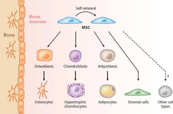
Figure 1.2 Mesenchymal stem cells can give rise to osteocytes, hypertrophic
chondrocytes and adipocytes, or self-renew to produce identical cells that may later differentiate toward lineages that form the skeleton and bone marrow stroma. Trans-differentiation of MSCs into other non-mesodermal cell types has been also reported in vitro but remains controversial in vivo 25.
9
1.2.2 ISOLATION OF MESENCHYMAL STEM CELLS
Mesenchymal stem cells can be isolated from many different sites of the body including the superior iliac crest of the pelvis in humans, the tibial and femoral marrow compartments and the thoracic and lumbar spine 26-29. MSCs can be isolated and enriched with standard cell culture methods in which samples are fractionated on a density gradient and plating the isolated cells at densities from 1.0 x 10⁴ to 0.4 x 10⁶ cells/cm230.
1.2.3 DIFFERENTIATION OF MESENCHYMAL STEM CELLS
It was demonstrated in many studies that MSCs can differentiate into bone, cartilage and fat tissues 31-33. β-glycerophosphate, ascorbic acid-2-phosphate, dexamethasone and fetal bovine serum are required for the activation of osteogenic differentiation. When cultured with these supplements, mesenchymal stem cells increase their alkaline phosphatase activities and deposit a calcium-rich mineralized extracellular matrix, in addition to showing osteoblastic morphology. Chondrogenic differentiation of mesenchymal stem cells requires a three-dimensional culture configuration, a serum free nutrient medium and a member of the TFG-β superfamily. In the presence of these factors, MSCs upregulate the expression of cartilage specific extracellular matrix components and start to lose their fibroblastic morphology. Adipogenic differentiation can be initiated by culturing MSCs in a monolayer culture in the presence of isobutylmethylxanthine, and confirmed visually by the formation of large lipid vacuoles in differentiating cells. Peroxisome
10
proliferator-activated receptor-gamma and fatty acid synthetase also play important roles in the induction of adipogenic differentiation in MSCs 34.
1.3 EXTRACELLULAR MATRIX AND ITS COMPONENTS
Extracellular matrix (ECM) is the material that surrounds the cells. It is paramount for cell-to-cell communication and the maintenance of tissue integrity. The ECM is highly complex and consists of a great variety of proteins, primarily in the form of proteoglycans, glycoproteins and long nanofibers. Natural extracellular matrix components provide structural and biochemical support for cells, and direct cellular behaviors such as adhesion, migration, proliferation, and differentiation. The extracellular matrix microenvironment therefore has an indispensable role for the survival and the regeneration of the cells. The composition, topology and physicochemical characteristics of the extracellular matrix show differences according to cell and tissue types, wound- and disease-states, and age. In addition to these, the ECM has a very dynamic structure even within a single type of tissue, as it is constantly being remodeled by enzymatic and non-enzymatic processes 35. Extracellular matrix receptors, such as integrins and syndecans, take roles in the adhesion of the cells 36-37. The adhesion of the cells to the ECM triggers cytoskeletal changes and can facilitate the migration of these cells through the ECM 38. Collagens, elastins, fibronectins and laminins are the main fibrous extracellular matrix proteins 39. From these fibrous proteins, collagen is the most abundant one found in the extracellular matrix and it gives tensile strength, helps cell adhesion and support migration and chemotaxis 40. Collagens are defined by the triple helical
11
organization of component pro-α-chains that is formed as a result of the glycine amino acid being used in every third residue. There are now 28 known collagen types 41
. Fibronectin takes roles in extracellular matrix organization, cell attachment and function 42. Fibronectin also has important functions in cell migration during embryonic development, tumor metastasis and cardiovascular disease 43. Mechanical and biochemical changes in the extracellular matrix are perceived and acted upon by the cells through the crosstalk between integrins and the actin cytoskeleton 44.
1.4 NATURAL GLYCOSAMINOGLYCANS AND THEIR ROLES IN TISSUE REGENERATION
Glycosaminoglycans (GAGs) are highly polar, negatively charged, unbranched polysaccharides which play important roles in various biological processes, and are vital for the regeneration of damaged tissues. They have extremely heterogeneous and complex structures due to degree of substitution and positioning of sulfate/sulfonate and acetyl groups 45. GAGs are mainly composed of repeating disaccharide units (Figure 1). The repeating disaccharide unit always contains an amino sugar; either N-acetylglucosamine or N-acetylgalactosamine, along with a uronic acid, usually D-glucuronic or L-iduronic acid 46. GAGs can be classified into four basic groups according to their repeating disaccharide structures and functions in the body. These four basic groups are: (1) Heparin/Heparan sulfate, (2) Chondroitin sulfate/Dermatan sulfate, (3) Keratan sulfate, and (4) Hyaluronic acid/Hyaluronan 47. Except for hyaluronan, they contain sulfate/sulfonate groups, form proteoglycans by substituting protein cores, and are synthesized in the
12
endoplasmic reticulum and Golgi bodies 48. GAGs have diverse intracellular and extracellular actions, and play crucial roles in different biological processes (Table
1) including growth, proliferation and differentiation of the cells, regeneration of
nerve, cartilage and bone tissues, angiogenesis, and tumor metastasis through specific interactions with growth factors, growth factor receptors, extracellular matrix proteins, and other chemokines 49-54.
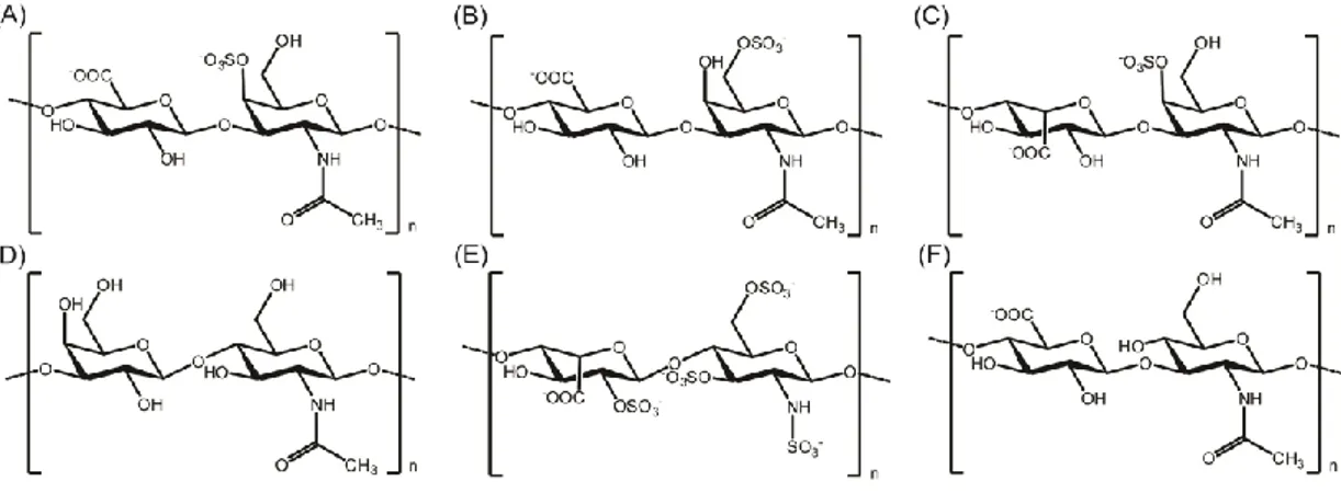
Figure 1.3 Chemical representations of the disaccharide units of glycosaminoglycans. (A) Chondroitin 4-sulfate, (B) Chondroitin 6-sulfate, (C) Dermatan sulfate, (D) Keratan sulfate, (E) Heparin, and (F) Hyaluronan.
Hyaluronan (HA), which is composed of alternating units of β-1,4-linked D-glucuronic acid-β-1,3-N-acetyl-D-glucosamine, is the only non-sulfated glycosaminoglycan 55. Unlike other glycosaminoglycans, it is synthesized in the plasma membrane and its primary structure does not contain a peptide component 48. It especially forms the extracellular matrix of soft connective tissues, and interactions between hyaluronan receptor and extracellular polysaccharides have effects on
13
locomotion and cell migration 56. HA also has important biological functions in wound healing and remodeling, and serves to initiate inflammatory responses, maintain structural cell integrity and promote recovery from tissue injuries 57.
Chondroitin sulfate (CS) is composed of β-1,3-linked D-glucuronic acid-β-1,4-N-acetyl-D-galactosamine. CS is a sulfated glycosaminoglycan commonly found in the cartilage extracellular matrix, where it serves to increase the resistance of this tissue against frictional and compressive stress 58-60. The CS disaccharides are sulfated on either C-4 or C-6 position of the N-acetyl-D-galactosamine residues and classified according to their sulfation patterns 61. Besides cartilage extracellular matrix, it is also the major element of the brain extracellular matrix and takes roles in plasticity, neural development, and regeneration. In adults, chondroitin sulfate proteoglycans also play roles in learning and memory processes 62.
Dermatan sulfate (DS) is another natural and sulfated glycosaminoglycan. DS is composed of β-1,3-linked L-iduronic acid-β-1,4-N-acetyl-D-galactosamine units and is also known as chondroitin sulfate B 63. In the cornea, it plays a crucial role in optical clarity by regulating fibril spacing 64. It also has roles in bone metabolism in pathological conditions by regulating osteoclast formation through interaction with receptor activator of NF-κB ligand and inhibition of signal transduction in osteoclast progenitor cells 65. DS can selectively inhibit thrombin without interfering with platelets. Therefore, it can also be used as anticoagulant for patients with acute renal failure after a major cardiovascular surgery 66.
Heparan sulfate (HS) is composed of alternating units of α-D-glucosamine and uronic acid, either β-D-glucuronic acid or α-L-iduronic acid, and linked by 1,4 glycosidic bond. The α-D-glucosamine can be either N-sulfated or N-acetylated 67.
14
Bearing one of the highest natural complexities among glycosaminoglycans, HS performs a wide range of biological activities including roles in blood coating, angiogenesis, and the protection of proteins from degradation 68. It also takes role in regulation of bone morphogenetic proteins' activities and induction of morphogenesis. For instance, cell surface heparan sulfate proteoglycans can intercede BMP-2 internalization and it was shown that these heparan sulfate supramolecules lead to transdifferentiation of C2C12 myoblasts into osteoblasts through direct regulation of BMP-2 activities 69.
Keratan sulfate (KS) is composed of β-1,3-linked D-galactose-β-1,4-N-acetyl-D-glucosamine units and sulfated on C-6 of either or both hexose moieties 70. KS is associated with a vast variety of biological functions in different tissue types and developmental stages. It is mainly found in cornea, cartilage, and brain. It is the major GAG in cornea and takes role in corneal hydration and maintenance of ECM structure 71. Due to its highly hydrated nature, KS suppresses cartilage damage in joints and exogenous KS application can be used for treatment of inflammatory arthritis 72. KS is also synthesized in central nervous system and takes an important role in modulating axonal growth after spinal cord injury 73.
In the extracellular matrices of all mammalian tissues, GAGs are found covalently attached to proteins, and classified as proteoglycans except for hyaluronan 74
. Proteoglycans are classified according to their GAG composition, localization and core proteins. Their molecular sizes, abundances, and sulfation and acetylation patterns change based on tissue types and developmental stages. Proteoglycans can be separated into three main families which are small leucine-rich proteoglycans, modular proteoglycans and cell-surface proteoglycans 75. Small leucine-rich
15
proteoglycans take roles in various signaling pathways and regulation of inflammatory response reactions. To illustrate, they can bind to and activate epidermal growth factor receptor, insulin-like growth factor 1 receptor, and TGF-β 76-77
. Modular proteoglycans take roles in mainly cell adhesion, migration and proliferation. Cell-surface proteoglycans can act as co-receptors facilitating ligand encounters with signaling receptors 35.
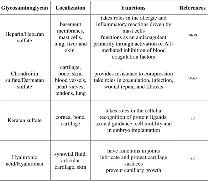
Table 1.1 The localization and primary functions of the natural glycosaminoglycans
in the body.
Glycosaminoglycan Localization Functions References
Heparin/Heparan sulfate
basement membranes,
mast cells, lung, liver and
skin
takes roles in the allergic and inflammatory reactions driven by
mast cells
functions as an anticoagulant primarily through activation of
AT-mediated inhibition of blood coagulation factors 78-79 Chondroitin sulfate/Dermatan sulfate cartilage, bone, skin, blood vessels, heart valves, tendons, lung
provides resistance to compression take roles in coagulation, infection,
wound repair, and fibrosis
60,63
Keratan sulfate cornea, bone, cartilage
takes roles in the cellular recognition of protein ligands, axonal guidance, cell motility and
in embryo implantation 70 Hyaluronic acid/Hyaluronan synovial fluid, articular cartilage, skin
have functions in joints lubricate and protect cartilage
surfaces
prevent capillary growth
16
1.5 PEPTIDE AMPHIPHILE MOLECULES AND THEIR USE IN REGENERATIVE MEDICINE
Peptide amphiphiles (PAs) are peptide-based molecules that can self-assemble into high-aspect ratio nanofibers. A typical peptide amphiphile macromolecule consists of four main regions. The hydrophobic domain that consists of a long alkyl tail forms the first region. A short peptide sequence which takes role in the formation of intermolecular hydrogen bonds forms the second region. The third region is formed from charged amino acids to enhance solubility of the macromolecule in water and to design pH and salt-responsive nanostructures and networks. The fourth region contains bioactive signals that is usually designed by mimicking epitopes and used for providing special signals that take roles in nanostructure-cell interactions and differentiation of the cells 81.
The amphiphilic nature of the peptide amphiphile molecules comes from the incorporation of a hydrophobic alkyl tail after the peptide sequence. This hydrophobic alkyl tail that forms the first region of the macromolecules as previously mentioned allows the presentation of peptide signals specifically on the periphery of self-assembled nanostructures. The hydrophobic tails can also be adjusted by using different alkyl chain lengths or by using different hydrophobic components 82. The short peptide sequence that forms the second region of the macromolecules usually consists of hydrophobic amino acids. This short peptide sequence allows formation of intermolecular hydrogen bonds generally in the form of β-sheets and leads for the presentation of bioactive signals on the nanofiber surface at a high density. Charged amino acids that form the third region of a peptide amphiphile molecule come after
17
the short peptide sequences and their number and identity determine the solubility of the peptide amphiphile molecules under physiological conditions. The bioactive peptide sequence formed by mimicking various epitopes constitutes the last and the most important region of the peptide amphiphile molecules if they will be used in a biological study. This region may consist of different biological signals such as one that promotes cell adhesion 83-84 or one that generates a pronounced therapeutic effect 85-86
.
Peptide amphiphile molecules were first presented approximately a decade ago as a regenerative tool for bio-mineralization. In this study, it was shown that peptide nanofibers with phosphoserine residues self-assembled into nanofibers that can nucleate thin hydroxyapatite crystals on their surfaces in vitro as observed in bone formation 87. The importance of phosphoserine residue for nucleation of hydroxyapatite was also proved in vivo using an orthotopic rat femoral critical-size defect model. In this study performed by Mata et al., the combined effects of fibronectin epitope RGDS and the phosphoserine residues on bone regeneration were determined by the analysis of hydroxyapatite nucleation, and it was concluded that the presence of phosphoserine residues on the peptide nanofibers enhances formation of biomimetic bone crystals 88.
Recently, various peptide amphiphile nanofibers that contain bioactive signals have been used to mimic natural sulfated glycosaminoglycans and to constitute a suitable microenvironment for the survival and differentiation of the osteogenic cells. In one study, a heparin-mimetic peptide amphiphile scaffold system was used to analyze osteogenic differentiation of Saos-2 cells. The BMP-2 binding capacity of these heparin-mimetic peptide scaffold was shown by using an ELISA based growth
18
factor binding assay. The nanofiber scaffold showed osteoinductive properties by stabilizing BMP-2 and providing a convenient microenvironment for bone regeneration with their sulfonate and carboxylate groups. These nanofiber networks also enhance the viability, proliferation and mineralization of the osteogenic cells by binding to BMP-2 89.
Ustun et al. used heparin mimetic PA macromolecule to analyze differentiation of chondroprogenitor ATDC5 cells. Heparin mimetic peptide scaffold was found to give both structural and functional assistance for the cells, causing rapid aggregation of the cells in insulin-free medium, which culminate in the formation of cartilage-like nodules. The sulfated glycosaminoglycan deposition was shown with Safranin-O staining, and the bioactive role of the nanofiber system was also revealed with qRT-PCR which was used to show expression levels of collagen II and aggrecan genes 90.
In a different study, the effect of heparin-mimetic PA on neurite outgrowth was analyzed in combination with laminin-derived PA. It was revealed that hydrogels composed of self-assembly of a laminin-derived PA and heparin mimetic PA increase the efficiency of the neurite outgrowth compared to laminin-derived PAs alone. It was also revealed that the inhibitory effect of chondroitin sulfate proteoglycans on the central nervous system can be overcome by using this scaffold 91
19
1.6 MOTIVATION AND GOALS
Despite vast advancements in biology and its sub-disciplines, it is still not possible to fully mimic sophisticated biological materials in either form or function. As such, regenerative medicine often aims to partially replicate the effect of biological structures by identifying and utilizing functional regions that contain crucial biological signals. Peptide amphiphile macromolecules allow direct incorporation of these functional regions into their primary sequences. By using peptide amphiphile molecules, nanostructures with high aspect ratios, complex architectures and desired biochemical characteristics can be easily produced under physiological conditions. These peptide nanostructures can then be used in various regenerative medicine applications, where they are expected to enhance the natural regeneration process due to their resemblances to the native microenvironments of their target tissues.
It was previously shown that synthetic matrices containing small-molecule chemical functional groups can be used for controlled induction of differentiation to multiple mesenchymal stem cell lineages 92. In this study, peptide amphiphile molecules modified with different small functional groups were used to determine the effect of GAG composition on rat mesenchymal stem cell fate. Our hypothesis was that the functions of natural glycosaminoglycans can be mimicked using a combination of various small functional groups; and we tested this hypothesis under in vitro conditions by investigating the differentiation tendency of rat mesenchymal stem cells over functionalized bioactive nanofibrous networks towards mesodermal lineages.
20
CHAPTER 2
21
2.1 CHEMICALS AND SOLUTIONS
Solid State Peptide Synthesis Reagents:
9-Fluorenylmethoxycarbonyl (Fmoc) and tert-butoxycarbonyl (Boc) protected amino acids except glyco amino acid, [4-[α-(2',4'-dimethoxyphenyl) Fmoc-aminomethyl]phenoxy] acetamidonorleucyl-MBHA resin (Rink amide MBHA resin), Fmoc-Glu(OtBu)-Wang resin, and 2-(1H-Benzotriazol-1-yl)-1,1,3,3-tetramethyluronium hexafluoro-phosphate (HBTU) were purchased from NovaBiochem and ABCR. Fmoc-Ser[β-Glc(OAc)4]-OH was purchased from AAPPTec. N,N-diisopropylethylamine (DIEA) and lauric acid were purchased from Merck. Piperidine, acetic anhydride, dichloromethane (DCM), dimethylformamide (DMF), trifluoroacetic acid (TFA) and triisoproplysilane (TIS) were purchased form Sigma-Aldrich. The remaining chemicals were purchased from Fisher and/or Alfa Aesar; all chemicals were used as provided. Deionized water with a resistance of 18 MΩ.cm (Millipore Milli-Q) was used in all experiments.
Cell Culture Reagents:
Dulbecco’s Modified Eagle Medium (DMEM), Penicillin/Streptomycin (PS) antibiotic combination and Fetal Bovine Serum (FBS) were purchased from Gibco, Life Technologies. Alamar Blue was purchased from Invitrogen. BrdU (colorimetric) was purchased from Roche. Safranin-O, Oil Red-O and Alizarin Red-S were purchased from Sigma-Aldrich.
22
2.2 SYNTHESIS OF PEPTIDE AMPHIPHILE MOLECULES
Peptide amphiphile molecules were synthesized by using a standard solid phase peptide synthesis method with Rink amide MBHA resin or glutamic acid loaded Wang resin. GLC-PA [Lauryl-VVAGKS(Glc)-NH2], SO3-PA [Lauryl-VVAGEK(p-sulfobenzoate)-NH2] and K-PA [Lauryl-VVAGK- NH2] were synthesized on Rink amide MBHA resin. E-PA [Lauryl-VVAGE] was synthesized on Fmoc-Glu-Wang resin. Prior to the first coupling reaction, the resins were swelled in DCM for 30 min and the solvent was exchanged to DMF, in which all remaining reactions were carried out. In the synthesis of every peptide amphiphile molecules, the solid state was washed three times with DMF, three times with DCM and finally again three times with DMF respectively between each step to remove unreacted chemicals. Each coupling reaction started with the removal of the Fmoc protecting group by using 20% (v/v) piperidine/dimethylformamide solution for 20 min. Amino acids were then prepared for the coupling reaction by dissolving in DMF and amino acid coupling reactions were performed by using 2 equivalents of amino acid, 1.95 equivalents of HBTU and 3 equivalents of DIEA in 10 mL DMF for 2.5 h. At the end of each coupling step, the completeness of the coupling reaction was determined with Kaiser test. If the reaction was complete, the amino acid chain was exposed to 10% (v/v) acetic anhydride/dimethylformamide solution for 30 min in order to acetylate unreacted amine groups prior to the next coupling cycle. These steps were repeated until the desired amino acid sequences were obtained (Table 2.1). The lauric acid tail was then added in a similar fashion to the amino acid coupling reaction, except that the coupling time was 4 h. After the synthesis procedure, peptide cleavage from the
23
solid phase was carried out for 2 h at room temperature with 95% cleavage cocktail (95:2.5:2.5 TFA:TIS:ddH2O) and excess TFA and DCM were subsequently removed with rotary evaporator. The remaining peptide amphiphile solution was then precipitated using overnight incubation in ice cold diethyl ether at -20 °C. The solution was then centrifuged at 8000 rpm for 15 min to completely precipitate peptide amphiphile molecules; diethyl ether was then decanted and the remainder in the flask was evaporated, and the peptide amphiphile molecules were dissolved with ddH2O. Finally, the peptide amphiphile solution was frozen at -80 °C for 4 h, lyophilized for 3 days and stored at -20 °C.
Table 2.1 Sequences, molecular weights, and charges of the synthesized peptide
amphiphile molecules.
Name Sequence Molecular
Weight Charge GLC-PA Lauryl-VVAGKS(Glc)-Am 903.12 +1 SO3-PA Lauryl-VVAGEK(p-sulfobenzoate)-Am 967.18 -2 K-PA Lauryl-VVAGK-Am 653.90 +1 E-PA Lauryl-VVAGE 655.82 -2
2.3 CHARACTERIZATION OF THE SELF-ASSEMBLED PEPTIDE NANOSTRUCTURES
High Pressure Liquid Chromatography (HPLC) was used to purify the synthesized peptide amphiphile molecules, while Liquid Chromatography-Mass
24
Spectroscopy (LC-MS), Circular Dichroism (CD), Scanning Electron Microscopy (SEM) and Transmission Electron Microscopy (TEM) were used to perform chemical and mechanical characterizations of peptide amphiphile molecules.
2.3.1 LIQUID CHROMATOGRAPHY-MASS SPECTROSCOPY
A quadruple time of flight (Q-TOF) mass spectrometer with electrospray ionization (ESI) source equipped with a reverse phase analytical high performance liquid chromatography was used to characterize the synthesized peptide amphiphile molecules. For the characterization of negatively and positively charged PA molecules, Agilent Zorbax Extend-C18 (2.1 x 50 mm) column and Zorbax SB-C8 (4.6 x 100 mm) column were used as stationary phases respectively. A gradient of water (0.1% formic acid or 0.1% NH4OH) and acetonitrile (0.1% formic acid or 0.1% NH4OH) was used as mobile phase during liquid chromatography.
High Pressure Liquid Chromatography:
Reverse phase preparative HPLC equipped with either Zorbax Extend-C18 (21.2 x 150 mm) column or Zorbax SB-C8 (21.2 x 150 mm) column as a stationary phase were used to purify negatively and positively charged PA molecules, respectively. A gradient of water (0.1% acetonitrile or 0.1% NH4OH) and acetonitrile (0.1% formic acid or 0.1% NH4OH) were used as a mobile phase during HPLC purification of the synthesized PA molecules. Furthermore; according to their synthesis purity, positively charged PA molecules were only treated with 0.1 M HCl
25
solution in dialysis bags, in which the remaining TFA removed through chloride ion exchange.
2.3.2 CIRCULAR DICHROISM
Circular dichroism analyses were performed by using a J-815 Jasco spectrophotometer in the far ultra-violet region. PA molecules were prepared and used at a final concentration of 200 μM during all analysis. Quartz cuvette with 1 mm pathlength was used for all measurements. Measurement parameters were selected as: digital integration time of 1 sec, band width of 1 nm, data pitch of 0.1 nm, and with standard sensitivity. The scanning results were the average of three readings in a spectral range between 190 nm and 300 nm. Following scanning, ellipticity data obtained from the measurements were converted to molar ellipticity (with the unit degree.cm2.mole-1) using the following formula:
[θ] =
θ
[θ]: Molar ellipticity, θ: Ellipticity, C: Concentration in Molar, l: Length in cm
2.3.3 SCANNING ELECTRON MICROSCOPY
Scanning electron microscopy samples were prepared by mixing 10 mM Glc-PA and 10 mM E-Glc-PA at 1:1 ratio, 10 mM Glc-Glc-PA and 10 mM SO3-PA at 1:1 ratio, 10 mM Glc-PA, 10 mM SO3-PA and 10 mM E-PA at 2:1:1 ratio, and finally 10 mM
26
K-PA and 10 mM E-PA at 1:1 ratio to have overall negatively charged hydrogels. Hydrogels were placed onto silicon wafers and incubated for 30 minutes before applying serial ethanol dehydration protocol. Hydrogels were dehydrated sequentially in 20% (v/v), 40% (v/v), 60% (v/v), 80% (v/v) and absolute ethanol, and subsequently dried by using a critical point dryer (Tousimis, Autosamdri-815B, Series C critical point dryer). Finally, samples were coated with 5 nm Au/Pd and analyzed by using a SEM (SEM, FEI Quanta 200 FEG) with an ETD detector at high vacuum mode at 10 keV beam energy.
2.3.4 TRANSMISSION ELECTRON MICROSCOPY
Lacey mesh ultrathin carbon coated copper grids were used for transmission electron microscopy analysis. TEM samples were prepared by mixing 2 mM Glc-PA and 2 mM E-PA at 1:1 ratio, 2 mM Glc-PA and 2 mM SO3-PA at 1:1 ratio, 2 mM Glc-PA, 2 mM SO3-PA and 2 mM E-PA at 2:1:1 ratio, and finally 2 mM K-PA and 2 mM E-PA at 1:1 ratio to have overall negatively charged hydrogels. The upper parts of grids were dipped for 1 min into samples that were previously diluted 100 times with distilled water; the grids were then stained with 2 wt% uranyl acetate for 40 seconds and kept in a fume hood until a dried film was obtained. Finally; samples were kept in a fume hood until a dried film was obtained, and analyzed by using a FEI Tecnai G2 F30 transmission electron microscope. STEM images were also obtained with a FEI Tecnai G2 F30 TEM working at HAADF mode. All TEM and STEM images were acquired at 300 kV.
27
2.4 PEPTIDE AMPHIPHILE NANOFIBER FORMATION
Peptide amphiphile nanofibers were formed by mixing different combinations of positively (Glc-PA and K-PA) and negatively (SO3-PA and E-PA) charged PA molecules at neutral pH. In this study, four different PA nanofiber network systems were used and these nanofiber networks were prepared by mixing: 1) 2 mM Glc-PA and 2 mM E-PA at a 1:1 ratio, 2) 2 mM Glc-PA and 2 mM SO3-PA at a 1:1 ratio, 3) 2 mM Glc-PA, 2 mM SO3-PA and 2 mM E-PA at a 2:1:1 ratio, respectively, and finally 4) 2 mM K-PA and 2 mM E-PA at a 1:1 ratio. The overall charges of the PA nanofiber networks were negative. All peptide amphiphile solutions were sterilized for 30 min under UV light shortly after they were prepared. Before the formation of nanofiber networks, pH of the peptide amphiphile solutions were checked by using pH-indicator strips and adjusted to neutral pH with NaOH or HCl. Finally, the solutions were sonicated for 30 min and mixed as above mentioned.
2.5 CELL CULTURE
Cell culture experiments were performed by using rat mesenchymal stem cells. Cells were incubated at 37 °C in a humidified atmosphere supplied with 5% CO2. Cell maintenance was done in low glucose DMEM (Dulbecco’s modified eagle’s medium) supplemented with 10% (v/v) Fetal Bovine Serum (FBS) and 1% (v/v) Penicillin/Streptomycin (PS). Cell culture was performed in 75 cm2 flasks and cells were seeded in these flasks as 3 x 10³ cells/cm2. Cell medium was replenished every 3 days and the cells were used in experiments or subcultured when they
28
reached ~90% confluency. Osteogenic differentiation medium was used in mineralization experiments including Alizarin Red-S staining and alkaline phosphatase activity assay. The growth medium of cells was changed to osteogenic medium after the cells reached ~90% confluency. Osteogenic medium contains low glucose DMEM, 10% (v/v) FBS and 1% (v/v) Penicillin/Streptomycin supplemented with 10 mM β-glycerophosphate, 50 μg/mL ascorbic acid and 10 nM dexamethasone.
In order to perform 2D cell culture experiments, well plates were coated with peptide amphiphile nanofiber networks which were formed as previously mentioned. Then, the well plates were incubated at 37 °C for 30 minutes and dried in a biological safety cabinet. After overnight drying, peptide coated well plates were additionally sterilized with ultraviolet light for 30 minutes and cells were seeded in different amounts according to the experiments.
2.5.1 CELL VIABILITY AND PROLIFERATION
Cell viability and proliferation analyses were performed by seeding rMSCs onto PA-coated and uncoated wells (TCP). 96-well plates were used for all analyses and cells were seeded at a density of 5 x 10³ cells/well. rMSCs were seeded in low glucose DMEM supplemented with 10% (v/v) FBS and 1% (v/v) Penicillin/Streptomycin under conditions of 5% CO2 at 37 °C in a humidified chamber.
Biocompatibilities of peptide amphiphile nanofiber networks were evaluated by using Alamar Blue assay (Invitrogen) at 24 h. At the end of 24 h, medium was
29
discarded and cells were washed with phosphate buffered saline (PBS). Then, cells were incubated with 10% (v/v) Alamar Blue in serum free media for 4 h. Absorbance at 570/600 nm excitation/emission was measured with a microplate reader (Molecular Devices Spectramax M5). Absorbance values that were showing the viability of the cells were normalized to uncoated wells.
The proliferation of the rMSCs on peptide amphiphile nanofiber networks was evaluated by using BrdU (colorimetric) at 72 h. At the end of 70 h, medium was discarded and then cells were incubated with standard cell culture medium supplemented with 100 μM BrdU labeling solution for 2 h. At the end of this incubation period, BrdU incorporation assay was performed as follows. First, cells were fixed with FixDenat solution for 30 minutes. Then, anti-BrdU-POD working solution was added to the wells and cells were incubated for 90 minutes. After that, the wells were washed with washing solution and finally substrate solution was added into the wells and proliferation rates of the cells were quantified by measuring absorbance (370 nm with 492 nm reference wavelength) with a microplate reader (Molecular Devices Spectramax M5).
2.5.2 ALIZARIN RED-S STAINING
Alizarin Red-S staining was performed by using 96-well plates that were PA-coated and unPA-coated. Cells were seeded at a density of 5 x 10³ cells/well under conditions of 5% CO2 at 37 °C in a humidified chamber either in low glucose DMEM supplemented with 10% (v/v) FBS and 1% (v/v) Penicillin/Streptomycin or in osteogenic differentiation medium (low glucose DMEM, 10% (v/v) FBS and 1%
![Figure 3.1 Chemical representations of peptide amphiphile molecules. (A) Glc-PA [Lauryl-VVAGKS(Glc)-Am], (B) SO 3 -PA [Lauryl-VVAGEK(p-sulfo benzoate)-Am], (C) E-PA [Lauryl-VVAGE], and (D) K-PA [Lauryl-VVAGK-Am].](https://thumb-eu.123doks.com/thumbv2/9libnet/6021825.127166/61.892.186.764.109.994/figure-chemical-representations-peptide-amphiphile-molecules-lauryl-benzoate.webp)