A SELF ASSEMBLED NANOFIBROUS STRUCTURE AS A NOVEL VACCINE ADJUVANT
A THESIS SUBMITTED TO
THE GRADUATE SCHOOL OF ENGINEERING AND SCIENCE OF BILKENT UNIVERSITY
IN PARTIAL FULFILLMENT OF THE REQUIREMENTS FOR THE DEGREE OF
MASTER OF SCIENCE IN
NEUROSCIENCE
By
Muhammed Burak Demircan January, 2017
A SELF ASSEBMBLED NANOFIBROUS STRUCTURE
AS A NOVEL VACCINE ADJUVANT
By Muhammed Burak Demircan January 2017
We certify that we have read this thesis and that in our opinion it is fully adequate, in scope and in quality, as a dissertation for the degree of Master of Science.
Ayşe Begüm Tekinay (Advisor)
Michelle Marie Adams
Asuman Bozkır
Approved for the Graduate School of Engineering and Science
Ezhan Karaşan
i
ABSTRACT
A SELF ASSEMBLED NANOFIBROUS STRUCTURE
AS A NOVEL VACCINE ADJUVANT
Muhammed Burak Demircan M.Sc. in Neuroscience Advisor: Ayşe Begüm Tekinay
JANUARY 2017
Vaccination is the most effective and cost-efficient way of protection against the major infectious diseases but ideal vaccine formulation has not been found. Recent vaccine systems are mainly composed of two major substitutes that are antigen and adjuvant. Recently it was demonstrated that widely used adjuvants exhibit some safety problems that affect the neural system such as neurotoxicity and autoimmune diseases. Therefore, there are increased concerns about side effects of the adjuvants and many researchers focus on developing new adjuvants that are effective and safe.
Peptide amphiphiles are chemically defined molecules that are able to self-assemble into nanofibrous structures. The nanofibrous structures are biocompatible, biodegradable, and biosafe and thereby they are ideal for vaccine systems. Also, nanofibrous structures don’t contain any substance that are potentially dangerous for neural system such as metals. Thus, nanofibrous structures are promising candidates to be alternative novel vaccine adjuvants.
In this thesis, I investigated the potential of a biotinylated nanofibrous structure as a novel vaccine adjuvant that is potentially safe. Briefly, biotinylated peptide amphiphiles were synthesized, purified and characterized to analyze the features of the novel material. The peptide amphiphiles were induced to form nanofibrous structures by self-assembly and antigens (ovalbumin) were bound to the biotinylated nanofibrous
ii
structures through streptavidin linkers. Splenocytes were treated with the nanofibrous structures to demonstrate the effects of the nanofibrous structures on the immune response. After the confirmation of efficient immune response that are induced by the nanofibrous structure in vitro, as enhancing release of stimulatory cytokines, inducing dendritic cell maturation and triggering the cross-presentation of the antigen, mice were immunized with the nanofibrous structure in the presence of antigen for further analysis of the nanofibrous structure efficiency as adjuvant in vivo. Both in vivo and in vitro results showed that the nanofibrous structure is able to effectively trigger the antigen specific immune response and thereby exhibit adjuvant properties.
Overall, I suggest that the nanofibrous structure is able to be used as a new vaccine adjuvant that induces effective antigen specific adoptive immune response and thereby it could be a good alternative of recently used adjuvants that are suspected to contribute some impairments in neural system.
Keywords: peptide amphiphile, self-assembly, nanofibrous structures, adjuvant, immune activation
iii
ÖZET
KENDİNLİĞİNDEN BİRARAYA GELEN NANOFİBRÖZ YAPININ
YENİ AŞI ADJUVANI OLARAK KULLANIMI
Muhammed Burak Demircan Nörobilimler, Yüksek Lisans Tez Danışmanı: Ayşe Begüm Tekinay
Ocak 2017
Aşılama, genel bulaşıcı hastalıklara karşı en etkili ve ucuz yoldur fakat ideal aşı formulasyonu hala bulunamamıştır. Günümüz aşı sistemleri temel olarak antijen ve adjuvan diye iki maddeden oluşmaktadır. Son bilimsel çalışmalar yaygın olarak kullanılan adjuvanların sinir sistemi üzerinde nörotoksisite ve otoimmün hastalıklar gibi yan etkilere neden olabildiğini göstermektedir. Bu sebeple adjuvanların yan etkileri hakkında artan bir endişe vardır ve birçok araştırma daha etkili ve güvenilir adjuvanlar bulmaya odaklanmaktadır.
Peptit amfifiller kimyasal olarak tanımlanabilen moleküller olup kendi kendine yapısallaşmayla nanofibröz yapıları oluşturabilmektedirler. Bu nanofibröz yapılar biyolojik olarak uyumlu, biyolojik olarak bozunabilir ve biyolojik olarak güvenli olduğundan aşı sistemleri için idealdirler. Ayrıca, nanofibröz yapılar, sinir sistemi için potansiyel olarak zararlı olabilecek (metaller) herhangi bir madde içermemektedirler. Böylece, bu nanofibröz yapılar alternatif, yeni bir adjuvan olarak umut vadeden adaylardır.
Bu tezde biyotinlenmiş nanofibröz yapıların yeni aşı adjuvanı olarak kullanımının potansiyelini araştırdım. Özetle biyotinlenmiş peptit amfifiller sentezlendi, saflaştırıldı ve bu yeni materyalin özelliklerini anlamak için karakterize edildi. Bu peptit amfifiller kendi kendine yapısallaşmayla nanofibröz yapıları oluşturması için yönlendirildi ve antijenler (ovalbuminler) streptavidin bağlayıcıları aracılığıyla biyotinlenmiş nanofibröz yapılara bağlandı. Splenositler bu nanofibröz yapılarla
iv
muamele edilerek bu yapıların immün yanıttaki etkileri gösterildi. Bu nanofibröz yapıların in vitro koşullarda uyarıcı sitokinlerin salımını arttırarak, dendritik hücre matürasyonunu indükleyerek ve antijenin çapraz sunumunu tetikleyerek etkili bir immün tepki oluşturduğunu doğruladıktan sonra bu nanofibröz yapıların adjuvan olarak etkisinin in vivo olarak ileri seviyede analizi için fareler bu nanofibröz yapılarla aşılandı. İn vivo and in vitro deney sonuçları bu nanofibröz yapıların antijen spesifik immun tepkiyi etkili bir şekilde artırdığı ve adjuvan özellik gösterdiğini doğruladı. Genel olarak, bu nanofibröz yapının, antijene özgü sonradan kazanılan immün yanıtı etkili bir şekilde indükleyen yeni bir aşı adjuvanı olarak kullanılabileceğini ve dolayısıyla, sinir sisteminde bazı bozukluklara katkıda bulunduğu düşünülen günümüz adjuvanlarına iyi bir alternatif olabileceğini öneriyorum.
Anahtar Kelimeler: peptit amfifil, kendiliğinden bir araya gelme, nanofibröz yapılar, adjuvan, bağışıklık aktivasyonu
v
ACKNOWLEDGEMENTS
Firstly. I would like thank to my advisors, Asst. Prof. Tekinay because she gave me the chance of being a volunteer student in her lab and it was the cause of my master acceptance. Also, she has provided me great guidance and support academically. I would like thank to Asst. Prof. Mustafa Guler for his contributions to my master researches. I would like thank to my jurors Prof. Dr. Asuman Bozkır and Asst. Prof. Michelle Adams for their contributions of this dissertation.
I would like to thank Neuroscience Graduate Program, UNAM and Bilkent University for providing pleasant facilities to me. I would like to thank to The Scientific and Technological Research Council of Turkey (TÜBİTAK) for BİDEB 2210E and TÜBİTAK grant (114Z562).
I would like to express my special thanks to Sehmus Tohumeken for his scientific contributions and valuable collaboration. I would like to express my thanks to Nuray Gündüz, Berna Şentürk, Gökhan Günay, İdil Uyan, Çağla Eren, Zeynep Orhan, Reshad Mamadov, Nurcan Haştar for their scientific contributions and helps. I would like to express my thanks to Özge Uysal, Mustafa Beter, Alper Devrim Özkan, Fatih Yergöz, Canelif Yılmaz, Melike Sever, İbrahim Çelik, Oya Ilke Senturk, Elif Aslan, Oguz Tuncay, Ahmet Emin Topal, Gülistan Tansık and Merve Şen for their help and friendships.
I would like to express my greatest thanks to my entire family members. Especially, my father Gürsel and my mother Zübeyde always support me and my brother Faruk gives me negative feedbacks. I want to thank to Muammer, Mustafa, Serkan and Selahaddin for their friendships.
I would like to express my appreciations to everybody who have a contribution to my thesis as experiences, knowledge, help, and emotional support.
vi
TABLE OF CONTENTS
TABLE OF CONTENTS ... vi
CHAPTER 1 ... 12
1.1Overview of the immune system ... 12
1.1.1 Innate immune system ... 12
1.1.2 Adaptive immune system ... 18
1.2Modulation of Immune Reactions ... 23
1.2.1 Pathogen-targeted immune reactions ... 24
1.2.2 Adjuvants ... 27
1.2.3 Adjuvants exhibit safety problem for neural system ... 30
1.3Materials for tuning immune response ... 32
1.3.1 Self-assembled peptide nanostructures ... 33
CHAPTER 2 ... 36
2.1 Introduction ... 36
2.2 Materials and Methods ... 40
2.2.1 Materials ... 40
2.2.2 Peptide synthesis ... 40
2.2.3 Preparation of antigen coupled nanofibrous complex ... 41
vii
2.2.5 Circular dichroism (CD) spectroscopy ... 41
2.2.6 Binding assay by ELISA ... 42
2.2.7 Cytokines determination using ELISA assay ... 43
2.2.8 The effect of nanofibrous complex on cell viability ... 43
2.2.9 The effects of nanofibrous complex on surface costimulatory markers and SIINFEKL-Kb+ complexes ... 44
2.2.10 Immunizations and determination of antibody responses ... 44
2.2.11 Effects of nanofibrous complex on splenocytes proliferation ... 45
2.3 Results and Discussion ... 46
2.3.1 Design and Characterization of the Nanofibrous Structure ... 46
2.3.2 Biotinylated Ovalbumins Bind to the Nanofibrous Complex ... 51
2.3.3 Nanofibrous Structure Has No Toxic Effect on Splenocytes ... 52
2.3.4 Nanofibrous Structure Promotes Cytokine Secretion ... 53
2.3.5 Nanofibrous Structure Promotes Cross-presentation of OVA ... 56
2.3.6 In vivo Effect of Nanofibrous Structure on Humoral Immunity ... 58
2.3.7 The Effect of Nanofibrous Structure on Splenocytes Proliferation ... 63
CHAPTER 3 ... 65
3.1 Conclusions and future perspectives ... 65
viii
LIST OF FIGURES
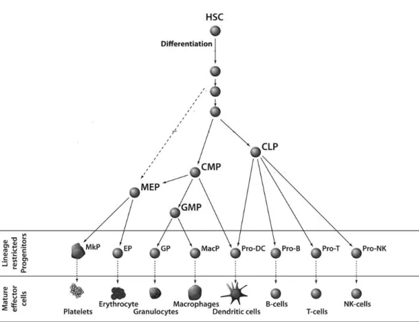
Figure 1.1 Differentiation of different kinds of blood cells from hematopoietic stem
cells ... 14
Figure 1.2 Innate immunity in tissue damage ... 17
Figure 1.3 Different antigen-processing pathways for the MHC class I and class II molecules ... 20
Figure 1.4 Overview of the adaptive immune responses after virus recognition by antigen presenting cells ... 22
Figure 1.5 Induction of adaptive immune responses to vaccines through PRR-mediated dendritic cell activation ... 26
Figure 1.6 Self-Assembly of Peptide Amphiphiles: From Molecules to Nanostructures ... 34
Figure 2.1 Chemical representation of biotinylated peptide amphiphiles... 47
Figure 2.2 Characterization of Biotinylated Peptide Amphiphiles ... 48
Figure 2.3 Structural Characterization of the Nanofibrous Structure ... 49
Figure 2.4 Morphological Characterization of Nanofibrous Structure. ... 50
Figure 2.5 Evaluation of Binding of Biotinylated Ovalbumin to the Nanofibrous Structure. ... 51
Figure 2.6 Evaluation Cytotoxic Effect of the Nanofibrous Structure... 52
Figure 2.7 Cytokine secretion profile of splenocytes treated with the Nanofibrous Structure ... 53
ix
Figure 2.8 Cytokine secretion profile of splenocytes treated with the Nanofibrous Structure ... 54 Figure 2.9 Cytokine secretion profile of splenocytes treated with the Nanofibrous
Structure ... 55 Figure 2.10 In vitro stimulation of adoptive immune responses by the Nanofibrous
Structure. ... 57 Figure 2.11 In vivo immunization with Nanofibrous Structure or CpG ODN. ... 59 Figure 2.12 IgG isotopes of immunized mice with Nanofibrous Structure or CpG
ODN ... 60 Figure 2.13 IgG isotopes of immunized mice with Nanofibrous Structure or CpG
ODN ... 61 Figure 2.14 IgG2a/IgG1 ratio of immunized mice with Nanofibrous Structure or CpG ODN. ... 62 Figure 2.15 Splenocytes proliferation of immunized mice with Nanofibrous Strucutre or CpG ODN ... 64
x
LIST OF TABLES
Table 1 Adjuvants in human vaccines………...28 Table 2 Clinically approved and late stage adjuvants………...29 Table 3 Estimates of daily and weekly intakes of aluminum in humans…………...31
xi
LIST OF ABBREVIATION
APCs : Antigen-presenting cells
CD : Circular Dichroism
BrdU : Bromodeoxyuridine
DAMPs : Damage-Associated Molecular Patterns
MHC : Major Histocompatibility Complex
CpG ODN : CpG Oligodeoxynucleotide
PAMPs : Pathogen-Associated Molecular Pattern
CTLs : Cytotoxic T Lymphocytes
TEM : Transmission Electron Microscopy
ASIA : Autoimmune Syndrome Induced by Adjuvants
LNs : Lymph Nodes
TLR : Toll Like Receptor
FDA : Food and Drug Administration
12
CHAPTER 1
1.1 Overview of the immune system
1.1.1 Innate immune system
Innate immunity is the organization consisting of cells and biological compounds, and they protect the body from invaders. These body defense system cells and molecules interact immediately and non-selectively to destroy bacteria or virus that cause infections. The immune system is present in every organism from the beginning and it is functional till the end of the life of organism. Innate immunity consists of surface barriers, cellular components and humoral molecules[1]. Surface barriers, as understood from the name are located on the surface and they firstly interact with invaders. There are several barriers protecting body and they can be mechanical, biological and chemical. For mechanical barriers the following can be examplified: exoskeletons, skin epidermis, lungs and gut. They compose a physical barrier between the organism and invaders. Internally mucus covered epithelial outer layer limit access of invaders to body. Another type of barriers that stop entering invaders to the body is the strong cellular membrane. Penetration of invaders is blocked by movement of cilia or peristalsis; this also prevents air passages. In the same way, microbes are stopped by tears in the eye and saliva in the mouth. First line of immunity highly consists of phospholipases, lysozymes and fatty acids and they cooperate in removing microbes from body. From previously stated chemicals, lysozyme and phospholipase can be found in saliva and tears and they resolve wall of microbes, thus prevent further spreading of infections. In the same manner sweat contains fatty acids in sweat and
13
stomach contains chemicals that reduce pH radically in stomach, further inhibit advance of microbes. Other parts of human body, like gut and skin microflora produce hazardous chemicals for microbes, too, and thus preserve entrance of pathogens to body. When the first surface barrier is down because of infections, the second defense mechanism, humoral molecules start to be synthesized[2]. Molecules to be secreted are interferons, cytokines, chemokines, complement system proteins and interleukins. These humoral factors have essential aspects in cell recruitment, inflammation, antibody secretion promotion, viral neutralization and attraction of other innate immune cells to the damaged place. Infected cells produce special chemicals that prompt inflammation. During this act, a mechanical barrier is formed in order to preserve further bacterial advancement as well as stimulation of infected tissues[3]. As a feedback to tissue infections, a multifactorial network of chemical signals start a host reaction in the damaged tissue. During this feedback leukocytes (monocytes, neutrophils and eosinophils) are activated and migrated from the venous system to the spots of infection. There are different types of immune cells (Figure 1.1). Leukocytes, which are named as white blood cells, compose only up to 1% of total blood, while red blood cells compose around 50%. There are main five categories of white blood cells: basophils, neutrophils, eosinophils, monocytes and lymphocytes. Lymphocytes consist of natural killer (NK) cells, T cells and B cells, and monocytes consist of macrophages and dendritic cells (DCs). Also, macrophages and dendritic cells are acknowledged as phagocytic[4].
14
Figure 1.1 Differentiation of different kinds of blood cells from hematopoietic stem cells. (Copyright © 2010 Wiley Periodicals, Inc. Reproduced with permission from.[5])
Cells of innate immunity are macrophages, DCs, neutrophils, eosinophils, mast cells and NK cells. Each of these cell groups have different action, in immunological mechanism. For example, eosinophils synthesize free radicals at the infected places to neutralize pathogens. Mast cells reside in mucosal membranes and bound tissues and play a part in curing damaged area in means of heparin and histamine secretion[3]. NK cells function in killing infected cells, and thus they are quite unique. Instead of swamping microbes, NK cells are focused in destroying infected cells by discriminating the normal vs infected cells via a general signal. Furthermore, NK cells do not require activation by other immune cells to perform their functions. NK cells
15
fight with parasitic infections and cancerous cells. They were initially known by their capability to eradicate susceptible cancerous cells and infected cells[6]. Their surface morphology is large granular lymphocyte like, however, they do not have T cell antigen receptors and CD3 complex that are definitive for T cells. Beyond this, they are able to secrete immunoregulatory and antibacterial cytokines such as TNF, GM-CSF and IFNγ. Their activities can be regulated by innate cytokines which direct biological purposes of NK cell reactions of proliferation, cytotoxicity, and interferon gamma (IFNγ) secretion[7].
Another type of innate immunity cells are known as monocytes. They are significantly bigger cells compared to other innate immune cells. Bone marrow is the place for maturation of these cells. They also can spread through the blood. Half of these cells are located throughout the body and the remaining half is found in the spleen. DCs and macrophages can be differentiated from monocytes. DCs are also essential cells of the innate immunity. DCs can engulf, process and present antigens to adaptive immune cells in lymphoid organs. Also, DCs have lymphocyte co-stimulatory molecules that give activation signals to adaptive immune cells and secrete cytokines to initiate immune activation[8]. Furthermore, they can also be found in tolerogenic state that inhibit self-antigen specific T cell activation and so decreasing autoimmune responses. DCs are also recognized as antigen presenting cells (APC)[9]. They can activate both B and T cells in an antigen specific manner. After introducing antigens on their peptide-binding proteins (MHC class I and MHC class II), they can specifically activate cytotoxic T cells and helper T cells accordingly. T cells cannot be stimulated without this commitment. Also, DCs are able to secrete IL-12 and TNF
16
alpha which take part in T cell stimulation and stimulation of another immune cells. So, DCs are very versatile in immune system manipulations[10]. Macrophages can be differentiated from monocytes that cross into tissue. They are very important for getting rid of cells that are damaged, excess, or dead and they can enable phagocytosis of bacteria, viruses and other invaders. Macrophages have various classes of pattern recognition receptors (PRRs) on their surfaces, which are essential for perception of danger signals[11]. The most essential PRRs are toll like receptor (TLRs). When TLRs are stimulated by invaders or other warnings, this activates the macrophages and they engulf the microbes or infected cells. Beyond that, TLR activation enable the induction of macrophages to secrete cytokines, which can trigger the activation of other immune cells and call the other cells to the damaged area (Figure 1.2)[12].
17
Figure 1.2 Innate immunity in tissue damage. (Copyright © 2006 BioMed Central Ltd. Reproduced with permission from ref.[13])
Macrophages are also one of the antigen presenting cells. One more influential function of macrophages is to activate process of inflammation. They are also recognized in the body as scavenger cells because they can remove debris and dead cells. TNFα is the main cytokine that is primarily secreted by macrophages. Innate immunity is strongly linked with adaptive immunity[14].
18 1.1.2 Adaptive immune system
Adaptive immunity is mainly based on the activity of T and B cells. There are distinct mechanisms that contain particular cells and unique procedures to get rid of from invaders or eradicate infected cells. Adoptive immunity is achieved after birth. Cells of adaptive immunity are focused to learn and remember by making an immunological memory. The adaptive immunity is highly specialized since the activation is made for only specific antigens. If an organism can be cured from an initial disease, he won’t be influenced by the same type of invader again, thanks to adaptive immune feedback which keeps it in memory and replies rapidly in the case of another disease[15]. Unlike innate immunity, the adaptive immunity acts in an antigen-specific manner. B and T cells express various antigen receptors via genetic recombination. Somatic rearrangement in the B and T cell receptors are the source of the variability, and so the capability of discrimination between self and non-self is thanks to the variability[16]. After T cells get activated, they directly recognize and destroy the infected cells. Also, T cells can give activation signals to B cells and B cells can secrete specific antibodies that target specifically encountered pathogens. The various kinds of antibodies that are produced by B cells are also mediated thanks to somatic rearrangement of immunoglobulins. Stimulation of B cells initiates hypermutation in genes that are associated with the immunoglobulins. The process maximizes the variety of the produced antibodies[17].
T cells are specified to remove cells affected from viruses or remove recognized tumor cells. T cells are additionally separated into various subgroups, including naive T cells which separate into T helpers and CTLs, regulatory T cells and
19
memory T cells. An efficient T cell-mediated immune response happens after the stimulation of T lymphocytes by APCs in secondary lymphoid tissues, mainly lymph nodes (LNs)[18].
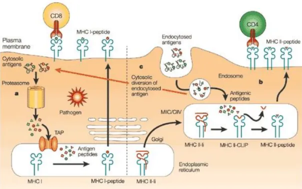
Naive T cells arise from bone marrow and found in the thymus. After positive and negative clonal selection, they turn into mature non-effector cells that are separately specialized against a given antigen, but can’t identify these antigens straightforwardly. Helper T cells (CD4+ cells) are fundamental for adaptive immune system. CD4+ cells are able to stimulate immune mechanism cells, taking into account B cells, macrophages and exterminator T cells. Primary CD4+ cells are Th1 and Th2[19]. They act as an essential line of protection system against intracellular and extracellular invaders and autoimmune and allergic reactions. Th17 is a subgroup of Helper T cells and plays a role in chronic inflammation, autoimmune disease and the elimination of extracellular microorganisms. Regulatory T cells are T cells that adjust the reactions of effector T cells and restrain pro-inflammatory pathways. Because of this self-correcting reaction, they also preserve immune cells from exterminating normal body cells. T helper cells are stimulated by APCs through antigen fragments that are bound to MHC class II molecules. MHC class II+antigen fragment complex can be recognized by T cell receptors on T cells (Figure 1.3). After this binding, there is another stimulation that is implemented through the effects of co-stimulatory chemicals: CD80 or CD86 on APCs[20]. These co-stimulatory chemicals attach to CD28 on T cells. A third signaling chemical, CD4, then binds to MHC-CD80/86 complex and improves their binding affinity. After MHC-TCR binding, the CD4+ T cell is stimulated and starts to proliferate.
20
Figure 1.3 Different antigen-processing pathways for the MHC class I and class II molecules. a) MHC class I molecules present peptides which are generally endogenous proteins or some cases pathogen derived proteins. b) Oppositely, MHC class II molecules present proteins that can come from endocytic route c) Dendritic cells are able to engulf antigens and present them to CD8+ cytotoxic T lymphocytes via cross-presentation. (Copyright © 2010 NPG. Reproduced with permission from ref.[21])
Stimulated helper T cells separate into Th1 or Th2 type that is mediated by presence of some cytokines. For instance, 12 activates T cells towards Th1 cells, while IL-4- activates T cells towards Th1 cells. Additionally, Th1 type cells secrete IFN-γ, TNF-α and IL-2 but Th2 type cells secrete IL-4, IL-5, IL-6 and IL-10. Few of the actively proliferating T cells may turn into memory CD4+ T cells. Memory CD4+ T cells support the immunological memory and react quickly when the body faces with the
21
same invader again. Effector T cell means activated T cell and they produce cytokines and enable to mediate apoptosis. Cytokines synthesized by CD4+ T cells have influence on B cells, cytotoxic T cells and macrophages[22].
T cell stimulation is linked with amount of antigenic peptides, affinity of T cell receptors to antigen fragments+MHC complexes, presence of cytokines and co-stimulatory signals in lymphoid organs. These all influence the effectiveness of CD8+ naive T cells as effector cytotoxic T lymphocytes (CTL). CTL should be activated by antigen fragment+MHC I complex and after it is activated, it mediates the apoptosis of the target cell that presents pathogenic peptides. This mechanism is also vital for removing tumor cells that have cancerous antigen fragment/MHC I complexes on their surface[22]. There is a distinct regulation via CD28 and CTLA-4 on T cells. The ligation of CD28 with CD80/86 on APCs mediates activation of T cells but the ligation of CTLA-4 with CD80/86 mediates inhibition for the activation of T cells. Traditional antitumor antibodies are utilized to prevent the attachment of CTLA-4 with CD80/86 to block inactivation of T cells. Programmed execution 1 (PD-1) inhibitory receptor may also mediate inhibition signal to T cells if it binds with PD-L2 and PD-L1[23]. B cells are also one of the APCs and sustain the capability to rescue antigens through their clonally made antigen receptors, which are named membrane immunoglobulins. B cells first identify antigens via its antigen receptor, and then engulf and process the antigen. Then antigen fragment/MHC class II complexes are presented onto the cell surface of the B cell. T cells can bind to the antigen fragment/MHC class II complexes on the B cell surface with the recruitment of CD4, TCR, and CD28. B cells’ CD40 ligand bind to T cells’ CD40-L that enable the activation of B cells. Then, CD4+ T
22
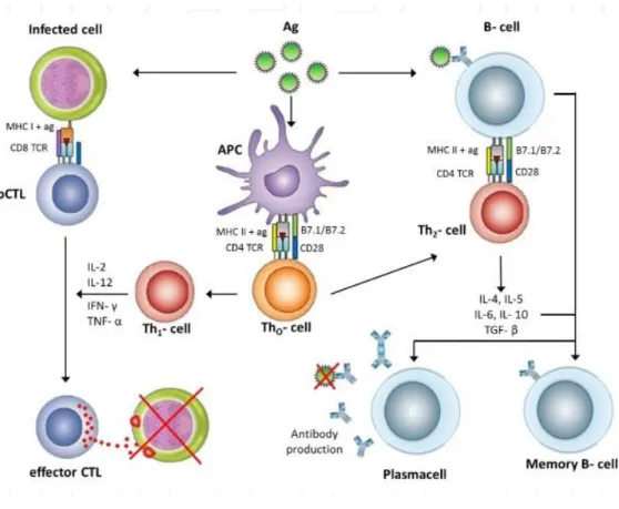
cells recognize the antigen fragment/MHC II complexes that are presented on the B-cell[24]. B cells are directed to clonal expansion, isotype switching and differentiation into memory cells after the binding with T cells. Both Th1 and Th2 cells are capable of stimulating B cells, and that cytokine exposure can identify what kind of immunoglobulin produced by the cells (Figure 1.4).
Figure 1.4 Overview of the adaptive immune responses after virus recognition by antigen presenting cells. (Copyright © 2012 InTechOpen. Reproduced with permission from ref.[25])
Th1 helper cells primarily activate B cells for IgG2a production, while Th2 helpers mediate B cells for the secretion of IgG1. Immunoglobulins are classified into five main types: IgE, IgG, IgA, IgM and IgD. They can be differentiated by the character
23
of heavy chain in the molecule. IgGs are mostly obtained in serum: almost 75% of serum IGs belong to subcategories like IgG1 and IgG2a[26].
1.2 Modulation of Immune Reactions
Vaccination was first used for treating smallpox disease and it was a successful treatment of smallpox using cowpox. Since that example `vaccination` term has been widely used as a general name for protection against infections[27]. Vaccines has usually contain infectious microbes like agents, and they are usually made of inactivated cells, weakened or inactivated form of pathogens or their proteins and toxins. Main idea behind this procedure is to make familiar the immune systems with these microbes to be remembered afterwards. Due to expensiveness of some vaccines, they cannot be obtained in third world countries. There is continuing issue of efficient vaccines against modern diseases like cancer, AIDS, Ebola, Ziko viruses, and HIV[28]. Despite their use for more than 200 years, vaccines are made by using an outdated technology in terms of rapid response in emergency situations. Obvious example for this was in 2009 during epidemy of Influenza A pandemic or so called H1N1[29]. The prime reason for this outbreak was stated limitations in making vaccines in appropriate time and their delivery to other world countries. As it was stated above, modern vaccines are generally made by using live-attenuated or inactivated viruses, but along with these, serious safety issue comes along[30]. Deactivated virus or bacteria can gain their activity back in body and cause serious infections. In order to make efficient vaccines key factors in immunology should be considered. Additionally, recent vaccine advancement aims to target tumor cells by
24
using the immunological mechanisms. Neurodegenerative and autoimmune diseases are also being tried to be treated by new vaccine systems.
1.2.1 Pathogen-targeted immune reactions
Different kinds of immune reactions are required to protect an individual from different kinds of invaders. Also, while immune mechanisms target various types of microbes and their components, they should not attack to body itself (being tolerant). Therefore, various distinct regulations are associated to maintain this tolerance. Invaders that can penetrate through barriers such as skin are targeted by innate immune mechanisms. Innate immune cells sense the microbes’ entry points and destroy microbes by engulfing them or producing antimicrobial chemicals that kill the microbes[31]. Innate immune cells recognize the invaders through their germline encoded receptors and these receptors are found on these cells during their whole life unlike adaptive immune cells which have alterations in their receptors. Main type of these receptors are pattern recognition receptors (PRRs) that sense certain general patterns of invaders such as unmethylated CpG of bacterial DNA and so PRRs mediate the recognition of pathogens. PRRs have various types and can be found on transmembrane, cytosol or can be released from cells. For instance, toll-like receptors (TLRs) can be located on cell-membrane or endosomal membrane[32].
Activation of PRRs are also important for inducing adaptive immune response because multiple signaling pathways are activated with the signals that come from PRRs (danger signal) and the activation induces the release of some cytokines and expression of some co-stimulatory receptors. Cytokines are proteins that can bind
25
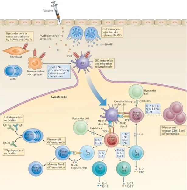
distinct types of receptors of other immune cells that induce the activation of the other immune cells[33]. APCs are required for the activation of co-stimulatory receptors to be able to stimulate adaptive immune cells. Therefore, adaptive immune response are required for many factors to be initiated and TLR-activation is accepted to be appropriate for providing these factors Figure (1.5). Thus, besides being the first line of defense, innate immunity also regulate the stimulation, duration and types of adaptive immunity.
Adaptive immune system mediates protection against invaders via two major cell types as B and T cells. These adaptive immune cells include various antigen receptors to be able to recognize all kinds of antigens in the environment. Random arrangement of the genes of the adaptive immune cells provide the variety of the receptors. Also, after infection, memory adaptive immune cells are produced and so there could be very fast activation and protection against the same pathogen by re-inducing adaptive immune response if the body face with the pathogen again[34].
26
Figure 1.5 Induction of adaptive immune responses to vaccines through PRR-mediated dendritic cell activation. Vaccines can contain pathogen-associated molecular patterns (PAMPs) or can trigger the local production of damage-associated molecular patterns (DAMPs). Pattern-recognition receptors (PRRs), which are expressed by DCs, recognize the PAMPs and DAMPs that trigger to DC activation, maturation and migration to the LNs. (Copyright © 2012 NPG. Reproduced with permission from ref.[35])
27
APCs mediate a link between innate and adaptive immunity. The cells engulf antigens from outside and process them to present on their surface to be able to be recognized by B or T cells. Besides the recruitment of APCs, also cytokines and co-stimulatory molecules are regulated by the immune response[36].
To sum up, the direction of adaptive immunity is strongly related with the presence of cytokines and co-stimulatory molecules that are produced by APCs and the interaction of invaders with innate immune cells mediate the expression of these molecules that direct adaptive immunity. Therefore, understanding of the interactions could provide us a chance to induce adaptive immunity by using synthetic and defined formulations without presence of invaders themselves.
1.2.2 Adjuvants
Vaccination is an efficient method to protect from microbe invasions. The prime purpose of vaccination is mediating a powerful immune reaction to invaders’ antigens and maintain long-lasting defense against disease that are caused by these invaders (Table 1). Pure antigens show weak immunogenicity and can’t increase a significant immune reaction. The compounds that are utilized to improve a particular immune reaction towards co-delivered antigens are named adjuvants.
28
Table 1 Adjuvants in human vaccines (Reproduced with permission from ref.[37])
Immune cells identify microorganisms via pathogen-associated molecular patterns (PAMPs) through their PRRs. These PAMPs can be utilized as adjuvants in a very controlled manner because they can be hazardous if they are not purified enough. For this reason, less hazardous types of PAMPs are specifically more proper as candidates to be adjuvants[38]. Some parts of microbes have a good potential to stimulate immune system. For instance, bacterial DNA portions (CpG motifs) are good activators of immune system as adjuvants. Normally, CpG motifs are not present in the mammals, and are only located in bacterial DNA. The CpG motif can bind and activate TLR9 that leads to the activation and proliferation of distinct immune cells. In addition, it mediates a Th1 type immune response. Adjuvants are utilized for various meanings for improving the immunogenicity of pure antigens, decreasing the total number of antigens applied for immunization and decreasing the administration numbers of vaccines[39].
Molecular mechanisms by which adjuvants function are not fully understood. But, there are some clues that adjuvants may use an assortment of mechanisms to
29
evoke immune response. These structures contain the depot effect (adjuvant enables to sustain presence of co-inoculated antigens at the injection site); enhancement of cytokines and chemokines; raising foreign antigen presentation to APCs; mediating maturation of the cells and activating APCs to move to the lymph nodes[40]. Many adjuvants have been identified up to now, however only some of them have been accepted by Food and Drug Administration (FDA) (Table 2). Accepted adjuvants are utilized from protection of various diseases by activating different immune reactions. Immune response can be manipulated towards a desired way by optimizing the type and applied dose of adjuvants.
Table 2 Clinically approved and late stage adjuvants (Reproduced with permission from ref.[41])
30
1.2.3 Adjuvants exhibit safety problems for neural system
Aluminum adjuvants are the most widely used adjuvants for vaccination formulations[42]. However, Aluminum is one of the most abundant metals that is linked to neurotoxicity and it mediates an alteration on the overall physiology of brain since different type of changes that are induced by aluminum tend to correlate with each other in human brain[43]. One study showed that aluminum-treated mice revealed dramatically enhanced apoptosis of motor neurons and induced the proliferation of reactive microglia in cortex and spinal cord. Also, they found the existence of aluminum in the cytoplasm of motor neurons and these aluminum positive neurons were also significantly positive for tau protein that may cause to Alzheimer's disease and the result is consistent with the result that showed aluminum accumulation in deformed proteins in Alzheimer's disease in human brain[44]. Also, mice were administrated with six doses of aluminum hydroxide for behavioral testing and these mice showed significant impairments for some motor functions including decreased spatial memory. They concluded that neurotoxicity of aluminum hydroxide that is widely used as an adjuvant, should be investigated with greater scrutiny and more research. Another study revealed that there is a significant increase in the level of apoptotic neurons with enhanced level of caspase-3 in lumbar spinal cord (250%) and primary motor cortex (190%) according to controls in mice after aluminum-injection. Aluminum-injection caused high motor neuron loss (35%) and enhanced astrocytes number (340%) in spinal cord. Thus, they suggested the possible effect for the aluminum adjuvant in many neurological disorders[45]. Also, negative impacts of aluminum on the nervous system are age-related. In adults, aluminum mediates
31
neurological deficits in age-related manner and has been linked to amyotrophic lateral sclerosis. Also, similar results were demonstrated in animal models. For instance, aluminum adjuvants were associated with model Gulf War syndrome and neurological deficits that cause amyotrophic lateral sclerosis symptoms in young mice. In developed countries, children receive many routine vaccine administrations with high amounts of aluminum adjuvants for different diseases, but they are much more vulnerable than adults for the toxicological risks of aluminum. US Food and Drug Administration vaccines safety assessments haven’t required appropriate toxicity testing because vaccines haven’t been recognized as inherently toxic. Thus, the appropriate amount of aluminum adjuvants that are administered to children is not examined by making appropriate toxicity testing in animals(Table 3).
Table 3 Estimates of daily and weekly intakes of aluminum in humans (Reproduced with permission from ref.[46])
32
Therefore, children are under most risk of aluminum adjuvant complications[47]. In adult humans, Aluminum vaccine adjuvant has been shown that it is linked to some serious autoimmune and inflammatory conditions such as autoimmune/inflammatory syndrome (ASIA) and also, children regularly take much higher amounts of aluminum from vaccines in comparison with adults. Thus, researches demonstrated that there is an increasing concerns for vaccination practices that may indeed be warranted[47]. There are new kinds of licensed adjuvants, but alum adjuvant still remains the major adjuvant in most of countries[37]. Consequently, it can be concluded that finding safer and more effective adjuvants is a major unmet need.
1.3 Materials for tuning immune response
Traditional vaccine systems have been based on administrating live-attenuated or deactivated forms of microbes into people in order to familiarize their immune systems about how to withstand against the microbe. Fundamentals of this approach have not been altered since discovery of vaccines. Even though this method creates favorable immune reaction against microbes, it has certain drawbacks. For instance, during a new pandemic, production of the vaccines is very problematic due to low expansion of new microbes or deficiency of resources. Also utilizing microbe itself is not a specific formulation, it may cause undesired side effects and there is a possibility for reactivation of the attenuated virus[48]. Because of these hard preparation process and safety concerns of the deactivated or live attenuated virus vaccines, it is vital to develop rational new methods for vaccines that will activate immune system in a controllable manner without safety problems. In that content, this demand could be met by designing novel materials that will not show the drawbacks.
33 1.3.1 Self-assembled peptide nanostructures
Self-assembly is a natural phenomenon and mimicking the self-assembly leads us to designing new materials that provide desired combinations of mechanical, biological and chemical characteristics. As an example, cell membrane is a self-assembled construction. Oppositely charged peptide amphiphiles in aqueous environment get together to form self-assembled peptide nanostructures. Peptide amphiphiles primarily consist of hydrophobic and hydrophilic parts. A classical peptide amphiphile molecule contains four key parts (Figure 1.6).
Figure 1.6 Self-Assembly of Peptide Amphiphiles: From Molecules to Nanostructures. (Copyright © 2010 Wiley Periodicals, Reproduced with permission from.[49])
34
A hydrophobic domain is required for self-assembly and an alkyl tail is used as hydrophobic domain. β-sheet forming domain enables the formation of secondary structures and various short peptide sequences can be used as β-sheet forming domains. Charged domain provides charge for the system and self-assembly can be directed by neutralizing the charge of differently charged peptide amphiphiles. Bioactive domain provides biofunctionality for the system. Different peptide amphiphiles can be designed for different biological purposes by using different bioactive epitopes[49]. Peptide amphiphiles have ability to self-assemble into a series of unique nanostructures that make them applicable for different intentions in biomedical engineering. Alteration of the bioactive part of peptide amphiphiles has been used for enhancing cell adhesion, differentiation, cell maintenance, cell growth and cell mobility.
Nanostructure forming by peptide amphiphiles is accomplished via noncovalent bindings. Self-assembled peptides are very suitable as biomaterial because they are easily designed, they are biocompatible for cells, and they have defined functional units, and low immunogenicity. Active regions of self-assembled peptide systems can also be manufactured for different intentions. For example, in one study, heparan-sulfate-mimicking peptide amphiphiles (HSM-PA) and laminin-mimicking peptide amphiphiles were designed and synthesized. HSM-PA and laminin-mimicking peptide amphiphiles were mixed and the formed nanofibers were used for neurite outgrowth[50]. It was shown that the mixing of the two peptide amphiphiles maximizes the neurite outgrowth rather than using them alone. One more widely utilized bioactive sequence is IKVAV which is a sequence derived from
35
laminin protein that enhances neural cell proliferation. Furthermore, IKVAV decreases astrocyte formation and reduces the risk of glial scars. It was discovered that self-assembled IKVAV peptide amphiphiles mediate a robust neuronal differentiation correlated to laminin. Accordingly, these data prove that logically manufactured, self-assembled peptide amphiphiles can be used as tools for manufacturing and advancing effective, controllable and practical materials for biological applications[51]. Rationally designed self-assembled peptide amphiphiles are promising new scaffolds that can be used for various biological applications including tuning immune response.
36
CHAPTER 2
2.1 Introduction
Vaccination is the most effective and cost-efficient strategy against infectious and intracellular pathogenic diseases, however ideal vaccine formulation remains to be a mystery[52]. The emergence of new pathogens, inadequate protection provided by the existing vaccines and finding an effective vaccine for cancer immunotherapy require more pathogen tailored and rationally engineered vaccines[53]. Therefore, synthetic subunit vaccines with one or a few selected recombinant proteins, which are normally present in the target pathogen, are receiving increased attention due to their high applicability for vaccine design for specific purposes, because of their precise chemical definition and being safer than live-attenuated or inactivated microorganisms with a lower cost of production[54, 55]. However, the low immunogenicity of recombinant proteins necessitate the usage of adjuvants in vaccine formulations for directing and maintaining the immune response against vaccine antigens[56]. Although many different types of adjuvants have been developed, ideal adjuvant formulation with good biocompatibility, biodegradability, and lower immunogenicity while activating both innate and adoptive immune system against using antigen is still sought[57].
Adjuvants are recognized as danger signals by DCs and enable the activation and maturation of these cells[58]. Therefore, adjuvants activating DCs have emerged as a successful strategy in order to develop effective vaccines[59]. DCs are effective antigen presenting cells which are specialized for antigen uptake, processing, presentation and activation of naïve T cells. In their immature form, DCs initially need
37
activation signals and subsequently maturation in order to perform their function[60]. After maturation, DCs migrate to lymphoid organs leading to T cell interaction through antigen-receptor binding and co-stimulatory molecules[61]. Captured exogenous antigens in DCs are normally processed and presented onto major histocompatibility complex (MHC) class II which activates CD4+ T cells. Then, the activated CD4+ T cells enable antigen-receptor binding and activation of B cells to provoke antibody formation[62]. However, effective response against infectious disease and cancer requires CD8+ T cells which are activated by associating with MHC class I. Cross presentation is a process which enables extracellular antigen presentation on MHC class I complex and thereby extracellular antigen specific CD8+ T cells are able to be activated in order to kill infectious and cancer cells[55]. Therefore, increasing need for the development of new adjuvants has lead research to find an ideal adjuvant which promotes cross-presentation of antigens and increases the level of the effector CD8+ T cells.
The engineering of materials, that are able to tune the immune system, is a potential candidate in order to overcome problems of recent vaccine formulations. They are able to enhance the stability of antigen, antigen delivery, stimulation of immature DCs, and cross-presentation[63]. Thereby, new adjuvants that can mimic pathogen cues in a reductionist fashion are being designed through manipulating materials. Moreover, a rational design considering intracellular pathways inside DCs can enable better control of antigen presentation to DCs by changing the resultant cytokine environment and/or directing the route of antigen delivery towards cross presentation[64].
38
Self-assembling peptides are good examples for biomaterials that have the potential to be used as vaccine adjuvants because of ease of design, their chemical and compositional definition, low immunogenicity, biodegradability and biocompatibility[56]. Also, these nanostructures provide multivalency which is the repetitive display of ligands with high affinity and avidity. However, the self-assembling peptides that have previously been reported to be acting as adjuvants have limitations due to their difficult synthesis or assembly methods and problematic conjugation of antigens to the adjuvant which requires covalent conjugation, which is not suitable for all antigens[65-68]. Moreover, previous studies failed to show promising results for the activation of cytotoxic CD8+ T cells that are necessary for specific elimination of infected cells by viruses or intracellular bacteria and tumours[69].
In this thesis, I present the development of a new nano-adjuvant called as biotinylated nanofibrous structure. These nanofibrous structures have a unique design which enables the presentation of biotin groups on their outer surface. Streptavidin (SA)-biotin affinity is one of the most strong interactions known so far[70]. Since SA has four binding sites for biotin, after mixing SAs with the nanofibrous structures, SAs bind these nanofibrous structures that allows binding of any other biotinylated molecules to the nanofibrous structures. Thereby, different kinds of antigens can be biotinylated with basic methods and these biotinylated antigens can bind to the nanofibrous structures through streptavidin linker[71]. In this system, the nanofibrous structures and antigens are in close proximity within a single construct, which is important for internalization of adjuvant and antigens into the same DC, inducing the
39
necessary antigen specific immune responses[72]. Also, the repetitive organization of antigens and biotin as biochemical signals provides the nanofibrous structures being a virus like nanostructure besides its shape, which is resembling rod-like viruses[73]. I demonstrated that the efficiency of the adjuvant system by using biotinlylated ovalbumin, which is a model antigen, that is able to enhance antigen specific humoral and cellular responses in in vitro and in vivo models. In this thesis, I suggest that the nanofibrous structure is a promising new type of adjuvant to boost humoral and cellular immune responses, which can be applied to vaccine development and immunotherapies.
40
2.2 Materials and Methods
2.2.1 Materials
[4-[α-(20,40-dimethoxyphenyl) Fmoc-aminomethyl] phenoxy] acetamidonorleucyl-MBHA resin (Rink amide acetamidonorleucyl-MBHA resin), 9-fluorenylmethoxycarbonyl (Fmoc), tert-butoxycarbonyl (Boc), and 2-(1H-benzotriazol-1-yl)-1,1,3,3-tetramethyluronium hexafluorophosphate (HBTU) were obtained from ABCR and NovaBiochem. Cell culture reagents were obtained from Life Technologies, while non-essential amino acid solution was from Sigma Aldrich. AlamarBlue cell viability reagent was purchased from Invitrogen. CD40, CD86 and OVA257-264 (SIINFEKL) peptide bound to H-2Kb APC antibodies for flow cytometry were purchased from eBioscience. ELISA reagents were from Life Technologies. IFNγ antibodies, IL-12 antibodies and IFNγ protein, IL-12 protein were purchased from R&D systems. IL-6 antibody and protein were from eBioscience. Ovalbumin specific antibodies IgG2a (Abcam), IgG1, IgG2b (eBioscience) and anti-mouse IgG –Peroxidase antibody (Sigma Aldrich) were provided from Southern Biotechnologies (USA). BrdU (5-bromo-2-deoxyuridine) was obtained from Thermo Scientific.
2.2.2 Peptide synthesis
Synthesis of lauryl- VVAGKK-Biotin-Am (B-PA) was performed on Rink Amide MBHA resin. 2 equivalents of Fmoc-protected amino acid, 1.95 equivalents of HBTU and 3 equivalents of N,N-diisopropylethylamine were used for amino acid couplings (two hours for each aminoacid). 5% TFA solution in DCM was used for removing of Mtt group of Fmoc-Lys(Mtt)-OH and then biotin conjugation was carried out by using the same protocol for amino acid couplings. 20% (v/v) piperidine in DMF was used
41
for removing of Fmoc group (twenty minutes). Then, 10% (v/v) acetic anhydride solution in DMF was used for blocking of the remaining free amine groups (thirty minutes). The resin was washed with the mixture that contains DMF, dichloromethane (DCM) and DMF after each step. Removal of the peptide from the resins was accomplished by using the trifluoroacetic acid/triisopropyl silane/ H2O/DCM mixture
(with the ratio of 5:2.5:2.5:90).
2.2.3 Preparation of antigen coupled nanofibrous complex
Nanofibrous complexes were prepared through spontaneous self-assembly of 0.023% (w/v) biotinylated peptide amphiphile molecules in the aqueous environment. Self-assembled nanofibers were mixed with Streptavidin (Sigma-Aldrich), and after 40 min, biotinylated ovalbumin (OVA) (Galab) was added to formed nanostructure (1:1:1 exact molar ratio for nanofibrous complexes, Streptavidin and ovalbumin)
2.2.4 Transmission electron microscopy (TEM) imaging
Nanofibrous complexes were imaged by TEM. 15 μL of nanofibrous complex was prepared on parafilm as described above. TEM grids were exposed to the solutions. Grids were put away from solutions and then 2% (w/v) uranyl acetate (Ted Pella) was used for staining the nanofibrous complex. After washing with ddH2O twice, grids
were dried for 1 h at RT. Images were taken by a FEI Tecnai G2F30 instrument in STEM mode.
2.2.5 Circular dichroism (CD) spectroscopy
0.2 mM solutions of nanofibrous complex were measured from 300 to 190 nm. (Scanning speed= 100 nm/min, data pitch= 1 nm, DIT= 4 s and bandwidth= 1 nm) All
42
measurements were assessed with three data accumulations and sensitivity was selected as standard. A JASCO J815 CD spectrometer was used for data accumulations.
2.2.6 Binding assay by ELISA
ELISA was used to show the binding of biotinylated OVA to the nanofibrous. Plates (Nunc MaxiSorp) were coated with two fold serially diluted self-assembled nanofibrous complex that was prepared as described above and stored at 4 °C overnight. After three washes with washing buffer (1% Tween 20 in 0.9% (w/v) NaCl solution in PBS pH 7.4) plates were dried by tapping and blocked with 1x assay buffer (Thermo Scientific) for 1 h room temperature. After washing and drying, the wells were incubated with SA (80 ng/mL in ddH2O) for 1 h at RT. After five washes with washing buffer, the wells were incubated with OVA (60 ng/mL in ddH2O). After five washes with washing buffer and drying, OVA specific antibody (0.25 μg/mL) was added into the wells and stored at 4 °C overnight. After five washes with washing buffer and drying, mouse IgG HRP was added into the wells for 2 h at room temperature. After five washes with washing buffer and drying, TMB (3,3′,5,5′-Tetramethylbenzidine) substrate was added to the wells for 30 minutes and the reaction was stopped by 1.8 N H2SO4. Absorbance was measured by microplate
reader (Spectramax M5, Molecular Devices) at 450 nm (subtracted reference value at 650 nm). All treatments were performed with at least three replicates and shown as mean +/− standard deviation.
43
2.2.7 Cytokines determination using ELISA assay
Splenocytes were seeded on 96-well culture plates (5 × 10⁵ cells/well) and CpG ODN (1 μg/mL)+OVA (0.1 μg/mL) and nanofibrous complex (prepared as described above) were added into the wells. After 48 h, supernatants were collected and transferred to the MaxiSorpTM plates (Thermo Scientific, NUNC) that were coated with 6, IL-12 or IFN-γ capture antibodies and blocked with 1x assay buffer and stored at 4 °C overnight. After five washes with washing buffer and drying, biotin-labeled detection antibodies were added into the wells and incubated for 2 h at room temperature. After five washes with washing buffer and drying, HRP (horse radish peroxidase)-conjugated streptavidin was added into the wells and incubated for 1 h at room temperature. After five washes with washing buffer and drying, TMB substrate was added into the wells and incubated for 20 minutes and after adding stop solution (1.8 N H2SO4), absorbances were measured by microplate reader (Spectramax M5,
Molecular Devices) at 450 nm (subtracted reference value at 650 nm). All treatments were performed with at least three replicates and shown as mean +/− standard deviation.
2.2.8 The effect of nanofibrous complex on cell viability
The cytotoxicity of nanofibrous complex was analyzed on splenocytes with alamar blue assay. Splenocytes were seeded in 96-well plates at 5 × 10⁵ cells/well for 3 h and CpG ODN and nanofibrous complex (prepared as described above) were added into the wells and incubated for 24 h at 37 °C. 10 % Alamar blue (Invitrogen) solution was added into wells and incubated for 4 hour at 37 °C. Absorbance was measured by microplate reader (Spectramax M5, Molecular Devices) at 570 nm (subtracted
44
reference value at 600 nm). Absorbances indicated the cell viability percentage relative to untreated cells.
2.2.9 The effects of nanofibrous complex on surface costimulatory markers and SIINFEKL-Kb+ complexes
For the analyses of the expression levels of CD40 and CD86 co-stimulatory molecules and the levels of the SIINFEKL presentation, specific mAbs were used for the staining of CD40, CD86 and SIINFEKL complexed with H-2Kb. Splenocytes were seeded into 96-well culture plates (5 × 10⁵ cells/well) and CpG ODN (1 μg/mL)+OVA (0.1 μg/mL) and nanofibrous complex (prepared as described above) were added into the wells. After 24 hour, cells were transferred to the eppendorfs, centrifuged and washed with 1x PBS. After blocking with FACS buffer (3% BSA + 1% sodium azide in 1x PBS) for 10 minutes, specific mAbs were added into the eppendorfs and incubated for 1 h at 4 °C. After washing with 1x PBS, the cells were sorted by flow cytometry (Accuri C6-Cytometer- equipment with BD Accuri C6 Software). The number of events were 20,000 for each samples. The experiments were performed with at least tree replicates and the results of two different experiments were shown.
2.2.10 Immunizations and determination of antibody responses
Male Balb/c mice (10–13 weeks old) were immunized with 500 μL vaccine formulations (in isotonic sucrose solution) subcutaneously. Mice were divided into 2 groups (n = 5) and immunized with CpG ODN (10 μg)+OVA (6.8 μg OVA) as positive control and the nanofibrous complex (biotinylated peptide amphiphile (150 μg), SA (9.2 μg), OVA (6.8 μg OVA)) in 500 μl sucrose solutions. Mice were immunized two times at day 0 and day 15 and sacrificed on day 21. Also, at 21, animals were bled, and
45
sera were obtained. The amounts of IgGs in sera were assessed with ELISA. OVA antigen was coated onto MaxiSorpTM plates and incubated at 4 °C overnight. The next day, wells were blocked with 1x assay buffer and 100 fold diluted sera were added into the wells and stored at 4 °C overnight. After five washes with washing buffer and drying, IgGs were detected with HRP conjugated anti-IgG, anti-IgG1 and anti-IgG2a. After substrate (TMB) addition, absorbances were calculated with microplate reader (SpectramaxM5) at 450nm (subtracted reference value at 650 nm).
2.2.11 Effects of nanofibrous complex on splenocytes proliferation
Immunized mice splenocytes were stimulated by OVA and splenocytes proliferations were analyzed by using BrdU assay (Roche). Cells were seeded into 96-well culture plates (5 × 10⁵ cells/well). 10 µM BrdU labeling solution was added into the wells and
incubated for 24 h. The next day, cells were fixed with FixDenat for 30 minutes. Then anti BrdU-POD solution was added into wells and incubated for 90 minutes. Absorbances were calculated with microplate reader (SpectramaxM5 at 370nm (subtracted reference value at 492 nm). Absorbances indicated the proliferation rates of the cells relative to untreated cells.
2.2.12 Statistical analysis
Statistical analysis was performed by using Graphpad Prism 6. One-way ANOVA, Two-way ANOVA and student t test was used to compare the significance between the groups. Error bars show SEM (standard error of mean) and p value less than 0.05 were accepted as statistically significant.
46
2.3 Results and Discussion
2.3.1 Design and Characterization of the Nanofibrous Structure
Biotinylated peptide amphiphiles were synthesized and nanofibrous structures were formed by self-assembly of the biotinylated peptide amphiphiles (Figure 2.1). The different groups in the peptide sequences of the biotinylated peptide amphiphile have distinct functions. Self-assembly of the biotinylated peptide amphiphiles are driven by lauryl group via hydrophobic collapse in aqueous environment. Glycine functions as a spacer. Lysine residues contribute positive charge which increases the solubility of the molecules in aqueous environment and also one of them is to graft biotin. β-sheet formation is facilitated by the Val-Val-Ala group during self-assembly. According to the configuration, there are two main functional regions for self-assembled nanofibrous structure; a hydrophobic inner region (alkyl tail and β-sheet forming sequence) and a hydrophilic outer region (biotins) [49, 73, 74].
47
Figure 2.1 Chemical representation of biotinylated peptide amphiphiles. Exact mass of biotinylated peptide amphiphile is 1007.60.
The purification of biotinylated peptide amphiphile molecules were acquired by high performance liquid chromatography (HPLC) and the quality of the purity was investigated by using liquid chromatography-mass spectrometry (LC-MS). The results showed that biotinylated peptide amphiphiles were highly pure and the observed mass has a consistency with expected mass (Figure 2.2).
48
Figure 2.2 Characterization of Biotinylated Peptide Amphiphiles. (A) LC chromatogram of biotinylated peptide amphiphile molecules at 220 nm. (B) Mass spectrometry analysis of biotinylated peptide amphiphile molecules. [M+H]+
49
Figure 2.3 Structural Characterization of the Nanofibrous Structure. CD spectra of nanofibrous structure
The pure biotinylated peptide amphiphiles form nanofibrous structures in aqueous environment by self-assembly. Circular dichroism (CD) spectroscopy was used for analyzing the secondary structures of the nanofibers. We observed a negative peak at around 220 nm and a positive peak around 200 nm that is indicative of β-sheet formation. As expected, there was no peak for only CpG ODN (Figure 2.3). CD spectroscopy results confirmed that the nanofibrous structures form β-sheets and that ODNs did not interrupt β-sheet formation.
50
Figure 2.4 Morphological Characterization of Nanofibrous Structure. (A) STEM images of the nanofibrous structures. Scale bar is 500 nm.
In order to make morphological characterization of the nanofibrous structure in aqueous solutions, 0.023% (w/v) of biotinylated peptide amphiphiles in water was used. Then, the nanofiberd were exposed on a carbon grid after uranyl acetate staining, the nanofibrous structures were observed by transmission electron microscopy (TEM). The TEM results showed that the nanofibrous structures were formed as uniform cylindrical bundles.
51
2.3.2Biotinylated Ovalbumins Bind to the Nanofibrous Complex
Biotinylated ovalbumin was used as model antigen for studying the nanofibrous structure mediated immune responses. Since SA has four binding sites, after mixing them with the biotinylated nanofibrous structures, SAs bind these nanofibrous structures that allows binding of any other biotinylated molecules to the nanofibers. Thereby, we predicted that biotinylated ovalbumins can bind to the nanofibrous structures through streptavidin-biotin linkages.
Figure 2.5 Evaluation of Binding of Biotinylated Ovalbumin to the Nanofibrous Structure. ELISA results of biotinylated ovalbumin to diluted nanofibrous structures.
Wells of ELISA plate were coated with serially diluted nanofibrous structure and then treated with SA and biotinylated OVA, respectively. The results showed that OVA could bind to the nanofibrous structures and OVA binding decrease proportionally with dilution of the nanofibrous structures (Figure 2.5A). Also, more than 60% of total added OVA could bind to the nanofibrous structures and then the OVA release from the nanofibrous structures gradually (Figure 2.5B).
52
2.3.3 Nanofibrous Structure Has No Toxic Effect on Splenocytes
Figure 2.6 Evaluation of Cytotoxic Effect of the Nanofibrous Structure. Cytotoxic effects of CpG ODN and the nanofibrous structure on splenocytes analyzed with Alamar Blue assays. (Statistical analysis was done with one-way ANOVA with Tukey’s multiple comparison test. No significant difference was observed)
CpG ODN had proliferative effect on splenocytes. Cytotoxicity results showed that the nanofibrous structure is highly biocompatible with splenocytes (Figure 2.6).
53
2.3.4 Nanofibrous Structure Promotes Cytokine Secretion
Spleen cells were isolated in order to analyze the immune response against the nanofibrous structure in vitro. Cytokine production profiles were analyzed in order to find directions of the immune response of the nanofibrous structure compared to CpG ODNs. B-class CpG ODN was used and it triggers a Th1-biased immune response by elevating IFNγ, TNFα and IL-12 levels.27
Figure 2.7 Cytokine secretion profile of splenocytes treated with the Nanofibrous Structure. Mice splenocytes treated with the compounds and IFNγ cytokine concentration of culture were calculated with ELISA (* :p<0.05, *** :p < 0.001 by One-way ANOVA with Tukey’s multiple comparison test)
Nanofibrous structures significantly promoted the levels of IFNγ compared to CpG ODN (Figure 2.7).
54
Figure 2.8 Cytokine secretion profile of splenocytes treated with the Nanofibrous Structure. Mice splenocytes treated with the compounds and TNFα cytokine concentration of culture were calculated with ELISA (** :p < 0.01 by One-way ANOVA with Tukey’s multiple comparison test)
Nanofibrous structures significantly promoted the levels of TNFα compared to CpG ODN (Figure 2.8).
55
Figure 2.9 Cytokine secretion profile of splenocytes treated with the Nanofibrous Structure. Mice splenocytes treated with the compounds and IL-12 cytokine concentration of culture were calculated with ELISA (*** : p < 0.001, **** : p<0.001 by One-way ANOVA with Tukey’s multiple comparison test)
Nanofibrous structures significantly promoted the levels of IL-12 according to CpG ODN (Figure 2.9). These results showed that both CpG ODN and the nanofibrous structure trigger Th1-biased immune response but the nanofibrous structure has a robust effect according to CpG ODN.
56
2.3.5 Nanofibrous Structure Promotes Cross-presentation of OVA
Cross-presentation mediates the presentation of exogenous antigens onto MHC class I molecules that is vital for CD8+ T cell activation[75]. Thereby, an ideal adjuvant should promote cross-presentation of co-delivered antigens in order to eradicate infected or cancer cells through the activation of CD8+ T cells.
![Figure 1.2 Innate immunity in tissue damage. (Copyright © 2006 BioMed Central Ltd. Reproduced with permission from ref.[13])](https://thumb-eu.123doks.com/thumbv2/9libnet/6011370.126678/19.892.180.774.201.621/figure-innate-immunity-copyright-biomed-central-reproduced-permission.webp)
![Table 1 Adjuvants in human vaccines (Reproduced with permission from ref.[37])](https://thumb-eu.123doks.com/thumbv2/9libnet/6011370.126678/30.892.167.832.205.342/table-adjuvants-human-vaccines-reproduced-permission-ref.webp)
![Table 2 Clinically approved and late stage adjuvants (Reproduced with permission from ref.[41])](https://thumb-eu.123doks.com/thumbv2/9libnet/6011370.126678/31.892.174.784.724.1063/table-clinically-approved-late-stage-adjuvants-reproduced-permission.webp)
![Table 3 Estimates of daily and weekly intakes of aluminum in humans (Reproduced with permission from ref.[46])](https://thumb-eu.123doks.com/thumbv2/9libnet/6011370.126678/33.892.174.781.715.941/table-estimates-weekly-intakes-aluminum-humans-reproduced-permission.webp)


