European Journal of Radiology 49 (2004) 179–181
Quiz case
˙Ibrahim Can Kürkçüo˘glu
a,∗, Atilla Ero˘glu
a, Nurettin Karao˘glanoglu
a,
Pınar Polat
b, Fazlı Erdo˘gan
caDepartment of Thoracic Surgery, School of Medicine, Ataturk University, Gogus Cerrahisi ABD., 25240 Erzurum, Turkey bDepartment of Radiology, School of Medicine, Ataturk University, Radyologi ABD., 25240 Erzurum, Turkey
cDepartment of Pathology, School of Medicine, Ataturk University, Patologi ABD., 25240 Erzurum, Turkey
Received 28 February 2003; received in revised form 1 March 2003; accepted 5 March 2003
1. Patient
1.1. Clinical history
An 18-year-old girl presented with a slowly growing mass on the left pectoral area close to anterior axillary line. There was no trauma history. The lesion was learned to show moderate enlargement during the last 3 years. There was also a pain on her left shoulder extending to the arm in recent months. On physical examination, a tender, soft tissue mass, 5 cm in greatest diameter was detected deep into the left pectoralis muscle. It was also palpable in axillary fold. Laboratory data and chest X-ray were normal.
1.2. Sonographic findings
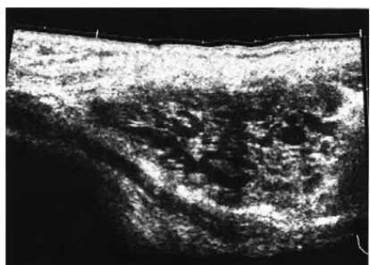
The patient referred to ultrasound (US) examination. US examination revealed an anechoic lesion with relatively thick septations within it (Fig. 1). The lesion was located within the left pectoralis major muscle.
1.3. Computed tomography findings
Computed tomography (CT) revealed a well circum-scribed mass lesion with heterogeneous density in the left pectoralis muscle. There was a marked decreased at-tenuation at the anterior portion of the lesion relative to the other parts suggesting fatty tissue. But density mea-surement revealed 23–25 HU is consistent with fluid or
∗Corresponding author. Tel.:+90-442-316-6333; fax:+90-442-316-6340.
E-mail address: [email protected] (˙I.C. Kürkçüo˘glu).
Fig. 1. US section shows an anechoic lesion intermixed with echogenic thick septations within the fibrils of pectoralis major muscle.
semisolid nature of the lesion. There was a small periph-eral hyperdense area consistent with calcification (arrow) (Fig. 2).
Fig. 2. Axial CT section reveals a well-circumscribed lesion within the pectoralis major muscle. Note anteriorly located low attenuation area relative to the other components of the lesion at small calcification (arrow). 0720-048X/$ – see front matter © 2003 Elsevier Ireland Ltd. All rights reserved.
180 ˙I.C. Kürkçüo˘glu et al. / European Journal of Radiology 49 (2004) 179–181
Fig. 3. (a) Axial Flash T1-weighted section shows a homogeneous mass lesion at the left upper thorax isointense relative to neighboring muscle tissue. (b) Axial HASTE T2-weighted MR image homogeneous hyperin-tense lesion. (c) T1-weighted sagital image after contrast medium injec-tion shows marked contrast enhancement at the septainjec-tions.
1.4. Magnetic resonance (MR) imaging findings
The lesion was homogeneous on T1-weighted spin-echo magnetic resonance (MR) imaging. The signal intensity of the lesion was similar to neighboring muscle tissue (Fig. 3a). T2-weighted images showed homogeneous increased
sig-nal intensity within the lesion (Fig. 3b). T1-weighted
MR images were taken after the injection of contrast medium. We detected contrast enhancement within the septations.
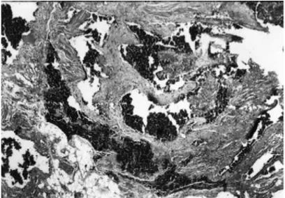
Fig. 4. Pathologic examination revealed congestion of vascular structures and dilated vessels that were lined by endothelial cells. The vessels were surrounded with fat cells, which composed large vacuolated cytoplasm and eccentric nucleus (HE× 40).
2. Diagnosis
Fine needle aspiration biopsy revealed normal blood cells. Excision biopsy was performed with the presumed diagnosis of hemangioma, skin incision made on anterior axillary hair line. Pectoralis muscle fibers separated with blunt dissection, and there after from the lateral to medial 4×3×1.5 cm mass were extirpated. Pathologic examination revealed congestion of vascular structures and dilated vessels lined by endothelial cells. The vessels were surrounded with fat cells, which were composed of large vacuolated cytoplasm and eccentric nucleus (Fig. 4).
3. Discussion
Soft tissue hemangioma is a common soft tissue neo-plasm. Indeed it is the most frequent tumor of infants and children and constitutes 7% of all benign tumors. These le-sions more commonly affect women and are usually discov-ered in the first three decades of life. Clinical abnormalities include pain, a mass that may intermittently change size, and occasionally a bluish discoloration of the overlying skin[1]. Cavernous hemangiomas of skeletal muscle are rare. Al-though previous reports described localization of intercostal muscle and ribs this is the second case of pectoralis muscle localization[2–6].
Radiographs of soft tissue hemangiomas are often normal, although a nonspecific mass may be apparent. Phleboliths represent a characteristic calcification and are most com-monly associated with cavernous hemangioma (30–50% of cases).
US evaluation of soft tissue hemangioma typically reveals a complex mass. Phleboliths are best evaluated by radio-graph or CT, however, acousting shadowing may be detected at large calcifications on US.
Soft tissue hemangiomas often show an ill-defined mass on non-contrast enhanced CT studies and marked post con-trast enhancement[1]. Serpentine vascular structures and fat
˙I.C. Kürkçüo˘glu et al. / European Journal of Radiology 49 (2004) 179–181 181
overgrowth may be apparent. These features are better de-picted by MRI. CT may identify Phleboliths not seen on ra-diographs and MR imaging. Lesions are often heterogeneous on all MR pulse sequences because of the combination of tissues present, although low intensity usually predominates T1-weighted images. Vascular components of the heman-giomas typically show high signal intensity on T2-weighted MR images whereas fat shows intermediate intensity. Soft tissue hemangiomas demonstrate prominent enhancement after intravenous administration of gadolinium.
Although hemangioma is the most common soft tissue neoplasm, it is uncommon at the chest wall. Differential diagnosis should be taken into consideration with the other
most common chest wall lesions[5]. The most common
be-nign soft tissue tumors of the chest wall are neurilemoma, fibroma, lipoma and fibrolipoma. Lipoma and fibrolipoma can be easily differentiated by their characteristic radiologic findings both on CT and MR. Neurilemoma reveals hetere-goneous signal intensities within it and shows marked con-trast enhancement after concon-trast medium injection on CT or
MR imaging [1]. Fibrosarcoma, desmoid tumor, malignant
fibrous histiocytoma, leiomyosarcoma, hemangiosarcoma are other possible malignant chest wall soft tissue tumors that can be confused with hemangiomas. But their aggres-sive growth pattern and more heterogeneous appearances can be useful in differentiating these tumors.
Fine needle aspiration or cut biopsies are not diagnostic in those cases. The biopsy material is generally insuffi-cient in differential diagnosis due to normal tissue ele-ments and vascular structures that form the hemangioma. Literature results showed generally normal blood cells and tissue diagnosis in most of the cases in fine needle
biopsies [3,4]. The histopathologic evaluation of our bio-psy material also revealed normal blood cells. This diag-nosis did not exclude the diagdiag-nosis of neoplasm and the choice of surgical procedure. The appearance of the mass during surgery and the absence of malignant cells at the biopsy are sufficient to exclude the radical surgery. To-tal surgical resection of tumor is the treatment of choice for cavernous hemangiomas of the chest wall muscle. The appropriate procedure for the total excision is dic-tated by two considerations: the mass may reform at least the size of the original lesion which often follows partial excision; radical surgery is not necessary because cav-ernous hemangiomas do not have malignant behaviors or metastasize.
References
[1] Castellote A, Vázquez E, Vera J, Piqueras J, Lucaya J, Garcia-Peña P, Jiménez JA. Cervicothoracic lesions in infants and children. Radio-graphic 1999;19:583–600.
[2] Kara M, Dikmen E, Sak DS, Sevket K. Intercostal arteriovenous hemangioma. Eur J Cardiothorac Surg 2000;18:622–4.
[3] Buckwalter JA. Musculoskeletal neoplasms and disorders that resem-ble neoplasms. In: Weinstein SL, Buckwalter JA, (Eds.), Turek’s Or-thopaedics. Philadelphia: J.B. Lippincott, 1994:315.
[4] Winchester DJ, Victor TA, Fry WA. Intercostal hemangioma presenting as a chest wall tumor. Ann Thorac Surg 1992;54:145–6.
[5] Athanassiadi K, Kalavrouziotis G, Rondogianni D, Loutsidis A, Hatzimichalis A, Bellenis I. Primary chest wall tumors: early and long term results of surgical treatment. Eur J Cardio-Thoracic Surg 2001;19:589–93.
[6] Perugini G, Bonini G, Giardina C, Mapelli L. Cavernous hemangioma of the pectoralis muscle mimicking a breast tumor. Am J Radiol 1994;162:1321–2.