RESEARCH
ARTICLE
Ismail Dogan Konuk1 Ilknur Suidiye Yorulmaz2 Onur Ozlu3Derya Ozcelik4 Fatih Alper Akcan5 Pelin Cetin6
1
Anesthesiology and
Reanimation Depth., Derince Training and Research Hospital, İzmit, Turkey 2
Anesthesiology and Reanimation Depth., Düzce University, Medical School, Düzce, Turkey
3
Anesthesiology and Reanimation Depth., TOBB Economy ve Technology University, Medical School, Ankara, Turkey
4
Plastic and Reconstrictive Surgery, İstanbul, Turkey 5
Ear, Nose and Throat Diseases Depth, Düzce University, Medical School, Düzce, Turkey
6
Anesthesiology and Reanimation Depth., Akçakoca Goverment Hospital, Düzce, Turkey Corresponding Author:
İlknur Suidiye Yorulmaz Anesthesiology and Reanimation Depth., Düzce University, Medical School, Düzce, Turkey
mail: [email protected] Phone:+90 5055428555
Received: 26.08.2020 Acceptance:25.09.2020 DOI: 10.18521/ktd.785571
Konuralp Medical Journal
e-ISSN1309–3878
[email protected] [email protected] www.konuralptipdergi.duzce.edu.tr
Relationship between Trigeminocardiac Reflex, QT, QTc and
Anesthesia in Septorhinoplasty Surgeries
ABSTRACT
Objective: Trigeminocardiac reflex is a reflex characterized by hypotension, bradycardia,
gastric hypermotility or asystole that develops as a result of stimulation of the trigeminal nerve. In our retrospective study, in septorhinoplasty operations performed under general anesthesia, trigeminocardiac reflex development was investigated primarily during the periods where the reflex was surgically stimulated. Secondly, the effect of different inhalation anesthetic agents on the emergence of this reflex was investigated.
Methods: Anesthesia notes and Datex Ohmeda icentral central monitor records of
septorhinoplasty cases operated between 01 / January / 2016 - 30 / November / 2016 were retrospectively examined and detected through the Hospital Information Management System software. Induction, application of local anesthesia, surgical incision, initiation of incision suturing and 5 minutes after extubation were recorded from the records. It was determined that two different inhalation anesthetics were administered in 60 patients who met the criteria, and analyzes were performed in 2 separate groups as group sevoflurane and group desflurane, and the development of QT, QTc and Trigeminocardiac reflex was investigated.
Results: Although there was no difference between the groups, when the basal values were
compared with the other periods, it was found that the development of TKR and QT and QTc experts were mostly observed in the periods of local anesthesia, surgical incision and incision suturing. (p <0.001).
Conclusions: We think that the inhalation anesthetic agents used mostly in the sevoflurane
group play a facilitating role in the development of TKR, especially by creating a cumulative effect during periods when the trigeminal nerve is maximally stimulated.
Keywords: Trigeminocardiac Reflex, QT Intervale, QTc, Electrocardiogram, Anesthesia,
Septorhinoplasty
Septorinoplasti Ameliyatlarında Trigeminokardiyak Refleks,
QT, QTc ve Anestezi İlişkisi
ÖZET
Amaç: Trigeminokardiak refleks trigeminal sinirin uyarılması sonucu gelişen hipotansiyon,
bradikardi, gastrik hipermotilite veya asistoli ile karakterize bir refleksdir. Genel ve lokal anestezi altında yapılan septorinoplasti operasyonları sırasında trigeminokardiyak refleks gelişebildiği gözlenmiştir. Retrospektif çalışmamızda genel anestezi altında yapılan septorinoplasti operasyonlarında birincil olarak cerrahi olarak refleksin uyarıldığı düşünülen periyodlarda trigeminokardiyak refleks gelişimi araştırılmıştır. İkincil olarak da farklı inhalasyon anestezik ajanların bu refleksin ortaya çıkışında etkisi araştırılmıştır.
Gereç ve Yöntem: 01/ Ocak/2016- 30/Kasım/2016 tarihleri arasında opere edilmiş
septorinoplasti vakalarının anestezi notları ve Datex Ohmeda icentral merkezi monitör kayıtları ve MİA-MED Hastane Bilgi Yönetim Sistemi yazılımı üzerinden retrospektif olarak incelenerek saptandı. İndüksiyon, sınırlı uyuşturma uygulaması, cerrahi insizyon, insizyon sütürasyonu başlangıcı ve ekstübasyondan 5 dakika sonra değerleri kayıtlardan saptandı. Kriterlere uyan 60 hastada 2 farklı inhalasyon anestezik madde uygulandığı tespit edilerek grup sevofluran ve grup desfluran olarak 2 ayrı grupta analizler yapıldı ve QT, QTc ve Trigeminokardiyak refleks gelişimi araştırıldı.
Bulgular: Gruplar arasında fark olmamasına rağmen bazal değerler ile diğer periyodlar
karşılaştırıldığında TKR gelişiminin ve QT, QTc uzmanlarının en fazla lokal anestezi uygulaması, cerrahi insizyon, insizyon sütürasyonu periyotlarında izlendiği saptandı. (p<0,001).
Sonuç: En fazla sevofluran grubunda olmak üzere kullanılan inhalasyon anestezik ajanların
özellikle trigeminal sinirin maksimal uyarıldığı periyodlarda kümülatif etki yaratarak TKR gelişiminde kolaylaştırıcı bir rol oynadığını düşünmekteyiz.
Anahtar Kelimeler: Trigeminokardiyak refleks, QT intervali, QTc, elektrokardiyogram,
INTRODUCTION
Trigeminocardiac reflex (TCR) is a type of reflex that is encountered in the practice of anesthesia, and which could even develop to a cardiac arrest and asystole. Controlled, randomized, or retrospective trials are very few in the literature regarding this reflex, which is induced by the stimulation of the trigeminal nerve and its sensory branches in some procedures that involve facial regions, though there are abundant case reports in the literature. Our study aimed to make a contribution to the literature in this regard. Our primary objective is to examine the development of the Trigeminocardiac reflex (TCR) in patients who underwent septorhinoplasty, and our seconder objective is to investigate the contribution of the anesthetic medications to the occurrence of this reflex through the QT and QTc intervals.
MATERIAL AND METHODS
Our study was established through examining retrospectively the anesthesia records of the patients who underwent septorhinoplasty between January 01, 2016, and November 30, 2016, as well as the central monitor records of Datex Ohmeda Icentral, and the software of Hospital Information Management System. Ethical approval of the Local University Clinical Research Ethics Committee was obtained with the decision number 2016/10, and dated January 2nd, 2017.
Patients who have ASA 1-2 physical status concerning the anesthetic risk classification, aged between 18 and 65 years, who were planned to undergo an elective septorhinoplasty surgery and the patients who were induced by propofol, rocuronium, remifentanil and inhaled anesthetic agents as anesthetic agents were included in the reviewing.
Patients; who have cardiac pathology, congenital, and acquired long QT syndrome, who receive medications that lead to prolongation of the QT-interval, who receive antiarrhythmic drugs, who have electrolyte disorders, renal failure, thyroid hormone disorders, liver failure, and pregnant women were excluded from the study.
Given the fact that the challenges that might be experienced throughout the intubation and the number of multiple intubation attempts might impact the heart rate adversely, patients who were recorded to had been intubated in more than 2 attempts were excluded from the research.
Data acquisition: The values of all patients who underwent septorhinoplasty between January 1st, 2016, and November 30th, 2016 that includes the data related to induction, local anesthesia, surgical incision, the initiation of incision suturing and 5 minutes after extubation were filled in the anesthetic follow-up forms routinely. Concurrently, the vital parameter and electrocardiogram records of the patients were retrospectively obtained from the patient
registry, which is recorded incessantly on to the external memory device from the Datex- Ohmeda Icentral L-NET (C) 05, software system, version 5.1 of the main computer that is located at in the operating room. The values of QT and QTc were evaluated and calculated based on these records. Bazett's formula was used manually for the calculation of QTc. The amount of administered medication, durations of anesthesia and surgery were determined through anesthesia follow-up forms, and the software of the MIA-MED Hospital Information Management System.
Heart rates and non-invasive blood pressure values, as well as peripheral oxygen saturation, and end-tidal carbon dioxide (EtCO2) values of the patients throughout the related durations were obtained from the anesthetic records. Upon examining the data retrospectively, it was determined that 2 different inhaled agents were administered in all patients who underwent septorhinoplasty. The patients were divided into 2 groups regarding these inhaled anesthetic agents, and these groups were also compared statistically among themselves in addition to the general assessment. Patients were subdivided as Desflurane Group (The group administered with Desflurane) and Sevoflurane Group (the group administered with sevoflurane) based on the administration of the inhaled anesthetic agents. It was determined from the surgical records and through the software of the Hospital Information Management System that the solution of adrenaline at a concentration of 1: 80000 with prilocaine 2% was administered to all groups as a local anesthetic solution during the mucosal injection.
Identification of TCR: Having a decrease in heart rate below 60 beats/min or 20% or more of baseline measurement values, namely bradycardia, and/or development of asystole was considered as TCR. Meanwhile, a decrease of 20% or more in the mean values of arterial blood pressure from the baseline measurement values were considered to be hypotension (1).
Statistical Analysis: Descriptive statistics
(values of mean, standard deviation, median, minimum, maximum, percentage) of all variables, which were involved in the study, were computed. The normality assumption of quantitative variables was analyzed through the Shapiro-Wilk test. Independent samples t-test and Mann-Whitney U-test were conducted for group comparisons. In order to compare the time-dependent variables, Repeated Measures ANOVA (either post hoc Tukey' HSD test or Dunn's test) was used. Relationships between qualitative variables were analyzed through the Pearson's Chi-Square test. The software of SPSS 22 was used for statistical analyses, and results were considered statistically significant at p <0.05.
RESULTS
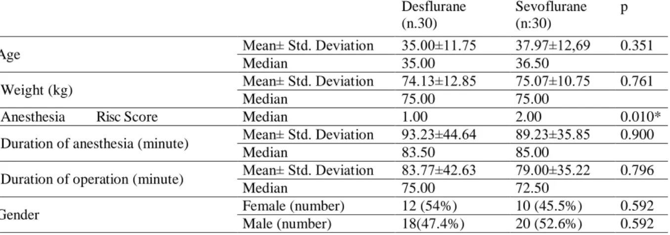
It was found that a total of 60 patients had undergone a septorhinoplasty procedure. There is no statistically significant difference between the groups regarding gender (p=0.562) (Table 1).
In addition to that, there was no significant difference between the groups in terms of age,
weight, duration of anesthesia and surgery. The median value of the anesthesia score in the sevoflurane group was significantly higher compared to the value measured in the desflurane group (p=0.010) (Table 1).
Table 1. Demographic data. Anesthesia risc score was found to be higher in the desflurane group. All other
features are homogeneously distributed between the 2 groups.
Desflurane (n.30)
Sevoflurane (n:30)
p
Age Mean± Std. Deviation 35.00±11.75 37.97±12,69 0.351
Median 35.00 36.50
Weight (kg) Mean± Std. Deviation 74.13±12.85 75.07±10.75 0.761
Median 75.00 75.00
Anesthesia Risc Score Median 1.00 2.00 0.010*
Duration of anesthesia (minute) Mean± Std. Deviation 93.23±44.64 89.23±35.85 0.900
Median 83.50 85.00
Duration of operation (minute) Mean± Std. Deviation 83.77±42.63 79.00±35.22 0.796
Median 75.00 72.50
Gender Female (number) 12 (54%) 10 (45.5%) 0.592
Male (number) 18(47.4%) 20 (52.6%) 0.592
There was no significant difference between the groups regarding total propofol dose, total rocuronium dose (mg), and total remifentanil dose (mcg) (p> 0.05) (Table 2).
There was no significant difference between the groups in terms of systolic blood pressure (SBP) values that were measured at different periods (p=0.311). However, it was determined that SBP values in both groups significantly changed depending on time (p <0.001).
Table 2. Except for sevoflurane and desflurane
drug amounts, there is no difference between drug doses used. (Mean± Std. Deviation)
Desflurane (n:30)
Sevoflurane
(n:30) p
Total propofol dose
(mg) 186.33±.12 184.50±22.45 0.516
Total rocuronium dose
(mg) 54.67±10.90 57.17±9.26 0.536
Total remifentanyl
dose (µg g) 350.30±178.44 332.43±116.64 0.935 Total inhalated
Anesthetic agent dose (mL)
93.34±71.78 50.35±30.42 0.396
In the desflurane group, the basal SBP value was significantly higher compared to the values, which were measured following the mucosal injection and mucosal incision, as well as the values during the incision suturing (p <0.001). The measurement value 5 minutes after intubation is significantly higher than the measurement values after mucosal incision, during the incision suturing (p=0.049), and 5 minutes after extubation (p <0.001). Whereas, SBP measurement value following the mucosal injection is significantly
lower than the value that was measured 5 minutes after extubation (p <0.001). Furthermore, the SBP value, which was measured following the mucosal incision, is significantly lower than the value that was measured 5 minutes after extubation (p <0.001). Yet, the SBP value that was measured 5 minutes after extubation is significantly higher than the value measured during the incision suturing (p <0.001).
Meanwhile, it was detected that the basal SBP value in the sevoflurane group was significantly lower compared to the values that were measured after mucosal injection and mucosal incision, as well as the value, measured during incision suturing (p <0.001). The SBP value of 5 minutes after intubation was determined to be significantly higher than the values that were measured during the incision suturing and after the mucosal incision (p<0.001). Moreover, It was determined in the sevoflurane group that value of SBP following the mucosal injection was significantly higher compared to the values measured after mucosal incision and after incision suturing (p <0.001), whereas it was significantly lower than the value, which was measured 5 min after extubation (p=0.002). Besides, it was found that SBP value among the sevoflurane group, which was measured 5 minutes after extubation, was significantly higher compared to the values of SBP that were measured following the mucosal incision and during incision suturing (p <0.001).
There was no significant difference between the groups regarding diastolic blood pressure (DBP) values that were measured in different periods (p=0.312). However, it was determined that DBP values in both groups changed significantly over
time (p<0.001). Basal DBP value in the desflurane group was significantly higher compared to the values, which were measured after the mucosal injection and mucosal incision, as well as the values during the incision suturing (p <0.001). The value, which was measured 5 min. after intubation, was significantly higher than the value measured during incision suturing as well as the values that were measured after mucosal injection, and mucosal incision (p <0.001); however, it was significantly lower than the value, which was measured 5 min. after extubation (p=0.021). Meanwhile, DBP value, which was measured after mucosal injection, was significantly lower than the value measured 5 minutes after extubation (p<0.001). Furthermore, DBP value, which was measured after mucosal incision, was significantly lower compared to the value that was measured 5 minutes after extubation (p<0.001). The DBP value, which was measured 5 minutes after extubation, was significantly higher than the value that was measured during the incision suturing (p<0.001). Moreover, it was determined that the basal value of DBP in the sevoflurane group was significantly higher than the values that were measured following the mucosal injection and after the mucosal incision, as well as the value, measured during the incision suturing (p <0.001). It was detected that the DBP value of the sevoflurane group, which was measured 5 minutes
after intubation, was significantly higher than the values measured following the mucosal injection and after mucosal incision, as well as during incision suturing (p <0.001). In the sevoflurane group, DBP value that was measured after mucosal injection was significantly higher than the value, which was measured after mucosal incision (p <0.001), whereas it was significantly lower than the value, which was measured 5 minutes after extubation (p<0.001). Besides, the DBP value in the sevoflurane group, which was measured 5 min. after extubation was significantly higher compared to the DBP values that were measured after the mucosal incision and during incision suturing (p<0.001).
There was no significant difference between the groups in terms of CO2 measurement values that were measured at different durations (p=0.595). However, it was determined that CO2 values in both groups changed significantly over time (p<0.001).
Basal CO2 value in the desflurane group was significantly higher compared to the values that were measured after the mucosal injection and during the incision suturing (p<0.001). Moreover, the value, which was measured 5 minutes after intubation, was significantly higher than the value that was measured during the incision suturing (p<0.001) (Table 3).
Table 3. Mean blood pressure, heart rate, oxygen saturation QT and QTc values in the desflurane group.( Mean± Std. Deviation)
Desflurane (n=30) Basal After 5 minutes of intubation After mucosal injection After mucosal incision After incision suturation 5minutes After extubation Mean arterial pressure (mmHg) 95.17±10.04 89.03±15.09 80.27±11.74 p<0.001 α 72.30±9.84 p<0.001α 77.40±14.97 p<0.001α 100.3±9.48 Heart rate
(beat per minute ) 72.93±6.63
87.13±12.23 p<0.001 80,03±15,77 p<0.001 75.43±12.74 65,03±10,76 p<0.001 Ʃ 81.07±11.97 p<0.001 QT (msn) 374.43±27.48 364.97±47.67 381.97±41.25 p<0.001& 386.30±32.33 p<0.001& 390.00±38.90 p<0.001& 365.43±26.81 QTc 383.70±23.63 388.57±38.38 401.20±21.16 405.87±31.97 402.97±41.08 388.23±18.91 End-tidal carbondioxide (mmHg) 39.47±2.67 38.60±11.65 36.63±3.48 35.57±3.46 p<0.001 34.03±3.52 p<0.001 37.13±2.99 Oxygen saturation (%) 97.73±1.31 98,50±0.94 98.40±0.72 98.07±0.78 98.37±1.00 97.87±1.20
& According to the baseline values, there are statistically significant QT prolongations at a statistically high level of values after mucosal injection, mucosal incision, and incision suturing.
α According to the baseline values, there is a statistically significant mean blood pressure in the values after mucosal injection, mucosal incision, and incision suturing.
Ʃ It is statistically significantly lower than basal values. Meanwhile, it was detected that the basal CO2 value in the sevoflurane group was significantly higher than the values that were measured after mucosal injection and during the incision suturing (p<0.001). The value of CO2 in the sevoflurane group, which was measured during the incision suturing, was significantly lower compared to the values that were measured 5 minutes after intubation, and mucosal injection, as
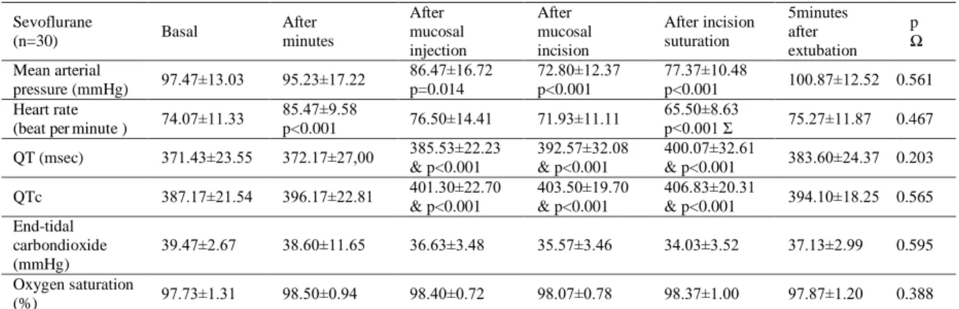
well as the values measured after mucosal incision, and 5 minutes after extubation (p<0.001) (Table 4). There was no significant difference between the groups regarding the values of heart rate (HR) that were measured in different periods (p=0.467). It was found out that HR values in both groups varied significantly throughout the period (p<0.001) (Table 4).
Table 4. Mean blood pressure, heart rate, oxygen saturation QT and QTc values in sevoflurane group. (Mean± Std. Deviation) Sevoflurane (n=30) Basal After 5 minutes of intubation After mucosal injection After mucosal incision After incision suturation 5minutes after extubation p Ω Mean arterial pressure (mmHg) 97.47±13.03 95.23±17.22 86.47±16.72 p=0.014 72.80±12.37 p<0.001 77.37±10.48 p<0.001 100.87±12.52 0.561 Heart rate
(beat per minute ) 74.07±11.33
85.47±9.58 p<0.001 76.50±14.41 71.93±11.11 65.50±8.63 p<0.001 Ʃ 75.27±11.87 0.467 QT (msec) 371.43±23.55 372.17±27,00 385.53±22.23 & p<0.001 392.57±32.08 & p<0.001 400.07±32.61 & p<0.001 383.60±24.37 0.203 QTc 387.17±21.54 396.17±22.81 401.30±22.70 & p<0.001 403.50±19.70 & p<0.001 406.83±20.31 & p<0.001 394.10±18.25 0.565 End-tidal carbondioxide (mmHg) 39.47±2.67 38.60±11.65 36.63±3.48 35.57±3.46 34.03±3.52 37.13±2.99 0.595 Oxygen saturation (%) 97.73±1.31 98.50±0.94 98.40±0.72 98.07±0.78 98.37±1.00 97.87±1.20 0.388
& According to the baseline values, there are statistically significant QT prolongations at a statistically high level of values after mucosal injection, mucosal incision, and incision suturing.
Ω These are the p values for the comparison of the values in the sevoflurane and desflurane groups according to the recording periods. Accordingly, there is no significant difference between the groups in all recording periods.
Ʃ Statistically significantly lower than basal values.
Basal HR value in the desflurane group was significantly lower than the values that were measured 5 minutes after intubation, after mucosal injection and 5 minutes after extubation; whereas, it was significantly higher than the value that was measured during the incision suturing (p<0.001 for each). The value, which was measured 5 min after intubation, was significantly higher than the value that was measured throughout the other periods (for each of them, p<0.001). Besides, the value, which was measured after mucosal injection, was significantly higher compared to the value of incision suturing. The post-mucosal incision value was significantly higher compared to the value that was measured during the incision suturing, whereas it was lower than the value which was measured 5 min after extubation (for each of them, p<0.001). The HR value that was measured 5 minutes after extubation is significantly higher compared to the value measured during the incision suturing (p<0.001) (Table 3).
The basal HR value in the sevoflurane group was significantly lower than the value that was measured 5 minutes after intubation, whereas it was significantly higher compared to the value which was measured during the incision suturing (for each of them, p<0.001). The HR value of the sevoflurane group, which was measured 5 minutes after the intubation, was significantly higher compared to the values that were measured after mucosal injection, after mucosal incision, 5 minutes after incision suturing, and extubation (p<0.001 for each of them) (Table 4).
It was determined that the HR value after mucosal injection was significantly higher than the value that was measured during the incision suturing (p<0.001). In this group, the HR value, which was after mucosal incision, was found to be significantly higher than the value that was measured during the incision suturing (p=0.010). The HR value of the sevoflurane group, which was measured during the incision suturing, was
significantly lower compared to the value that was measured 5 minutes after extubation (p=0.010).
There was no significant difference between the groups regarding the QT values that were measured at different periods (p=0,203). However, it was found out that the QT values of both groups varied significantly in the course of time (p<0.001).
The basal QT value of the desflurane group was significantly lower compared to the values that were measured following the mucosal incision and during the incision suturing (For each of them, it was detected to be significantly higher than the value of post-mucosal injection) (p=0.010) (Table 3).
The basal QTc value of the desflurane group was significantly higher compared to the values that were measured after the mucosal injection, and mucosal incision, as well as the value during the incision suturing (p<0.001). Whereas, the value of post-mucosal injection was significantly lower than the post-mucosal incision value and the value that was measured during the incision suturing (for each of them, p<0.001). The QTc value, which was measured 5 min after intubation, was significantly lower compared to the values that were measured after mucosal injection, following the mucosal incision, and during the incision suturing (for each of them, p<0.001). Furthermore, the QTc value, which was measured following the mucosal incision, was significantly higher than the value that was measured 5 minutes after extubation (p<0.001). Besides, the value, which was measured after mucosal injection, was significantly higher compared to the value of incision suturing. The post-mucosal incision value was significantly higher than the value that was measured during the incision suturing, whereas it was lower than the value which was measured 5 min after extubation (for each of them, p<0.001). The QTc value, which was measured 5 minutes after extubation, was significantly lower than the value that was measured during the incision suturing (p<0.001).
The basal QTc value of the sevoflurane group was significantly lower than the values that were measured after mucosal injection and during the incision suturing, whereas it was found to be significantly higher than the value which was after mucosal incision (for each of them, p<0.001). The QTc value of the sevoflurane group, which was measured 5 min after intubation, was determined to be significantly higher compared to the values that were measured following the mucosal injection, after mucosal incision, 5 min after incision suturing and extubation (for each of them, p<0.001). The QTc value of this group, which was measured after mucosal injection, was found to be significantly higher than the value that was measured after mucosal incision (p<0.001), whereas it was significantly lower than the value that was measured during the incision suturing (p=0.022). QTc value that was measured after mucosal incision was found to be significantly lower compared to the value, which was measured during the incision suturing and 5 minutes after the extubation (p=0.010). The QTc value of the sevoflurane group, which was measured during the incision suturing, was detected to be significantly higher than the value that was measured 5 minutes after extubation (p=0.010).
DISCUSSION
The trigeminocardiac reflex is a type of brainstem reflex. Trigeminocardiac reflex (TCR) is the sudden onset of parasympathetic dysrhythmia, sympathetic hypotension, apnea, or gastric hypermobility during the stimulation of the trigeminal nerve itself or its sensory branches by a mechanical or thermal stimuli (2).
As defined by Schaller et al.; TCR is the sudden decrease in heart rate and mean arterial blood pressure by more than 20% compared to basal values, which occurs due to the physical (mechanical, electrical) or chemical stimulation of the trigeminal nerve and its branches. (3- 5).
Whereas, other sudden autonomic responses that come into existence without any hemodynamic fluctuation were considered as trigeminovagal reflex (TVR), which occurs in response to stimulation of the trigeminal nerve at any point (6-7).
The stimuli that increase the trigeminocardiac reflex most substantially are the stimuli in the form of distension (8). Chowdhury et
al. revealed that a moderate stimulus such as suturing the skin could induce a temporary asystole (9). Moreover, it has also been put forward that the bradycardia that is induced by vagal stimulation, which occurs due to the stimulation of nasopharyngeal receptors, is associated with the shortening of the QT interval, which is a measure of simultaneous and ventricular repolarization. It has been disclosed in the literature that simultaneous sympathetic activation, which is combined with parasympathetic, is more effective on cardiac functions (10). Thus, in order to examine the trigeminocardiac reflex that was caused by the stimulation of the trigeminal nerve and its sensory branches, patients who had a single type of surgical intervention, namely septorhinoplasty, were included in this study. Through this methodology, it was intended to prevent the potentially misleading and possible deviations that might occur during diagnosis due to the different types and duration of the stimulus as well as because of the different medications. Hence, it was attempted to uniform any result that might arise in various formations and to clarify the results. It was investigated through reviewing the records of routine anesthetic follow-up registry retrospectively, designing the inclusion criteria limited and exclusion criteria comprehensive that whether the incidence of TCR in these procedures and the inhaled anesthetic agents, which had been administered, would increase the incidence rate of this reflex. The reason behind the exclusion of fluctuations in specific episodes when performing the reviewing is to try to focus on the incidence of the examined reflex through considering the maximal stimulation points generated by this surgical procedure.
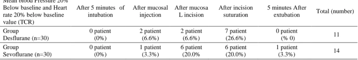
The periods, which are our review based on, are baseline, 5 minutes after intubation, post- mucosal injection, post-mucosal incision, after incision suturing, and 5 minutes after extubation. TCR was detected in 17 of 60 patients through our review. TCR was determined in 6 of 17 patients who developed TCR in 2 separate periods and for 1 of the TCR was detected in 3 separate periods. Of these 17 patients, 8 were from the sevoflurane group, while 9 were from the desflurane group. Of the patients in the sevoflurane group, TCR was determined in one of them in 3 separate periods and 5 of them in 2 periods; while of the 9 patients in the desflurane group, TCR was determined in 1 patient in 2 separate periods (Table 5,6,7).
Table 5. The distribution of those whose mean blood pressure is 20% below the baseline value and whose heart
rate is 20% below the baseline value are monitored according to the groups and periods. Mean blood Pressure 20%
Below baseline and Heart rate 20% below baseline value (TCR) After 5 minutes of intubation After mucosal injection After mucosa L incision After incision suturation 5 minutes After
extubation Total (number) Group Desflurane (n=30) 0 patient (0%) 2 patient (6.6%) 2 patient (6.6%) 7 patient (26.6%) 0 patient (% 0) 11 Group Sevoflurane (n=30) 0 patient (0%) 1 patient (3.3%) 6 patient (20.0% 6 patient (20.0%) 1 patient (3.3%) 14
Table 6. The distribution of values with heart rate below 20% of the baseline value according to groups and periods is monitored. Heart Rate 20% below baseline After 5 minutes of intubation After mucosal injection After mucosal incision After incision suturation 5 minutes after extubation Group Desflurane (n=30) 0 patient (0%) 3 patient (10.0%) 2 patient (6.6%) 10 patient (33.3%) 1 patient (3.3%) Group Sevoflurane (n=30) 0 patient (0%) 5 patient (16.6%) 5 patient (16.6%) 8 patient (26.6%) 3 patient (10.0%)
Table 7. Only cases with mean blood pressure below 20% of the baseline value are monitored according to
groups and periods. Mean arterial pressure 20% below baseline
After 5 minutes of intubation After mucosal injection After mucosal incision After incision suturation 5 minutes after extubation Grup Desflurane (n=30) 9 patient (% 30.0) 11 patient (% 36.6) 18 patient (60.0%) 15 patient (50%) 1 patient (% 3.3) Grup Sevoflurane (n=30) 7 patient (23.3%) 10 patient (33.3%) 18 patient (60.0%) 13 patient (43.3%) 1 patient (3.3%) In a prospective study conducted in
rhinoplasty procedures in our clinic in 2013, it was revealed that stimulation of the columellar region gave rise to the development of TCR under general anesthesia. In this study, unlike our scan, the periods were recorded based on the milestones including; just before (basal) mucosal injection, during mucosal injection into the columella and nasal dorsum, and at the 1st, 5th, 10th, 30th, and 60th seconds after the mucosal injection. In this study, local anesthetic was injected into the columella area in one group, while local anesthetic was injected into the nasal dorsum in the second group. Meanwhile, for the third group, 2 ml of local anesthetic solution was injected into the mid- columella and anterior maxillary cleft 10 minutes after the injection into the nasal dorsum, and the related data were recorded. TCR was detected in 8 patients in group 1 and 1 patient in group 2. TCR did not occur in group 3. And consequently, it was revealed that an increased incidence of TCR was determined due to the stimulation of the columellar region. It was determined from the surgical records that the same local anesthetic injection protocol was performed routinely in our health care center, similar to the previous study, and it was injected into the columellar region and nasal mucosa (11).
One of our drawbacks for these retrospective reviewing records is that there are no records regarding the depth of anesthesia; and the other one is that the volume of the gas, which was used in the procedures, is not contained in the records.
In a systematic review of the trigeminocardiac reflex, it was reported that the incidence of asystole increased by 1.2 times in
surface analgesia; however, when the surface analgesia was compared with deep anesthesia, the risk of asystole increased by 4.5 times (12). It has been revealed in the literature that some medications, which are administered in the practice of anesthesia, might lead to the shifts in the QT interval. It has given rise to the question that whether life-threatening arrhythmias and death cases could occur in the perioperative period due to the prolonged QT interval (13). These drugs include inhaled anesthetic agents, opioids, and dexmedetomidine (14-15). Previous studies have put forward that the administration of sevoflurane could also prolong the QT. Hence, the impacts of 2 anesthetic agents, which had been administered in the procedures, on QT and the occurrence of TCR was also intended to be investigated in the research. Fluctuations might occur in the QT interval due to sympathetic activation. The duration of induction, laryngoscopy, tracheal intubation, and inadequate anesthesia during the practice of anesthesia, might give rise to increase in the sympathoadrenal activity, hence, the prolongation of the QT interval even in healthy adults (16).
Nathanson et al. revealed that the mean arterial pressure values after induction of anesthesia dwindled during the period, which lasted until the incision of the skin, in the both group who had been administered with sevoflurane and desflurane, and the values of heart rate reduced; however, these reductions were greater in the sevoflurane group. Mean arterial pressure and heart rate values did not surpass the 20% of baseline values throughout the maintenance of anesthesia. It has been underscored
that both inhaled anesthetic agents manifest similar characteristics regarding hemodynamics (17).
Even though both groups exhibited analogous characteristics regarding hemodynamics in our study, reductions in the values of blood pressure values were observed, notably in sevoflurane, during the periods when the trigeminal nerve was stimulated intensely. Viskin et al. revealed in their research about the assessment of QT prolongation that automatic measurements could be used in selected patients, but manual measurements were safer in normal patients (18). Luo et al. concluded that there is a significant correlation between the interval of QTc and R-R values, which were calculated through the formula, and so the manual measurement is reliable (19). In order to find the related values of QTc, we used Bazzet's formula and calculated QT manually in our study. Whyte et al. determined a dose- dependent prolongation of QTc between 28 and 55 milliseconds among children whose induction and maintenance of anesthesia had been performed with the administration of sevoflurane (20). In our study, it was detected to be a prolongation of 29 ± 9 milliseconds compared to baseline in the sevoflurane group, while a prolongation of 16 ± 12 milliseconds was detected in the desflurane group. Since it has been revealed that it does not impact the RTD (transmural dispersion of repolarization), sevoflurane is considered as safe in torsadogenic terms; though it is well-known in the literature that sevoflurane might lead to prolongation in QTc through causing prolongation in the duration of action potential and repolarization (21). Furthermore, there is information on that it generates a downward trend in QTd and QTc. QTd was not studied in our research. Considering all this information, it is noteworthy that prolongations in QT and QTc can facilitate the development of TCR. From this point of view, it is evident that prospective studies are needed.
Kuenzberg et al. have revealed in their study, which is based on the administration of sevoflurane that the QTc interval was prolonged progressively compared to the duration of sevoflurane administration. Even though there was no significant difference between the two groups in our reviewing, compared to basal values, the intervals of QT and QTc were significantly prolonged in both groups, particularly in the sevoflurane group, after mucosal injection, after mucosal incision and after incision suturing; and it
was observed that it reached its longest interval during the incision suturing (Table 3,4). These periods were also found to be periods when the trigeminal nerve was stimulated maximally and the most frequent development of TCR occurred. It was determined that a mean volume of 50.35 ±30.42 mL sevoflurane was administered in the sevoflurane group, and a mean volume of 93.34 ± 72.78 mL desflurane was administered in the desflurane group (Table 2). No statistically significant difference was found between the groups regarding the duration of procedures (Table 1). Given that all these findings, we consider that the inhaled agents, which were administered predominantly in the sevoflurane group, particularly sevoflurane play a facilitating role in the development of the TCR, through generating a cumulative impact during the maximal stimulation of the trigeminal nerve (22).
Leung et al. revealed that narcotics, which are used during induction, could suppress sympathetic stimulus effects of desflurane (23).
It was determined in our reviewing that remifentanil had been administered as a narcotic in the procedures. There is no significant difference between the two groups regarding the administration of remifentanil (Table 2).
Since they could affect the subsequent measurement values, the patients who had been administered with atropine for bradycardia in the reviewing were excluded from the study. It was detected that there were merely 2 patients whose characteristics conformed to the above-mentioned criteria. One of them occurred at the end of extubation and the other one after the induction, which was due to bronchospasm.
CONCLUSION
It was found that there was no difference between the two groups regarding the development of TCR; however, there were longer prolongation values in QT and QTc during the periods with maximal trigeminal nerve stimulation in sevoflurane, compared to basal values. This finding is also in line with the literature. We are of the opinion that the choice of anesthetic agent is a facilitating factor regarding the development of trigeminocardiac reflex. We also consider that more elaborate studies, which are randomized, controlled, prospective, and examine the depth of anesthesia as well, should be performed related to the development of TCR and the impact of anesthetic agents.
REFERENCES
1. Meuwly C, Chowdhury T, Sandu N, Reck M, Erne P, Schaller B. Anesthetic influence on occurence and treatment of the trigemino-cardiac reflex. Medicine.2015;94(18):e807)
2. Chowdhury T, Belachew A, Sandu N, Meuwly C, Schaller B. The trigeminocardiac reflex- An example of reflexive heart rhythm change. Abnormal cardiac ritm.Ed. Francisco R. Brejio-Marquez. 2015. Chapter 5. Intech Open Publication. ISBN:978-953-51-2148-0
3. Schaller B, Probst R, Strebel S, Gratzl O. Trigeminocardiac reflex induced by an intraorbital foreign body. Ophtalmica. 2006;220:348.
4. Schaller B. Trigemino-cardiac reflex during transsphenoidal surgery for pituitary adenomas. Clin Neurol Neurosurg. 2005;107:468-474.
5. Schaller BJ, Weigel D, Fillis A, Buchfelder M. Trigemino-cardiac reflex during transsphenoidal surgery for pituitary adenomas: methodological descriptionof a prospective Skull base study protocol. Brain Res 2007;1149:69-75.
6. Arasho B, Sandu N, Spiriev T, Prabhakar H, Schaller B. Management of the trigeminocardiac reflex: facts and own experience. Neurol India. 2009;57:375-380.
7. Schaller B, Cornelius JF, Prabhakar H, et al. The trigemino-cardiac reflex: an update of the current knowledge. J Neurosurg Anesthesiol.2009;21:187-195.
8. Schaller B, Probst R, Strebel S, Gratzl O. Trigeminocardiac reflex during surgery in the cerebellopontine angle. J Neurosurg 1999;90:215-220.
9. Chowdhury T, Sandu N, Meuwly C, Cappellani RB, Schaller B. Trigeminocardiac reflex:Differental behaviour and risk factors in the course of the trigeminal nerve, Future Neurology 2014;9:41-47.
10. Paton JF, Boscan P, Pickering AE, Nalivaiko E. The yin and yang of cardiac autonomic control: vago-sympathetic interactions revisted. Brain Res. Rev.2005;49:455-565.
11. Özçelik D, Toplu G, Türkseven A, Sezen G, Ankaralı H. The importance of the trigeminocardiac reflex in rhinoplastic surgery. Ann Plast Surg. 2015. Aug;75(2):213-218.
12. Meuwly C, Chowdhury T, Sandu N, Reck M, Erne P, Schaller B. Anesthetic influence on occurance and treatment of the trigemino-cardiac reflex: a systematic literature review. Medicine (Baltimore). 2015;94(18):e807.
13. Schmeling WT, Warltier DC, McDonald DJ, Madsen KE, Atlee JL, Kampine JP. Prolongation of the QT interval by enflurane, isoflurane, and halothane in humans, Anesth Analg 1991; 72(2): 137–144
14. Bond AN, Beerle BJ, Arnold RW. Dexmedetomidine nasal sedation procedures more Oculocardiac reflex during strabismus surgery. J Pediatr Ophthalmol Strabismus. 2016;53(5):318.
15. Arnold RW, Biggs RE, Beerle BJ. Intravenous dexmedetomidine augments the oculocardiac reflex. J AAPOS. 2018;22(3):211-213.e211.
16. Lindgren L, Yli-Hankala A, Randall T, Kirvela M, Scheinin M, Neuvonen P.J. Haemodynamic and catecholeamine responces to induction of Anaesthesia and tracheal intubation: comparison between propofol and thiopentone. Br J Anaesth 1993;70(3):306-310.
17. Nathanson MH, Fredman B, Smith I, White PF. Sevoflurane versus desflurane for outpatient anesthesia: A comparison of maintance and recovery profiles. Anesth Analg. 1995;81:1186-1190.
18. Viskin S, Rosovski U, Sands A.J, Chen E, Kistler P.M, Kalman J.M, Rodriguez Chavez L, İtturale Tores P, Cruzz F.F, et al. İnaccurate electrocardiographic interpretation of long QT: The majority of phsysicians cannot recognize a long QT when they see one, Heart Rhythm 2005; 2(6): 569-574
19. Luo S, Michler K, Johnston P, Macfarlane P.W. A comparison of commonly used QT correction formulae: The effect of heart rate on the QTc of normal ECGs . J Electrocardiol, 2004;37:81-90.
20. Whyte SD, Sanatani S, Lim J, Booker PD. A comparison of the effect on dispersion of repolarization of age-adjusted MAC Values of sevoflurane in children. Anesth Analg, 2007;104(2):277-282.
21. Stamelos CM, Stavroulakis E. Impact of anesthetic drugs and adjuvants on ECG markerin of torsadogenisity. Br J Anaesth. 2014;112(2):217-230.
22. Kruenzberg E, Loeckinger A, Kleinsasser A, Lindner KH, Puehringer F, Hoermann C. Sevoflurane progresively prolongs the QT interval in unpremedicated female adults, Eur J Anaesthesiol, 2000;17:662-664.
23. Leung J, Pastor M, Darwin A. Dissociation between hemodinamics and sympathetic activation during anaesthetic induction with desflurane. Can J Anaesth. 1998;45:533-540.