Address for correspondence: Dr. Kaan Okyay, Fevzi Çakmak Caddesi No. 45, Bahçelievler, Ankara-Türkiye
Phone: +90 312 203 68 68 Fax: +90 312 223 86 97 E-mail: [email protected] Accepted Date: 02.10.2015 Available Online Date: 26.11.2015
©Copyright 2016 by Turkish Society of Cardiology - Available online at www.anatoljcardiol.com DOI:10.5152/AnatolJCardiol.2015.6645
Kaan Okyay, Aylin Yıldırır, Mutlu Çiçek, Alp Aydınalp, Haldun Müderrisoğlu
Department of Cardiology, Faculty of Medicine, Başkent University Ankara Education and Research Hospital; Ankara-Turkey
Serum cystatin C and neutrophil gelatinase-associated lipocalin in
predicting the severity of coronary artery disease in diabetic patients
Introduction
Cardiovascular disease (CVD) is the leading cause of mor-tality and morbidity in diabetic patients (1). In diabetes mel-litus (DM), obstructive coronary artery disease (CAD) is more common, and diabetic patients have more extensive disease than patients with other risk factors for CAD (2). Thus, it has clinical importance to find out simple and robust biomarkers that predict the presence and severity of CAD in this high-risk population.
Neutrophil gelatinase-associated lipocalin (NGAL) belongs to the lipocalin superfamily of proteins that accumulate in gran-ules of neutrophils (3). It has been demonstrated to be released in case of renal tubular damage due to ischemia and/or neph-rotoxins and suggested as an early marker of acute kidney in-jury (4). Furthermore, NGAL is expressed in endothelial cells and macrophages in atherosclerotic plaques (5). It enhances the activity of matrix metalloproteinase-9, which plays important role in plaque instability (6). NGAL modulates inflammation and
promotes development and progression of atherosclerosis (3–6). Previously, serum NGAL levels have been shown to be associ-ated with the extent of stable CAD (7) and to be elevassoci-ated in acute coronary syndromes (8). However, despite the putative role of inflammation in pathogenesis of type 2 DM and CVD (9, 10), NGAL has not yet been elucidated in predicting the severity of CAD in diabetic patients.
Cystatin C is a low-molecular-weight (13kD) protein be-longing to the cystatin super- family of cysteine proteinase inhibitors. It is synthesized by all nucleated cells, filtered from glomeruli, and is almost completely reabsorbed by proximal renal tubules (11). When compared to serum creatinine, it is less influenced by age, gender, race, muscle mass, exercise, and diet. Therefore, serum cystatin C is accepted as a superior blood parameter than creatinine clearance for estimating renal functions (12). Cystatin C has been shown to be related with increased coronary atherosclerotic burden (13, 14), probably due to contribution to inflammatory processes. Inflammatory cytokines stimulate the production of lysosomal cathepsins,
Objective: Cystatin C and neutrophil gelatinase-associated lipocalin (NGAL) are biomarkers of renal functions. We evaluated their roles in pre-dicting the severity of coronary artery disease (CAD).
Methods: Fifty-two consecutive type 2 diabetic patients (32 males, 65.7±8.6 years) who underwent coronary angiography (CAG) for stable CAD were included in this single-center, prospective, cross-sectional study. Patients with an estimated glomerular filtration rate <60 mL/min/1.73 m2
and with a history of by-pass surgery and/or coronary stent implantation were excluded. The vessel score and Gensini score were calculated to assess the presence and severity of CAD. Mann–Whitney U test, Spearman test, and multiple linear regression analysis were used for the main statistical analyses.
Results: Serum cystatin C levels were higher in patients with multivessel disease than in those with single vessel disease [1260 ng/mL (953–1640) vs. 977 ng/mL (599–1114), p=0.017]. According to the median Gensini score, the higher score group also had higher cystatin C levels than the lower score group [1114 ng/mL (948–1567) vs. 929 ng/mL (569–1156), p=0.009]. However, serum NGAL levels were similar between these sub-groups. There was a positive correlation between cystatin C and Gensini score (r=0.334, p=0.016). Multiple linear regression analysis revealed serum cystatin C as an independent predictor of the Gensini score (β=0.360, t=2.311, p=0.026). These results may aid in defining cystatin C as a surrogate marker of the extent of CAD in further clinical trials.
Conclusion: Serum Cystatin C, but not NGAL levels, could predict the severity of CAD in diabetic patients. (Anatol J Cardiol 2016; 16: 756-61) Key words: coronary artery disease, cystatin C, neutrophil gelatinase-associated lipocalin, diabetes mellitus
which degrade extracellular matrix proteins; and concentration of cystatin C, a cathepsin inhibitor, is elevated to counterbal-ance this elastolytic overactivity (15). In metabolic syndrome, cystatin C has been found to be associated with severity of CAD (16). However, there are limited data evaluating this association in diabetic population.
We argued that these biomarkers could reflect the increased inflammatory status in diabetic coronary atherosclerosis. Ac-cordingly, in the present study, we aimed to determine serum cystatin C and NGAL levels for predicting the severity of CAD in diabetic patients.
Methods
Study population
This was a single-center, prospective, cross-sectional study. Power analysis of the study was performed using the program of G*Power power-and-sample size calculation Ver-sion 3.1.9.2. Düsseldorf Universität, Germany (17). Accordingly, with an alpha error value of 0.05 and Cohen’s effect size of 0.8 and power of 0.80, the projected sample size was found to be 52 subjects for the inferential statistics (t tests-means: difference between two independent groups). The study was conducted in accordance with the guidelines proposed in the Helsinki Declaration and approved by Başkent University Institutional Review Board and supported by Başkent University Scientific Research Fund. All the participants gave written informed con-sent before enrollment.
The subjects were consecutive type 2 diabetic patients who underwent elective CAG due to suspected diagnosis of stable CAD in our institute. Patients with a serum creatinine of >1.4 mg/ dL for men and >1.2 mg/dL for women and/or with an estimat-ed glomerular fi ltration rate (eGFR) <60 mL/min/1.73 m2, active or chronic liver diseases, active infectious conditions, chronic inflammatory or rheumatological diseases, and malignancies were not included. Also, patients with unstable angina, conges-tive heart failure, severe valvular disorders, and history of prior percutaneous coronary intervention /coronary artery by-pass graft operation were not included. Fifty-two eligible type 2 dia-betic patients were analyzed.
Weight and height of the patients were measured without heavy outer garments and shoes, after a 12-hour fasting peri-od. Office blood pressure was measured two times on the right arm in the sitting position, with a 5-min interval, and the average value was used. Hypertension was accepted as systolic blood pressure ≥140 mm Hg; diastolic blood pressure, ≥90 mm Hg, or being treated with anti-hypertensive medication. Diabetes mel-litus was defined as being treated with anti-diabetic medica-tions (oral anti-diabetics / insulin). Smoking was defined as the regular use of tobacco. Hyperlipidemia was defined as having total cholesterol level >200 mg/dL or use of hypolipidemic medi-cations. eGFR was calculated by Modification of Diet in Renal Disease (MDRD) equation (18).
Laboratory measurements
The venous blood samples for routine laboratory measure-ments were taken after 12-hours overnight fasting within 72 hours prior to CAG and analyzed in 1 hour. For determination of cystatin C and NGAL levels, the venous blood samples were drawn on the morning of CAG and stored at −20°C until analysis time, and were detected with sandwich enzyme immunoassay method using commercial Human Cystatin C kits (Biovendor Laboratory Medicine Inc., Brno, Czech Republic) and Human Lipocalin-2/NGAL kits (Biovendor Laboratory Medicine Inc.), re-spectively, by a microelisa analyser device (DSX Model; Dynex Technologies, Chantilly, VA, USA).
Coronary angiography
The coronary angiographies were performed using the stan-dard Judkins technique. The angiograms were reviewed by two independent experienced invasive cardiologists for the presence and severity of CAD. Coronary vessel score was ranged between 0 and 3 according to the number of the main coronary arteries— left anterior descending (LAD), left circumflex (Cx), and right cor-onary artery (RCA)—with a ≥50% stenosis. Significant CAD was defined as a vessel score ≥1. Gensini score (19) was calculated for evaluation of the CAD severity in each patient. This system scores the stenosis in the epicardial coronary arteries (1 point for 1–25% stenosis, 2 for 26–50% stenosis, 4 for 51–75% stenosis, 8 for 76–90% stenosis, 16 for 91–99% stenosis, and 32 for total occlusion) and multiplies this number by a constant number in term of the anatomical position of the lesion. Multipliers are 5 for the left main coronary artery; 2.5 for the proximal segment of the LAD and Cx; 1.5 for the mid-segment of the LAD; 1 for the RCA, the distal segment of the LAD, the posterolateral artery, and the obtuse marginal artery; and 0.5 for other segments. The score in every segment is calculated, and the total score gives the Gensi-ni score. The patients who have history of coronary revascular-ization (percutaneous coronary intervention or coronary artery bypass graft operation) were excluded from the study since the original Gensini’s scoring system is technically inappropriate for such cases. The median Gensini score was 23 (5–51, IQR: 46). The patients were divided into groups of severe and non-severe CAD according to this median score.
Statistical analysis
The normality of distribution was tested using Kolmogorov– Smirnov test. Continuous variables were presented as mean±SD if normally distributed otherwise as median with 25th and 75th per-centiles. Categorical variables were presented as numbers and percentages and were compared using chi-square test (Fisher’s exact test if needed). Student’s t-test or Mann–Whitney U test was used for comparison of continuous variables where ap-propriate. Correlation analysis was performed using Spearman test. A multiple linear regression analysis model was generated in order to investigate the independent predictors of the Gensini score. Receiver operating characteristic (ROC) analysis was
used to predict the cut off points for cystatin C in determination of multi-vessel disease. Then, sensitivity and specificity were calculated. Statistical analysis was performed by SPSS software (version 17; Statistical Package for Social Sciences, Chicago, IL). A p value of <0.05 was considered statistically significant.
Results
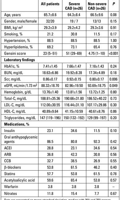
The mean age of the study population (20 females and 32 males) was 65.7±8.6 years. The clinical data and demographic features of the entire study population are presented in Table 1.
The patients were divided into subgroups of severe and non-se-vere CAD. Patients with higher Gensini score had higher serum creatinine and lower eGFR values compared with those having lower Gensini score. Other parameters were similar in regard to groups arranged with Gensini score (Table 1).
There was a trend toward higher cystatin C levels in patients with significant CAD (n=34) than in those without (n=18) [1101 (908–1378) vs. 915 (569–1162), respectively, p=0.09]. Regarding the patients with significant CAD, serum cystatin C levels were found to be higher in patients with multivessel disease (n=18) than in those with single vessel disease (n=16) [1260 ng/mL (953–1640) vs. 977 ng/mL (599–1114), respectively, p=0.017]. Se-rum cystatin C concentrations were increased across the vessel score [0 vessel, n=18: 915 ng/mL (569–1162); single vessel, n=16: 977 ng/mL (599–1114); two vessels, n=8: 1199 ng/mL (915–1476); and three vessels, n=10: 1393ng/mL (967–1724); p=0.03] (Fig. 1). The patients with higher Gensini score had also higher cys-tatin C levels than those with lower Gensini score [1114 ng/mL (948–1567) vs. 929 ng/mL (569–1156), respectively, p=0.009]. In all of the above-mentioned subgroups, there were no differences in serum NGAL levels (Table 2 summarizes cystatin C and NGAL levels compared between the groups).
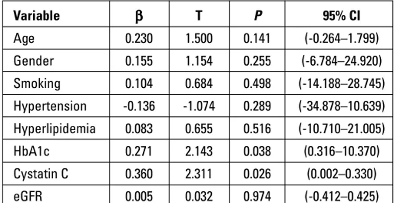
Cystatin C levels were positively correlated with Gensini score (r=0.334, p=0.016), and serum creatinine levels (r=0.501, p<0.001), and negatively with eGFR value (r=0.479, p<0.001). Mul-tiple linear regression analysis revealed that serum cystatin C (=0.360, t=2.311, p=0.026) and hemoglobin A1c (=0.271, t=2.143, p=0.038) were the independent predictors of the Gensini score (Table 3). ROC curve analysis (Fig. 2) revealed that serum cystatin C level of 1111 ng/mL has a 67% sensitivity and 75% specificity for estimating multivessel CAD [p=0.017, area under the curve (AUC): 0.740, 95% CI: 0.568–0.911].
Discussion
In the present study, we investigated the diagnostic value of serum cystatin C and NGAL levels for the prediction of CAD se-verity in type 2 diabetic patients. Our results showed that serum
Table 1. Demographics and baseline clinical parameters of the patients
All patients Severe Non-severe P
CAD (n=26) CAD (n=26) Age, years 65.7±8.6 64.3±8.4 66.5±9.6 0.08 Gender, male/female 32/20 19 / 7 13/13 0.15 BMI, kg/ m2 29.2±3.9 29.2±4.2 29.3±3.6 0.91 Smoking, % 21.2 30.8 11.5 0.17 Hypertension, % 88.5 88.5 88.5 1.00 Hyperlipidemia, % 69.2 73.1 65.4 0.76 Gensini score 23 (5–51) 51 (29–69) 4.75 (1–10) <0.001 Laboratory findings HbA1c, % 7.41±1.45 7.66±1.47 7.18±1.43 0.24 BUN, mg/dL 18.63±6.86 19.92±8.28 17.34±4.89 0.18 Scr, mg/dL 0.86±0.17 0.92±0.15 0.80±0.17 0.006 eGFR, mL/min /1.73 m2 88.32±18.70 82.96±19.50 93.69±18.75 0.049 Hemoglobin, g/dL 13.76±1.40 13.81±1.56 13.72±1.25 0.80 Total-C, mg/dL 188.81±35.36 190.66±31.80 186.52±40.22 0.73 LDL-C, mg/dL 112.00±30.55 116.44±31.10 107.17±29.86 0.30 HDL-C, mg/dL 40.89±9.64 41.15±10.59 40.61±8.76 0.86 Triglycerides, mg/dL 147 (119–196) 150 (132–192) 129 (99–197) 0.20 Medications, % Insulin 23.1 34.6 11.5 0.10 Oral antihypoglycemic agents 86.5 80.8 92.3 0.42 ACEI 28.8 23.1 34.6 0.54 ARB 36.8 42.3 30.8 0.56 CCB 32.7 38.5 26.9 0.55 β-blockers 53.8 61.5 46.2 0.40 Statins 57.7 53.8 61.5 0.78 Acetylsalicylic acid 59.6 65.4 53.8 0.57 Warfarin 3.8 3.8 3.8 – Nitrates 11.8 15.4 7.7 0.67
Data are presented as mean±standard deviation, median with 25th and 75th
percen-tiles, or number and frequencies where indicated
ACEI - angiotensin converting enzyme inhibitor; ARB - angiotensin receptor blocker; BMI - body mass index; BUN - blood urea nitrogen; C - cholesterol; CAD - coronary artery disease; CCB - calcium channel blocker; eGFR - estimated glomerular filtration rate; HbA1c - hemoglobin A1c; HDL-C - high-density lipoprotein cholesterol; LDL-C - low-density lipoprotein cholesterol; Scr - serum creatinine
Student’s t-test or Mann–Whitney U Test and chi-square test were used for statistical analyses Serum c ystatin C ng/mL Vessel score 1700.00 1500.00 1300.00 1100.00 900.00 700.00 500.00 0 1 2 3
Figure 1. Comparison of cystatin C levels according to the vessel score. Serum cystatin C concentrations were increased across the vessel score (P=0.03). Posthoc tests revealed significant differences in cystatin C levels between no-vessel and three-vessel disease (P=0.016) and between one-vessel and three-vessel disease (P=0.020)
cystatin C levels were higher in multivessel CAD and strongly predicted the disease severity. However, we did not observe any association between serum NGAL levels and the severity of CAD. To our best of knowledge, this is the first study investigating cystatin C and NGAL levels simultaneously in regard to coronary atherosclerotic burden in type 2 diabetic patients.
Cardiovascular diseases (CVDs) are the leading causes of mortality and morbidity in DM, and diabetic patients have more extensive disease than patients having other risk factors for CAD (1, 2). The increased prevalence of accompanying disorders such as hypertension and dyslipidemia also contribute to the higher CVD event rates in diabetes (20). The increased CVD risk in type 2 DM can be explained by a complex combination of various traditional and nontraditional risk factors. The pathogenesis of CVD in diabetes includes epigenetic, intracellular, and metabolic changes resulting in oxidative stress, inflammation, and endothe-lial dysfunction (9). In type 2 DM, chronic toxicity of glucose and free fatty acids is related with the induction of inflammatory pro-cesses within various tissues, particularly in vascular wall (10). Moreover, a low-grade inflammation plays an important role in the pathogenesis of the insulin resistance (21). Due to this strict relation, different biochemical markers reflecting inflammatory processes are used for screening CVD risk in diabetics.
It has been shown that impaired renal functions are related with increased all-cause and cardiovascular mortality, adverse cardiovascular events, and hospitalization rate in a large, com-munity-based population (22). NGAL and cystatin C have been proven as biomarkers of renal injury and functions and accepted being accurate parameters for estimating renal functions (4, 12). These biologic substances also have important contributions to inflammatory processes involved in atherosclerosis as dis-cussed earlier (3–6, 13–15).
In recent clinical trials, cystatin C has been found to be rela-ted with severity of CAD and a predictor of mortality and adverse cardiovascular events in patients with acute coronary events (23–25). Niccoli et al. (13) evaluated 70 patients with normal re-nal functions and stable CAD and reported a relation between increased cystatin C and coronary atherosclerotic burden. How-ever, in the study of Doğaner et al. (26), which included 88 non-diabetic patients with an eGFR of >60 mL/min/1.73 m2, cystatin C levels were lower in patients with CAD, and they showed an inverse association between number of involved coronary ar-tery and cystatin C levels. They speculated that higher levels of cystatin C might indicate the presence of vulnerable plaques. That study, however, did not show the plaque morphology. They
also found no correlation between cystatin C and eGFR in their study population. Subsequently, in two clinical trials including both diabetic and non-diabetic stable CAD patients without renal dysfunction (eGFR of >60 mL/min/1.73 m2), cystatin C was cor-related with severity of CAD quantified by Gensini score (14, 27). Qing et al. (16) provided that cystatin C levels were elevated in asymptomatic CAD in metabolic syndrome patients. They demonstrated positive correlations between cystatin C and num-ber of diseased vessel and the Gensini score, and they found cys-tatin C to be an independent predictor of CAD severity calculated by Gensini score. In this point, our results are in agreement with those studies demonstrating a close relation between cystatin C and severity of CAD. We showed that patients with multivessel disease and those with higher Gensini score had also increased serum cystatin C levels. In our study, there was a positive and modest correlation between cystatin C levels and the Gensini
Table 2. Cystatin C and NGAL concentrations according to extent and severity of coronary artery disease
Vessel score Gensini score
Single-vessel Multi-vessel P Lower gensini score Higher gensini score P Serum cystatin C, ng/mL 977 (599–1114) 1260 (953–1640) 0.017 929 (569–1156) 1114 (948–1567) 0.009 Serum NGAL, ng/mL 92 (60–133) 95 (65–152) 0.64 101 (63–139) 92 (62–143) 0.69
NGAL - neutrophil gelatinase-associated lipocalin. Mann–Whitney U test was used for statistical analyses
Table 3. Multivariate linear regression analysis model to investigate independent predictors of Gensini score
Variable β T P 95% CI Age 0.230 1.500 0.141 (-0.264–1.799) Gender 0.155 1.154 0.255 (-6.784–24.920) Smoking 0.104 0.684 0.498 (-14.188–28.745) Hypertension -0.136 -1.074 0.289 (-34.878–10.639) Hyperlipidemia 0.083 0.655 0.516 (-10.710–21.005) HbA1c 0.271 2.143 0.038 (0.316–10.370) Cystatin C 0.360 2.311 0.026 (0.002–0.330) eGFR 0.005 0.032 0.974 (-0.412–0.425)
CI - confidence interval; eGFR - estimated glomerular filtration rate; HbA1c - hemo-globin A1c Sensitivity ROC curve 1.0 0.8 0.6 0.4 0.2 0.0 0.0 0.2 0.4 0.6 0.8 1.0 1 - Specificity
Figure 2. Receiver operating characteristics curve analysis to find out predictive values of cystatin C levels in estimating multivessel CAD (P=0.017, AUC: 0.740, 95% CI: 0.568–0.911)
score and linear regression analysis revealed cystatin C as an independent predictor of the Gensini score. Furthermore, in our study, AUC of ROC analysis was 0.740. It was reported as 0.622 by Qing et al. (16) and 0.588 by Wang et al. (27). This indicates that cystatin C as a sole marker had a higher predictive value in our study. Nevertheless, our AUC value indicates that there could be some additional unknown predictors affecting atherosclerotic burden in this cohort, supporting the idea that atherosclerosis is a multifactorial and complex disease. When evaluating renal functions of the diabetic patients, the cut-off value of 1111 ng/ mL can be an acceptable level to effectively identify severe CAD.
Serum NGAL has been shown to be elevated in a variety of CAD such as coronary ectasia (28), coronary slow flow phenomenon (29), and unstable coronary syndromes, including acute myocar-dial infarction (30, 31). However, the role of NGAL in predicting severity of stable CAD has not yet been clearly elucidated. Zo-grafos et al. (7) evaluated 73 stable CAD patients (21 with diabe-tes) and reported that serum NGAL level was modestly corre-lated (r=0.356) with Gensini score. In our study, however, serum NGAL levels were not related with presence or severity of stable CAD in diabetic patients.
To our knowledge, there have been no data regarding simulta-neous measurements of cystatin C and NGAL for the evaluation of stable CAD in either diabetic or non-diabetic population. Still, possible mechanisms underlying the link between cystatin C and CAD need to be described. It can simply be argued that renal mechanisms are responsible for this relation. However, in our diabetic cohort, mean serum creatinine values were within the normal range, but mean eGFR value was relatively low; this sug-gested slightly impaired kidney functions. Patients with severe CAD had higher serum creatinine, lower eGFR values, and higher cystatin C levels. Importantly, cystatin C remained as a predictor for CAD severity, even after adjustment for eGFR value. Hence, we cannot exactly explain the association between cystatin C and CAD severity via solely impaired kidney dysfunction, which predicts severe cardiovascular outcomes. Based on previous clinical trials demonstrating positive correlations between se-rum cystatin C and highly sensitive C reactive protein and fibri-nogen (14, 27), it could be speculated that cystatin C participates in inflammatory processes that are involved in coronary athero-sclerosis. Further clinical investigations with larger cohorts are needed to explain exact mechanisms.
Study limitations
Ours was a single-center and relatively small-sized study. We did not determine relations between CAD and serum NGAL and cystatin C levels after graduating the patients into different stages of nephropathy. Use of complementary biomarker(s) of inflamma-tion and considering the durainflamma-tion of DM which may affect levels of the markers could render our results more contributory. Ultimately, in biomarker assays racial/ethnic differences should be regarded, and our results may not generalize to the all diabetic population.
Conclusion
Our fi ndings showed that serum cystatin C levels, but not NGAL levels, were significantly associated with severity of sta-ble CAD in type 2 diabetic patients. Serum cystatin C levels could be a marker of coronary atherosclerosis independent of kidney function. We believe that our study may inspire further trials to investigate cystatin C in this regard.
Conflict of interest: None declared. Peer-review: Externally peer-reviewed.
Authorship contributions: Concept – A.Y., H.M.; Design – K.O., M.Ç., A.Y.; Supervision – A.Y., H.M.; Resource – M.Ç., A.A.; Data collection and/or processing – K.O., M.Ç., A.A., HM; Analysis and/or Interpretation – K.O., A.Y.; Literature search – K.O.; Writing – K.O., A.Y.; Critical review – A.A., A.Y., H.M.
References
1. Standards of medical care in diabetes-2015 abridged for primary care providers. American Diabetes Association. Clin Diabetes 2015; 33: 97-111. Crossref
2. Tomiwaza N, Nojo T, Inoh S, Nakamura S. Difference of coronary artery disease severity, extent and plaque characteristics between patients with hypertension, diabetes mellitus or dyslipidemia. Int J Cardiovasc Imaging 2015; 31: 205-12. Crossref
3. Kjeldsen L, Bainton DF, Sengelov H, Borregaard N. Identification of neutrophil gelatinase-associated lipocalin as a novel matrix pro-tein of specific granules in human neutrophiles. Blood 1994; 83: 799-807.
4. Haase M, Bellomo R, Devarajan P, Schlattmann P, Haase-Fielitz A. NGAL Meta-analysis Investigator Group. Accuracy of neutrophil gelatinase-associated lipocalin (NGAL) in diagnosis and prognosis in acute kidney injury: a systematic review and meta-analysis. Am J Kidney Dis 2009; 54: 1012-24. Crossref
5. Hemdahl AL, Gabrielsen A, Zhu C, Eriksson P, Hedin U, Kastrup J, et al. Expression of neutrophil gelatinase-associated lipocalin in ath-erosclerosis and myocardial infarction. Arterioscler Thromb Vasc Biol 2006; 26: 136-42. Crossref
6. Bolignano D, Coppolino G, Lacquaniti A, Buemi M. From kidney to cardiovascular diseases: NGAL as a biomarker beyond the con-fines of nephrology. Eur J Clin Invest 2010; 40: 273-6. Crossref
7. Zografos T, Haliassos A, Korovesis S, Giazitzoglou E, Voridis E, Ka-tritsis D. Association of neutrophil gelatinase-associated lipocalin with the severity of coronary artery disease. Am J Cardiol 2009; 104: 917-20. Crossref
8. Şahinarslan A, Kocaman SA, Baş D, Akyel A, Ercin U, Zengin O, et al. Plasma neutrophil gelatinase-associated lipocalin levels in acute myocardial infarction and stable coronary artery disease. Coron Artery Dis 2011; 22: 333-8. Crossref
9. Fonseca V, Desouza C, Asnani S, Jialal I. Nontraditional risk factors for cardiovascular disease in diabetes. Endocr Rev 2004; 25:153-75. 10. Moreno PR, Fuster V. New aspects in the pathogenesis of diabetic
atherothrombosis. J Am Coll Cardiol 2004; 44: 2293-300. Crossref
11. Grubb A. Diagnostic value of analysis of cystatin C and protein HC in biological fluids. Clin Nephrol 1992; 38 Suppl 1: S20-7.
12. Shimizu-Tokiwa A, Kobata M, Io H, Kobayashi N, Shou I, Funabiki K, et al. Serum cystatin C is a more sensitive marker of glomerular function than serum creatinine. Nephron 2002; 92: 224-6. Crossref
13. Niccoli G, Conte M, Della Bona R, Altamura L, Siviglia M, Dato I, et al. Cystatin C is associated with an increased coronary athero-sclerotic burden and a stable plaque phenotype in patients with ischemic heart disease and normal glomerular filtration rate. Ath-erosclerosis 2008; 198: 373-80. Crossref
14. Dandana A, Gammoudi I, Chalghoum A, Chahed H, Addad F, Ferchi-chi S, et al. Clinical utility of serum cystatin C in predicting coronary artery disease in patients without chronic kidney disease. J Clin Lab Anal 2014; 28: 191-7. Crossref
15. Taglieri N, Koenig W, Kaski JC. Cystatin C and cardiovascular risk. Clin Chem 2009; 55: 1932-43. Crossref
16. Qing X, Furong W, Yunxia L, Jian Z, Xuping W, Ling G. Cystatin C and asymptomatic coronary artery disease in patients with metabolic syndrome and normal glomerular filtration rate. Cardiovasc Diabe-tol 2012; 11: 108. Crossref
17. Faul F, Erdfelder E, Lang AG, Buchner A. G* Power 3: a flexible sta-tistical power analysis program for the social, behavioral, and bio-medical sciences. Behav Res Methods 2007; 39: 175-91. Crossref
18. Levey AS, Bosch JP, Lewis JB, Greene T, Rogers N, Roth D. A more accurate method to estimate glomerular filtration rate from serum creatinine: a new prediction equation. Modification of Diet in Renal Disease Study Group. Ann Intern Med 1999; 130: 461-70. Crossref
19. Gensini GG. A more meaningful scoring system for determining the severity of coronary heart disease. Am J Cardiol 1983; 51: 606. 20. Leosdottir M, Willenheimer R, Persson M, Nilsson PM. The
asso-ciation between glucometabolic disturbances, traditional cardio-vascular risk factors and self-rated health by age and gender: a cross-sectional analysis within the Malmo Preventive Project. Car-diovasc Diabetol 2011; 10: 118. Crossref
21. Martin-Timon I, Sevillano-Collantes C, Segura-Galindo A, Del Canizo-Gomez FJ. Type 2 diabetes and cardiovascular disease: Have all risk factors the same strength? World J Diabetes 2014; 5: 444-70. Crossref
22. Go AS, Chertow GM, Fan D, McCulloch CE, Hsu CY. Chronic kidney disease and the risks of death, cardiovascular events, and hospi-talization. N Engl J Med 2004; 351: 1296-305. Crossref
23. Soylu K, Aksan G, Nar G, Özdemir M, Gülel O, İnci S, et al. Serum neutrophil gelatinase-associated lipocalin levels are correlated with the complexity and the severity of atherosclerosis in acute coronary syndrome. Anatol J Cardiol 2015; 15: 450-5. Crossref
24. Ristiniemi N, Lund J, Tertti R, Christensson A, Ilva T, Porela P, et al. Cystatin C as a predictor of all-cause mortality and myocardial infarction in patients with non-ST-elevation acute coronary syn-drome. Clin Biochem 2012; 45: 535-40. Crossref
25. Silva D, Cortez-Dias N, Jorge C, Marques JS, Carrilho-Ferreira P, Mag-alhães A, et al. Cystatin C as prognostic biomarker in ST-segment elevation acute myocardial infarction. Am J Cardiol 2012; 109: 1431-8. 26. Doğaner YC, Aydoğan U, Aydoğdu A, Aparcı M, Akbulut H, Nerkiz
P, et al. Relationship of cystatin C with coronary artery disease and its severity. Coron Artery Dis 2013; 24: 119-26. Crossref
27. Wang GN, Sun K, Hu DL, Wu HH, Wang XZ, Zhang JS. Serum cys-tatin C levels are associated with coronary artery disease and its severity. Clin Biochem 2014; 47: 176-81. Crossref
28. Akyel A, Şahinarslan A, Kızıltunç E, Yıldız U, Alsancak Y, Akboğa MK, et al. Neutrophil gelatinase-associated lipocalin levels in iso-lated coronary artery ectasia. Can J Cardiol 2011; 27: 773-8. 29. Aksan G, Soylu K, Aksoy O, Özdemir M, Yanık A, Yüksel S, et al. The
relationship between neutrophil gelatinase-associated lipocalin levels and the slow coronary flow phenomenon. Coron Artery Dis 2014; 25: 505-9. Crossref
30. Lindberg S, Pedersen SH, Mogelvang R, Jensen JS, Flyvbjerg A, Galatius S, et al. Prognostic utility of neutrophil gelatinase-associ-ated lipocalin in predicting mortality and cardiovascular events in patients with ST-segment elevation myocardial infarction treated with primary percutaneous coronary intervention. J Am Coll Car-diol 2012; 60: 339-45. Crossref
31. Kafkas N, Demponeras C, Zoubouloglou F, Spanou L, Babalis D, Makris K. Serum levels of gelatinase associated lipocalin as indi-cator of the inflammatory status in coronary artery disease. Int J Inflam 2012; 2012: 189797. Crossref