Introduction
Following the discovery of Wolf and Markiw (1) of the alternation of myxosporean and actinosporean phases in the life cycle of Myxobolus cerebralis, the causative agent of whirling disease and the most intensely studied myxozoan due to its economic importance in salmonid culture, the number of studies on the actinosporean parasites of aquatic oligochaetes has increased. Myxosporean spores start their infection process within the digestive tract of a tubificid worm, the shell valves opening to release the sporoplasm. In the case of
Myxobolus cerebralis, mature spores on ingestion evert their polar filaments to attach to the epithelium of the oligochaete gut wall, after which the spores open along their suture line, releasing the sporoplasm, which penetrates the gut wall (2). However, some species of actinosporean develop in the coelomic cavity or in both sites of oligochaetes (3), or in the epidermis of a freshwater polychaete (4).
After the binucleate sporoplasm has penetrated the gut epithelium of the oligochaete, both diploid nuclei usually undergo multiple divisions to produce
Ultrastructural Observations on the Development of Some Actinosporean
Types Within Their Oligochaete Hosts
Ahmet ÖZER
Ondokuz May›s University, Fisheries Faculty, 57000 Sinop - TURKEY
Rodney WOOTTEN
University of Stirling, Institute of Aquaculture, FK9 4LA, Stirling, Scotland - UK
Received: 14.09.2000
Abstract: Synactinomyxon type 1, aurantiactinomyxon type 3, echinactinomyxon type 5 and raabeia type 4 were studied at the TEM level in order to determine the developmental stages within their oligochaete hosts. All the actinosporean types studied had uninu-cleate cells as the earliest stage of development. The formation of a subsequent binuuninu-cleate cell stage was either due to the division of the nucleus in a uninucleate cell or the plasmogamy of two uninucleate cells. The earliest pansporocyst formation seen was two outer somatic cells surrounding two inner generative αand βcells in all the actinosporean types studied. However, the formation of an early pansporocyst followed a four-nuclei stage only in raabeia. Subsequently, the number of somatic and generative cells increased as a result of mitotic divisions and reached 8 αand 8 βcells at the end of the division stages. Echinactinomyxon had only four somatic cells in the pansporocyst, whilst synactinomyxon, aurantiactinomyxon and raabeia had eight. Following the copulation of each pair of αand βcells, eight zygotes were formed. Then, two mitotic divisions of each zygote resulted in a four-cell stage of each sporoblast. Valvogenesis and capsulogenesis was followed by the formation of eight mature spores inside each pansporocyst. Key Words: Ultrastructure, Actinosporea, Aurantiactinomyxon, Synactinomyxon, Raabeia, Echinactinomyxon, Myxozoa
Baz› Aktinospor Tiplerinin Konaklar› Olan Oligokitlerdeki Geliflimi Üzerine Elektron Mikroskobik Gözlemler
Özet: Synactinomyxon tip1, aurantiactinomyxon tip3, echinactinomyxon tip5 ve raabeia tip4’ün konakç›lar› olan oligoketlerdeki geliflim evreleri Transmisyon Elektron Mikroskobu (TEM) kullan›larak çal›fl›ld›. Çal›fl›lan tüm aktinospor tipleri geliflimlerinin en erken evresinde bir çekirdekli hücrelerdi. Bir sonraki erken evre olan iki çekirdekli hücre, ya tek nukleuslu hücredeki nukleusun bölün-mesiyle ya da iki tek nukleuslu hücrenin birleflbölün-mesiyle olufltu. Tüm aktinospor tiplerinde gözlenen en erken pansporokist oluflum evresinde, d›fltaki somatik hücrelerin 2 adet olan αve βgeneratif hücrelerini çevreledi¤i gözlendi. Ancak, erken sporokist oluflumu sadece raabeia’da 4 çekirdek içeren hücre evresinini takiben gerçekleflti. Ard›ndan, somatik ve generatif hücrelerin say›s› mitoz bölün-me ile artt› ve sonunda 8αve 8βhücreleri olufltu. Synactinomyxon, aurantiactinomyxon ve raabeia 8 adet somatik hücreye sahip olurken, echinactinomyxon sadece 4 adet hücreye sahipti. Her bir αve βçiftinin kopülasyonu sonucu 8 adet zigot olufltu. Her bir zigottaki iki mitoz bölünmenin ard›ndan her sporoblastta 4 hücreli evre ortaya ç›kt›. Valf ve kapsül oluflumu her bir pansporokistte-ki 8 adet ergin sporun oluflumuyla sonuçland›.
multinucleate cells. On the other hand, in Neoactinomyxum eiseniellae the nuclei fuse to complete autogamy (5). The earliest actinosporean stages found in the intestinal epithelium of the oligochaete are uninuclear or binuclear and are located intercellularly or, exceptionally, intracellularly (2,6,7). The next developmental stage is the formation of the pansporocyst in which the actinospores are produced within enveloping cells which are actively engaged in the transport of nutrients from the host to the enveloped sporoblast cells. The origin of the early pansporocyst is disputed and currently there are several hypotheses (2,5,8,9).
At the beginning of sporogony, each zygote undergoes two mitotic divisions to produce either a diploid four-cell stage, as observed in the M. cerebralis actinosporean stage (2) and Raabeia gorligensis (3), or one cell with four nuclei, as in N. eiseniellae (5). Following the mitotic divisions, the four sporoblast cells are joined by desmosomal-like structures. Subsequently, three of the sporoblast cells surround the fourth cell, which gives rise to the sporoplasm. The three surrounding cells undergo another mitotic division, resulting in six cells which surround the sporoplasm. The development of the eight zygotes is asynchronous. Subsequently, capsulogenic cells, valvogenic cells and the cover of the sporoplasm are completed.
Transmission electron microscope studies on the fine structure of actinosporeans have been limited and have dealt only with some species or types of actinosporeans (2,5,7,9,10-16). However, the fine structure and morphogenesis of actinosporeans is still far from being fully understood. The structure of the early developmental stages, the ultrastructural changes of the cytoplasm during development, and the details of the sporoplasmic cells remain to be elucidated (7).
In this study, four types of actinosporeans belonging to the collective groups echinactinomyxon, synactinomyxon, raabeia and aurantiactinomyxon were studied using TEM in order to elucidate the development stages from the earliest stages to the formation of mature spores inside pansporocysts using the members of four different collective groups.
Materials and Methods
Four types of actinosporeans, consisting of synactinomyxon type 1, aurantiactinomyxon type 3,
raabeia type 4 and echinactinomyxon type 5, were collected from an Atlantic salmon farm in Scotland (58°30’ N, 4°40’ W) and examined under TEM (see Özer and Wootten (17) for type description). Oligochaete worms (Tubifex tubifex and Lumbriculus variegatus) infected with the actinosporean stages to be examined were cut into small pieces, fixed in 1% osmium tetroxide in 0.1 M cacodylate buffer, dehydrated in ethanol, and embedded in Spurr’s resin. Ultrathin sections were double stained with saturated aqueous uranyl acetate and lead citrate and examined with a Philips 301 transmission electron microscope at 80 kV.
Results
In this study, the developmental stages, from the earliest uninucleate cell to formation of mature spores inside pansporocysts, were elucidated for all the actinosporean types studied. The different stages are described in developmental order using illustrations from the four actinosporean types.
Semi-thin sections of oligochaetes showed that in heavily infected cases, there was obvious hypertrophy of the gut epithelium with most of the epithelial cells infected by actinosporeans at different stages of development. In some sections, released pansporocysts, each containing eight spores, could also be seen in the gut lumen. These mature spores had telescopically folded caudal processes and darker sporoplasms.
Early stages
The earliest stage seen was the fusion of two uninucleate cells in plasmogamy in raabeia-type development. Both cells had distinctive nuclei with a centrally located nucleolus and were similar to one another in size, with a diameter of 2.8-3.0 µm (Figure 1). The cytoplasm of each cell contained many mitochondria and many electron-dense bodies, mostly concentrated in one of the cells. Both cells had some surface projections. Junctions between cells were clearly visible. In some sections these junctions were numerous, showing more advanced fusion of the two cells.
However, the earliest stage observed in echinactinomyxon-, synactinomyxon- and aurantiactinomyxon-type development was a uninucleate cell containing a voluminous nucleus (2.2 – 2.5 µm) with an eccentrically located nucleolus. In all types, the
cytoplasm contained numerous mitochondria, a weakly developed endoplasmic reticulum and some surface projections. In addition, in echinactinomyxon and aurantiactinomyxon many phagosomes were also observed. Some of the phagosomes were remarkably large in echinactinomyxon (Figure 2). The cell membrane of these uninucleate cells had cytoplasmic projections extending between and in contact with the surrounding host epithelial cells. Most of the phagosomes were located at the outer edge of the cytoplasm underlying projections into the host cells.
At a more advanced stage in synactinomyxon, the nucleus divided equally, each part with a dense nucleolus.
The cytoplasm consisted of mitochondria, occasional dense bodies, weakly developed endoplasmic reticulum and a number of vacuoles. Between the two nuclei, an area of dense chromatin was visible. The two membranes of the nuclear envelope and surface projections were also clear. However, in echinactinomyxon a very voluminous nucleus containing two compact nucleoli, possibly representing the early development of a binucleate stage, was very rarely observed.
The next developmental stage seen was a binucleate cell with two nuclei of similar size in a diplokaryon arrangement occupying nearly half the cell volume in all actinosporean types. Both nuclei contained a dense
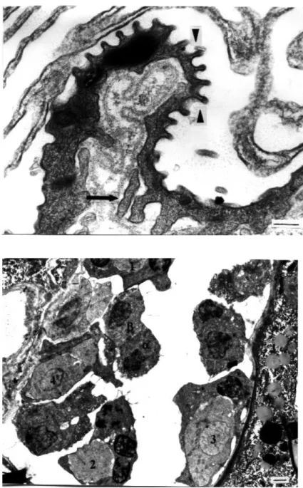
Figure 1. Two uninucleate cells at an early stage of fusion in raabeia. Arrow shows elec-tron-dense bodies and the junctions between the two cells are visible (arrow-heads). N1 and N2: nuclei, m: mito-chondria (uranyl acetate / lead citrate, x 7500) (Bar: 1 µm).
Figure 2. Uninucleate cell stage of echinactinomyx-on. There is a large nucleus (N) with a compact, eccentrically located nucleolus (n). Arrow indicates cytoplasmic projec-tions around the cell. m: mitochondria, Pg: phagosome (uranyl acetate / lead cit-rate, x 18000) (Bar: 0.5 µm).
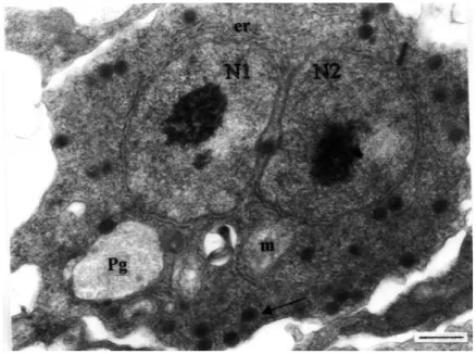
nucleolus that was located centrally. The presence of mitochondria and surface projections were common in all types of development. In addition, endoplasmic reticulum surrounded both nuclei and electron-dense bodies were scattered through the cytoplasm in echinactinomyxon (Figure 3). Electron-dense bodies were more concentrated in one half of the cytoplasm and each measured around 300 nm. The phagosomes were similar in size to those seen in the uninucleate stage.
Pansporocyst Formation
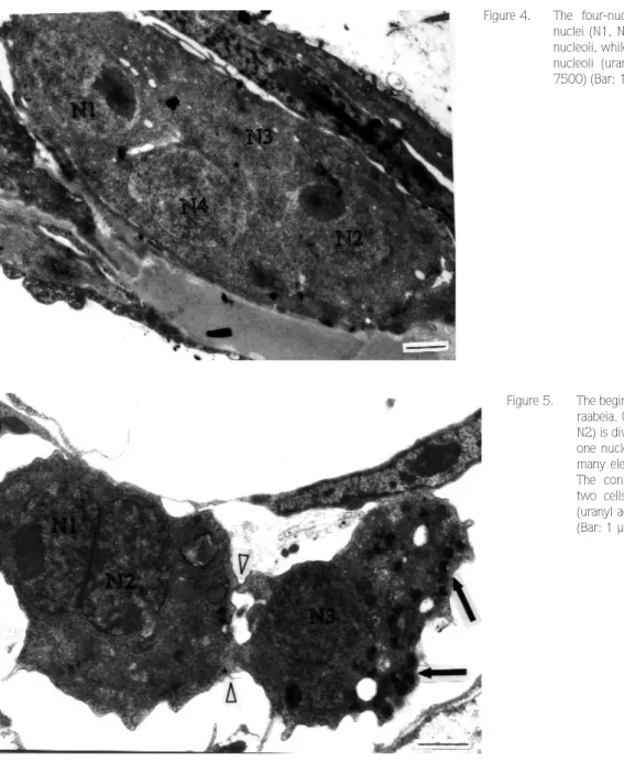
The very earliest stage of pansporocyst formation seen was a single cell containing four nuclei (Figure 4). The cell was oval and measured 11.3 µm in length and 4 µm in width. Nucleoli were only observed in two of the cells. This stage was observed only in raabeia.
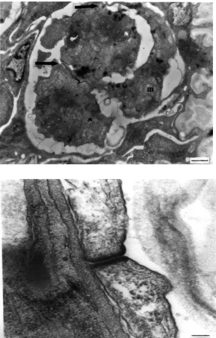
In the next developmental stage, seen only in raabeia and synactinomyxon, two cells, one with a dividing nucleus and the other with one nucleus, appeared to have junctions at the apposing sides of each cell (Figure 5). The division of nuclei in each cell in the binucleate stage was followed by the division of the cells themselves. One of the cells became flattened and surrounded the other, as seen in synactinomyxon (Figure 6). This stage of development was asynchronous and the peripheral cell remained intact while the other divided. Both cells had numerous small mitochondria. This stage was also seen to originate in raabeia as a result of plasmotomy, producing a four-cell stage developed from a four-nuclei stage. Even though only two nuclei were visible, the formation of four
cells was detected by the junctions present between each cell. Two of the four cells were larger in size and had large nuclei, one with a compact nucleolus. These two smaller cells were peripherally located (somatic cells) and contained more electron-dense bodies than the other two cells (generative cells). In some sections of raabeia, three cells connected by desmosomal junctions were also seen, possibly showing asynchronous formation of the four-cell stage. In the following stage of pansporocyst formation, two somatic cells, characterised by their electron-dense bodies, surrounded the two generative cells in echinactinomyxon, synactinomyxon, raabeia and aurantiactinomyxon. Junctions between two enveloping somatic cells were clearly seen and found to measure 13 nm (Figure 7).
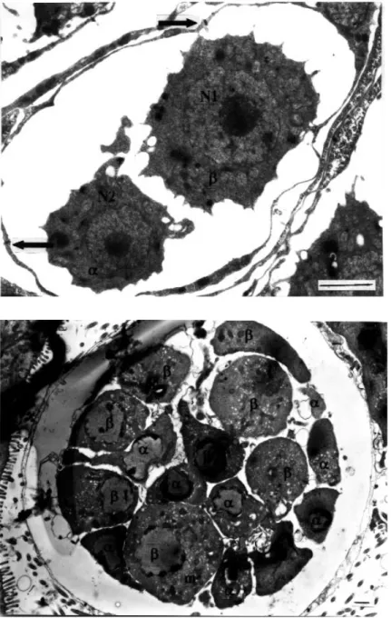
In raabeia, the somatic cells were very thin, although the more widened parts of each cell contained a nucleus. The two inner cells were different in diameter and formed a smaller α and a larger β cell (Figure 8). Both cells contained a very large nucleus with a centrally located nucleolus. The larger β cell measured 3 µm in diameter with a nucleus of 1.6 µm in diameter, while the smaller αcell measured 2.1 µm with a nucleus of 1.1 µm in diameter. Surface projections were present all around the inner cells. These surface projections from the larger
β cell seemed to be in contact with the surrounding somatic cells. Both cells contained many small mitochondria.
Echinactinomyxon and synactinomyxon showed some additional features at this stage. In echinactinomyxon, the
Figure 3. Binucleate cell stage of echinactinomyx-on. Two nuclei of similar size (N1, N2) occupy the centre of the cytoplasm. Endoplasmic reticulum (er) surrounds both nuclei. Arrow indicates electron-dense bodies. m: mitochondria, Pg: phagosome (uranyl acetate / lead cit-rate, x 18000) (Bar: 0.5 µm).
two enveloping somatic cells were to a great extent flattened and contained electron-dense bodies which were missing in the generative cells, while in aurantiactinomyxon, two nuclei were sometimes seen in one somatic cell, possibly reflecting the next division phase of the somatic cells into four.
In the next phase of development in all actinosporean types, the number of inner generative cells increased as a result of mitosis. These formed smaller α and larger β cells. The maximum complement of 8 αand 8βcells was observed only in synactinomyxon (Figure 9). As a result
of meiosis, several polar bodies (residual cells) were observed only in synactinomyxon and raabeia. They were very simple in structure and contained a large nucleus with microtubular bundles. At the same time, both somatic cells divided, to produce four enveloping cells in echinactinomyxon and eight in synactinomyxon, raabeia and aurantiactinomyxon. Desmosomal junctions between somatic cells were visible. In addition to the junctions, some surface extensions were also observed. They were most noticeable in aurantiactinomyxon (Figure 10). In raabeia, at this stage, the fusion of one αand one βcell to produce a zygote and the division of the zygotes into
Figure 5. The beginning of four-cell formation in raabeia. One cell with two nuclei (N1, N2) is dividing, and the other has only one nucleus (N3). The latter contains many electron-dense bodies (arrows). The connection points between the two cells can be seen (arrowheads) (uranyl acetate / lead citrate, x 9800) (Bar: 1 µm).
Figure 4. The four-nuclei stage of raabeia. Two nuclei (N1, N2) have eccentrically located nucleoli, while the other two (N3, N4) lack nucleoli (uranyl acetate / lead citrate, x 7500) (Bar: 1 µm).
four sporoblast cells in one pansporocyst was observed (Figure 11), indicating that these processes are asynchronous.
Sporogenesis
The process of sporoblast formation and cell differentiation was fully observed only in raabeia and aurantiactinomyxon, whilst they were partially observed in echinactinomyxon and synactinomyxon.
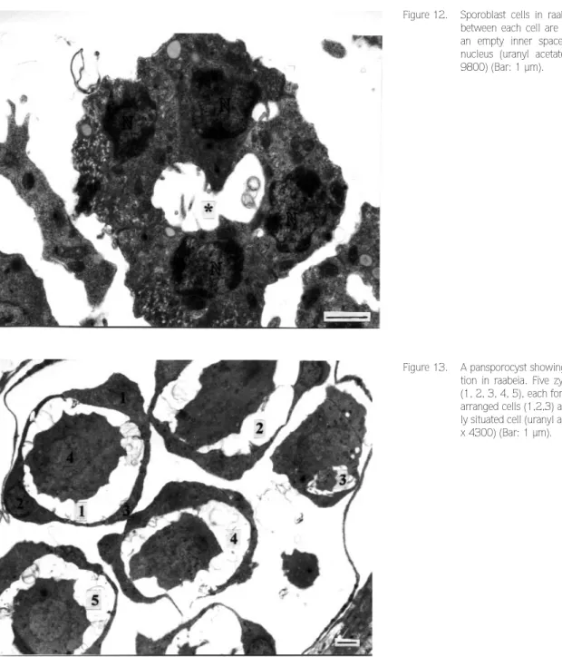
At the beginning of sporogenesis, each zygote divided twice and produced four sporoblast cells joined together by desmosomal junctions. In raabeia and
aurantiactinomyxon, the four sporoblast cells orientated in a circle with an empty space at the center (Figure 12). Junctions between each sporoblast cell became apparent. Each cell contained a remarkably large nucleus. Subsequently, three of the four sporoblast cells were arranged pyramidally surrounding the fourth centrally located cell, although their arrangement and appearance were very similar in raabeia (Figure 13) and aurantiactinomyxon. The nuclei of the surrounding cells were clear but the inner cells had large nucleoli of 2.3 µm in diameter. Subsequently, the three outer cells divided mitotically and produced six cells surrounding the
Figure 7. A desmosomal junction between two enveloping somatic cells in echinactinomyx-on. H: host cell (uranyl acetate / lead citrate, x 75000) (Bar: 0.1 µm).
Figure 6. Early stage of pansporocyst formation in synactinomyxon. One enveloping somatic and two inner generative cells can be seen. Arrows show the junctions between the cells. m: mitochondria (uranyl acetate / lead citrate, x7500) (Bar: 1 µm).
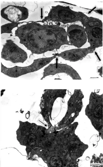
sporoplasm-originating cell, which was centrally located in raabeia development (Figure 14). This stage was asynchronous. At the same time, in both raabeia and aurantiactinomyxon, sporoplasm cells divided several times but the originating cell was easily differentiated from the daughter cells by its larger size. The originating cell had a centrally located very large nucleus. The cytoplasm of each sporoblast cell contained many mitochondria. Cell junctions were visible and each cell became thinner at the point of junction. Three of the six peripheral sporoblast cells remained round to oval in shape, whilst the other three were more elongated and
flattened. Possibly the first three cells were developing capsulogenic cells, while the other three were valvogenic cells (Figure 14).
The position of the capsulogenic and valvogenic cells started to become apparent as early as the seven-cell stage of sporoblast formation, and in more advanced stages the three capsulogonic cells were surrounded by more elongated valvogenic cells, as seen in raabeia (Figure 14). Both capsulogenic and valvogenic cells moved forward and left the sporoplasm at the posterior (Figure 15). The capsulogenic cell finally became
Figure 9. An advanced pansporocyst with 8αand 8β cells before zygote formation. Sur-rounding somatic cells are very thin. All inner generative cells contain many mito-chondria (m) (uranyl acetate / lead citrate, x3600) (Bar: 1 µm).
Figure 8. Early stage of pansporocyst formation in raabeia showing two somatic enveloping cells (α,β) and two generative enveloped cells. Junctions between the two somatic cells are visible (arrows). N1-N2: nuclei (uranyl acetate / lead citrate, x 7500) (Bar: 1 µm).
positioned ahead of the valvogenic cells. The junctions between valvogenic cells were clearly visible and these later surrounded the sporoplasm, which contained many nuclei. As the development progressed, the sporoplasm appeared to enter the space at the centre of the valvogenic and capsulogenic cells. The three capsulogenic cells became located in the anterior part of the developing spore body, and capsulogenic cells showed tubular formation in aurantiactinomyxon, presumably representing the very early stage of the polar capsule primordium.
In the early stage of polar capsule formation, the primordium was filled with an electron-dense material in all actinosporean types (Figure 16). The external tube first appeared as a circle in a transverse section in aurantiactinomyxon. At a more advanced stage, the cylindrical external tube was filled with an electron-dense granular substance which was also found in the capsular primordia in synactinomyxon.
As the polar capsule formation occurred, the external tube started to invert into the capsular primordium, and
Figure 11. An advanced stage of pansporocyst forma-tion in raabeia. While some of the cells are in the zygote formation stage (α,β), others are in early sporoblast formation (1, 2, 3) with several nuclei at the early sporoblast formation (uranyl acetate / lead citrate, x 4300) (Bar: 1 µm).
Figure 10. A part of an enveloping cell with surface projections on the inside (arrow) and out-side (arrowheads) of the cell in aurantiactin-omyxon (uranyl acetate / lead citrate, x 36000) (Bar: 0.2 µm).
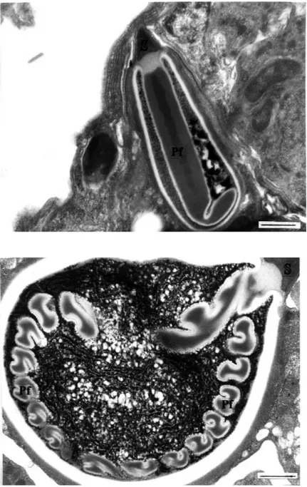
in the same sections some tubule formation was also observed at the distal part of the external tube in raabeia (Figure 17). Following the invagination of the external tube into the capsular promordia, helically coiled S-shaped filament windings were observed. There were 6, 9 and 6 coils in synactinomyxon, raabeia and aurantiactinomyxon, respectively. Polar filament windings with fibrils outside each fold were only observed in raabeia. However, in echinactinomyxon-type development, elongated polar filaments with a curve at
the distal end rather than filament coils were formed (Figure 18). Subsequently, a densely stained plug was produced at the apex of each polar capsule in all types of development. An electron-lucent layer made a turn underneath the stopper and formed the outer layer of the polar filament winding. There was an electron-lucent space between the stopper and the first polar filament winding in all types in raabeia (Figure 19). The stopper was much denser than the polar filament and was covered by the electron-dense layer of the polar capsule. The walls
Figure 13. A pansporocyst showing sporoblast forma-tion in raabeia. Five zygotes have divided (1, 2, 3, 4, 5), each forming 3 pyramidally arranged cells (1,2,3) and a fourth central-ly situated cell (uranyl acetate / lead citrate, x 4300) (Bar: 1 µm).
Figure 12. Sporoblast cells in raabeia. The junctions between each cell are clearly formed and an empty inner space (*) is visible. N: nucleus (uranyl acetate / lead citrate, x 9800) (Bar: 1 µm).
of the polar capsule consisted of an outer dense (200 nm) and inner lucent (180 nm) layer in all types. In all actinosporean types, each capsulogenic cell had a large nucleus located close to the polar capsules, and also contained many mitochondria of various sizes, which persisted until the complete formation of capsulogenic cells. When the polar capsule formation was almost complete, septate junctions between them became more apparent.
Malformations of the polar capsule formations were observed in raabeia and aurantiactinomyxon. Polar
capsules at the same developmental stage were sometimes not regular and round as normally observed, but nonetheless maintained normal cell junctions, as seen in raabeia (Figure 20). In other cases, the polar filament was accumulated at the apex of the polar capsule, and the electron-lucent and electron-dense layers which surrounded the entire polar capsule were not fully formed.
Valve development in all actinosporean types was completed by the valvogenic cells enclosing the three polar capsules and the sporoplasm by their overlapping
Figure 15. Sporoblast cell differentiation in raabeia. Two valvogenic cells (Vc) extend over the capsulogenic cells (Cc). Arrow indicates some of the sporoplasm which is enveloped by valvogenic cells while the rest remains uncovered. Gc: germ cells, N: nucleus (uranyl acetate / lead citrate, x 7500) (Bar: 1 µm).
Figure 14. The junctions between each dividing cell are visible (arrows) in raabeia. All cells con-tain many mitochondria (m). Note the large size of the nucleus in the sporoplasm orig-inating sporoblast cell (N). While the outer three capsulogenic cells (1-3) contain dense nuclei (N1, N2, N3), the three valvo-genic cells (4-6) lack nuclei in the plane of the section (uranyl acetate / lead citrate, x 7500) (Bar: 1 µm).
each other with septate junctions with a space of 8 nm. From the posterior of the maturing spores, the valvogenic cells extended into a mass of folded membranes. In the early stage of development, these projections were composed of two membranes, a large nucleus and some cytoplasm, which later disappeared in all actinosporean-type development as the projections became thinner. The shape of the caudal processes varied in different types of spore, e.g., elongated in aurantiactinomyxon (Figure 21), and rounded in raabeia. However, in synactinomyxon the valvogenic cells
extended into a mass of folded membranes which corresponded to the short caudal processes linking eight spores together.
The sporoplasm offered a rich variety of features in all types of actinospore development. Sporoplasmic plasmodia had numerous, mostly sub-spherical, nuclei with small eccentrically located nucleoli in synactinomyxon. In more advanced stages, sporoplasms had many elongated mitochondria and several nuclei surrounded by endoplasmic reticulum (Figure 22). The number of the germ cells and sporoplasmosomes
Figure 17. A transverse section of the external tube showing some microtubule forma-tion at the distal part (arrows) in raabeia (uranyl acetate / lead citrate, x 130000) (Bar: 0.1 µm).
Figure 16. An advanced capsular primordium in raabeia. A very electron-dense granulated substance occupies the inside of the pri-mordium and a longitudinal section through the external tube (Et) is also visible (uranyl acetate / lead citrate, x 22000) (Bar: 0.5 µm).
increased as the development progressed; they were 750 nm and 110 nm in diameter, respectively in echinactinomyxon. In the pansporocycts of all actinosporean types, near-mature spores were located at the centre, while caudal processes surrounded them, as observed in synactinomyxon (Figure 23).
Discussion
In the first phase, the earliest stage found in this study, all the individual types examined from the four
collective groups, echinactinomyxon, synactinomyxon, raabeia and aurantiactinomyxon, had uninucleate cells distributed intercellularly in the gut epithelium of Tubifex tubifex and Lumbriculus variegatus. This finding is in agreement with those of Lom and Dykova (9) in Triactinomyxon legeri and El-Matbouli and Hoffmann (2) in the triactinomyxon stage of Myxobolus cerebralis, although it differs from those of Ikeda (8) in tetractinomyxon, Ormieres (11) and Marques (14) in Neoactinomyxum eiseniellae, and Lom et al. (7) in aurantiactinomyxon and raabeia, where the earliest
Figure 18. A longitudinal section through a mature polar capsule in echinactinomyxon. A stop-per (S) is located at the apex of the polar capsule. The polar capsule has an elongat-ed polar filament (Pf) rather than filament coils (uranyl acetate / lead citrate, x 22000) (Bar: 0.5 µm).
Figure 19. A longitudinal section of a mature polar capsule in raabeia. Apart from the polar fil-ament windings, and electron-dense and lucent layers, a stopper (S) is located at the apex of the polar capsule (uranyl acetate / lead citrate, x 22000) (Bar: 0.5 µm).
stages found were binucleate cells. Binucleate cells were, however, very coµmonly observed as the second stage of development in the four actinosporean types in this study. The division of a nucleus in a uninucleate cell or the fusion of two uninucleate cells to form a binucleate cell was observed to be the transition stage between the uninucleate and binucleate cells. The formation of uninucleate and binucleate cells in the triactinomyxon stage of M. cerebralis was demonstrated by El-Matbouli and Hoffmann (2). According to their findings, after
epithelial penetration of the binucleate sporoplasm of M. cerebralis in the gut of T. tubifex, both diploid nuclei undergo multiple division to produce multinucleate cells. Plasmotomy of the multinucleate stage produces numerous uninucleate cells located intercellularly through the gut epithelia of T. tubifex. Some of these uninucleate cells undergo further multiple nuclear and cellular division (schizogony), while the others undergo plasmogamy to produce binucleate stages. In the present study, plasmogamy was observed only in raabeia.
Figure 20. A longitudinal section through a mal-formed polar filament in aurantiactino-myxon. Although there are several fila-ment windings, there is no polar cap-sule wall. N: nucleus, m: mitochondria (uranyl acetate / lead citrate, x 18000) (Bar: 0.5 µm).
Figure 21. A transverse section through a telescop-ically folded caudal process in auranti-actinomyxon (uranyl acetate / lead cit-rate, x 9800) (Bar: 1 µm).
The earliest uni- and binucleate cells generally occur intercellularly in triactinomyxon (2,9), in raabeia (7) and in the four actinosporean types in this study. Intracellular development has only been observed in Aurantiactinomyxon sp1. by Yokoyama et al., 1993 (7), but it did not occur in the aurantiactinomyxon type in the present study.
The two nuclei in the binucleate-cell stage were generally closely apposed to each other, but there was a gap between the two nuclear envelopes of the neighbouring nuclei of the four actinosporean types in the
present study and also in triactinomyxon (2,9), aurantiactinomyxon and raabeia (7).
However, both the uni- and binucleate stages exhibited similar features, such as numerous mitochondria, surface projections and phagosomes. It was proposed by Lom and Dykova (9) that phagosomes represent feeding by the invagination of folds of the plasmalemma and that they are evidence of a high metabolic rate within the cell. In addition to the phagosomes, surface projections around the uni- and binucleate cells also play a role in the food flux into the
Figure 22. Sporoplasm with several germ cells (Gc) and sporoplasmosomes (arrows) in echinactinomyxon (uranyl acetate / lead citrate, x 22000) (Bar: 0.5 µm).
Figure 23. A developed pansporocyst of synactino-myxon with three spores in different stages of development (uranyl acetate / lead citrate, x 2800) (Bar: 1 µm).
cell cytoplasm. The size of the phagosomes seemed to be different in all four actinosporean types studied here, and they were located at the periphery of the uni- and binucleate stages of the echinactinomyxon type examined. However, mitochondria did not exhibit any size difference between the earliest uni- and binucleate stages of the four actinosporean types examined, as was also found in the triactinomyxon stage of Myxobolus cerebralis (2). In the four actinosporean types in the present study, mitochondria had flat cristae in both stages. Rough endoplasmic reticulum was observed to surround the nuclei, as also described by El-Matbouli and Hoffmann (2), Lom et al. (7) and Lom and Dykova (9). Golgi bodies were observed only occasionally in the earliest stages of raabeia, as also found by El-Matbouli and Hoffmann (2) and Lom and Dykova (9). Electron-dense bodies were also observed in both the uninucleate and binucleate stages of the four actinosporean types in the present study. They were considered to be residual constituents of myxosporean-phase sporoplasm (7).
According to Lom and Dykova (9), the observation of simultaneous uni- and binucleate early stages as an advanced sporogony phase indicates the presence of dormant cells with inhibited development. These occur for more than 1 year in the triactinomyxon stage of M. cerebralis (2). Inhibited development might be important in determining the seasonal nature of spore formation and release seen in many actinosporeans. It was suggested by Leger (18) that autoinfections of oligochaetes may occur, and this would also explain the occurrence of early and late stages of development in the same worm, but there is no definite evidence that this process does actually occur.
In the development of actinosporeans, the next recognisable phase was the formation of the pansporocyst. However, the origin of the early pansporocyst with four cells is still disputed (2,3,9,19). In the present study, the early pansporocysts of echinactinomyxon, synactinomyxon, raabeia and aurantiactinomyxon were of similar appearance, with two enveloping somatic cells and two enveloped generative cells. This arrangement has been observed as a common feature of all actinosporeans studied to date (2,3,5,9,7,19,20).
The four-cell stage observed only in raabeia followed a similar developmental cycle to that seen in the triactinomyxon stage of M. cerebralis by El-Matbouli and
Hoffmann (2), even though the four-cell stage was not observed in the other types of actinosporean. Presumably, synactinomyxon-type pansporocyst formation follows a similar pattern to that of raabeia.
Earlier stages of pansporocyst formation were not observed in echinactinomyxon or aurantiactinomyxon. This has also been the case for most of the actinosporeans studied using TEM, possibly due to the fact that as a rule only advanced infections have been subjected to examination and that in this case the chance of finding true proliferative stages and their transformation into a pansporocyst is low (7).
The number of enveloping cells increased as a result of mitosis, becoming four in echinactinomyxon, as
observed in N. eiseniellae (5), and eight in
synactinomyxon, raabeia and aurantiactinomyxon, as seen in the triactinomyxon stage of M. cerebralis (2). The outer somatic cells had surface projections generally on the inner envelope in the echinactinomyxon, raabeia and synactinomyxon types, whilst in aurantiactinomyxon there were also projections on the outer surface. These projections seem a common morphological feature with minor differences in all types of actinosporean development (7,14), and they possibly have a role in mediating the flux of nutrients for the developing cells (7).
In all actinosporean types the encircling somatic cells were joined by desmosomal junctions at both ends. However, in the raabeia and aurantiactinomyxon types of Lom et al. (1997), an intercalated dense material, which was not observed in this study, was present at the cell junctions.
The encircled cells seen in raabeia had similar surface projections and lay in an amorphous matrix throughout the whole generative process until sporogenesis was completed, as was also observed in the triactinomyxon stage of M. cerebralis (2). However, in Triactinomyxon legeri, Lom and Dykova (9) did not observe such a matrix and there was no empty space between the inner and outer somatic cells.
Apart from the first mitotic division, yielding 2α and 2βcells, the second and third mitotic divisions were not observed fully in this study. Subsequent divisions were indicated by the presence of up to 16 cells in synactinomyxon.
At the end of gametogamy, eight zygotes occurred inside each pansporocyst surrounded by four or eight somatic cells according to the actinosporean type. This stage was seen in raabeia and aurantiactinomyxon in this study, and was seen in other actinosporean types studied ultrastructurally (7). The apparent absence of this stage in other actinosporean types in this study in all probability merely reflects the particular stage of development of the parasite in the worms examined.
In this study, it was found that there was no way of detecting infected oligochaetes without the release of mature spores. However, it was earlier noted by several authors (7,21,22,23) that infected worms often show discoloration, opaque areas and/or a degree of swelling, making it possible to identify infected worms within a population. In contrast, it was reported by Yokoyama et al. (24) that there were no such diagnostic signs in their actinosporean-infected worms, as was also the case in this study. El-Matbouli and Hoffmann (2) screened experimentally infected worms chronologically and, in this way, were able to observe all the stages of development of the triactinomyxon stage of M. cerebralis. In the present study, the asynchronous nature of development in the infected worms enabled the observation of several stages, but not all of them. Most stages of gametogenesis and sporogenesis were found in raabeia and aurantiactinomyxon in this study, in accordance with those shown by the hand drawings of Janiszewska (3) and the ultrastructural observations of El-Matbouli and Hoffmann (2).
Following the formation of the four-cell stage, the three outer cells each divide into one capsulogenic and one valvogenic cell and the fourth central cell also divides endogenously. In the next stage, the central cell divides mitotically several times and forms many germ cells, the number of which is characteristic for each actinosporean type (3). In the late sporoblast stage, two complexes, one comprising capsulogenic and valvogenic cells, and the second an endospore with sporoplasm germ cells, developed separately, as described for R. gorlicensis (3) and the triactiactinomyxon stage of M. cerebralis (2). When the outer valvogenic cells and capsulogenic cells reached an advanced state, the sporoplasm with its final number of germ cells (2), or with over 20 nuclei (25), started to penetrate into the spore cavity by an amoeboid movement of the sporoplasm, by a dehiscence of the envelope formed by the valvogenic cells, or by a combination of these factors, as observed by Janiszewska
(3) in Siedleckiella silesica. However, this forward movement of the sporoplasm seems common in actinosporeans (2,16,25).
The capsulogenic cells of all the actinosporean types studied here were easily distinguished at an advanced stage of sporogenesis, when they were situated at the apex of the spore and had a similar arrangement to that described for other actinosporeans (2,7). The first stage of polar capsule development observed in this study was a capsular primordium with a dense inner and lucent outer layer. However, an extremely dense club-shaped structure was observed as the origin of the primordium in T. legeri (9). This structure has not been described in other actinosporeans, including the types examined here. However, the unusual features in polar capsule formation observed in the raabeia and aurantiactinomyxon types studied here were also seen by Marques (5). The polar capsule formation in the four actinosporeans in the present study was similar to that described for the triactinomyxon stage of M. cerebralis by El-Matbouli and Hoffmann (2). The external tube of the raabeia type described here exhibited several microtubules surrounding the dense layer, which is identical to that in Neoactinomyxum eiseniellae (5). However, similar microtubules were also observed by Lom et al. (7) in the raabeia stage of Myxobolus cultus on the outer and inner surfaces. A relationship between the microtubules surrounding the external tube and the fibres running around the developing polar filament was suggested by Lom and Dykova (6).
In the mature polar capsule, two outer layers, one electron lucent and one electron dense, and a stopper at the apex have been common features in all actinosporeans studied to date (2,5,7). An empty or lucent space around the posterior part of the polar capsule was observed to be common in the raabeia and aurantiactinomyxon types in the present study. A similar feature was also observed in T. legeri (9), and the "empty space" was attributed to an artefact. However, it is now a common feature described in other actinosporeans such as aurantiactinomyxon and raabeia (7) and neoactinomyxum (5), suggesting that it is not an artefact but that the function of it is not known. Malformations in the polar capsule of the raabeia stage of M. cultus were described by Lom et al. (7) as being similar in appearance to those observed in raabeia- and aurantiactinomyxon-type actinosporean polar capsules in this study.
The numbers of coils of polar filaments are a species or type characteristic of actinosporeans. In echinactinomyxon there were no polar filament folds. The polar filament was observed to be straight and curved upwards at the base of the polar capsule, which may be a unique feature of this actinosporean type. Light-microscopic observations of the polar filament of mature spores released into the water showed that the length of the polar filament was not longer than the polar capsule itself. However, in other actinosporeans studied to date, the polar filaments have always been 2-3 times longer than the polar capsules. Another unique feature in the polar capsule formation of aurantiactinomyxon was the presence of virus-like particles in the polar capsule (7). Such particles were not seen in the present study.
Capsulogenic cells preserved their cytoplasmic constituents during development, including cisternae of RER, mitochondria, golgi and large nuclei. Lipid droplets and aggregates of glycogen granules were also reported in aurantiactinomyxon by Lom et al. (7). The cell junctions seen between both capsulogenic cells and those between capsulogenic and valvogenic cells in the actinosporean
types studied here and in T. legeri (9),
aurantiactinomyxon and raabeia (7), neoactinomyxum (5), and in the triactinomyxon stage of M. cerebralis (2) seem to be similar in arrangement. The final arrangement of the valvogenic cells in relation to the other cells of the
spores observed in this study was similar to that described by (2,5,9,7,26).
The sporoplasm was a multinucleate, simple, membrane-bound plasmodium (mother cell) with many inner (secondary) cells representing the actual infectious germs. Several sporoplasm cells were formed as a result of mitotic divisions and, thus, differed from endogenous cell division-forming germ cells observed by El-Matbouli and Hoffmann (2) and Lom et al. (7). The subsequent developmental stages were identical to those observed by the authors mentioned above for raabeia and the triactinomyxon stage of M. cerebralis, respectively. Many mitochondria, phagosomes and endoplasmic reticulum were major constituents of the sporoplasm in the four types of actinosporeans in the present study, as they were in T. legeri, Aurantiactinomyxon sp1., studied by Yokoyama et al. (1993), the raabeia stage of M. cultus, and the triactiactinomyxon stage of M. cerebralis. The close association of sporoplasm nuclei and germ cells seen in the aurantiactinomyxon type here was also described in the Aurantiactinomyxon sp1. of Lom et al. (1997) in addition to N. eiseniellae (5). The appearance of the sporoplasmosomes and their densely stained round-to-elongated shapes has been common in all actinosporeans studied to date, though their sizes have differed. The dense bodies seen in the early germ cell cytoplasm of T. legeri by Lom and Dykova (9) were not found in this study.
References
1. Wolf, K. & Markiw, M. E., Biology contravenes taxonomy in the Myxozoa: new discoveries show alternation of invertebrate and vertebrate hosts. Science 225: 1449-1459, 1984.
2. El-Matbouli, M. & Hoffmann, R. W., Light and electron microscopic studies on the chronological development of Myxobolus cerebralis to the actinosporean stage in Tubifex tubifex. Int. J. for Parasitol. 28: 195-217, 1998.
3. Janiszewska, J., Actinomyxidia I. Morphology, ecology, history of investigations, systematics, development. Acta Protozool. Polonica 2: 405-443, 1955.
4. Bartholomew, J. L., Whipple, M. J., Stevens, D. G. & Fryer, J. L., The life cycle of Ceratomyxa shasta, a myxosporean parasite of salmonids requires a freshwater polychaete as an alternate host. J. Parasitol. 83: 859-868, 1997.
5. Marques, A., Contribution a la connaissence des Actinomyxidies: ultrastructure, cycle biologique, systematique. PhD Thesis: Universite des Sciences et Techniques de Languedoc, Monpellier, France, 218 pp. 1984.
6. Lom. J. & Dykova, I., Ultrastructural features of the actinosporean phase of myxosporean (Phylum: Myxozoa): a comparative study. Acta Protozool. 36: 83-103, 1997.
7. Lom, J., Yokoyama, H. & Dykova, I., Comparative ultrastructure of Aurantiactinomyxon and Raabeia actinosporean stages of myxozoan life cycles. Arch. für Protistenkd. 148: 173-189, 1997. 8. Ikeda, J., Studies on some sporozoan parasites of sipunculids. I. The life history of a new actinomyxidian, Tetractinomyxon intermedium g. et sp. nov. Arch. für Protistenkd. 25: 240-242, 1912.
9. Lom, J. & Dykova, I., Fine structure of Triactinomyxon early stages and sporogony: myxosporean and actinosporean features compared. J. Protozool. 39: 16-27, 1992.
10. de Puytorac, P., L’ultrastructure des cnidocystes de L’ Actinomyxidie: Sphaeractinomyxon amanieui sp. nov. C. R. Acad Sci. Paris 256: 1594-1596, 1963.
11. Ormieres, R., Formation des capsules polaires dans la spore de L’ Actinomyxidie Aurantiactinomyxon eiseniellae Orm. Fre. (Etude Ultrastructurale). C.R. Acad Sci. Paris 271: 2326-2328, 1970.
12. Marques, A., Complexes synaptonemaux et stades initiaux chez les Actinomyxidies. C. R. Acad Sci. Paris 295: 501-504, 1982. 13. Marques, A., La sexualite chez les Actinomyxidies: Etude chez
Neoactinomyxon eiseniellae (Ormierez et Frezil, 1969), Actinosporea, Noble, 1980; Myxozoa Grasse, 1970. Annl Sci. Net., Zool., Paris 13e ser; 8: 81-101, 1986.
14. Marques, A., La fecondation et les premieres stades de la sporogenese d’une Actinomyxidie. C. R. Acad Sci. Paris 296: 717-720, 1993.
15. El-Matbouli, M., Hoffmann, R.W. & Mandok, C., Light and electron microscopic observations on the route of the triactinomyxon – sporoplasm of Myxobolus cerebralis from epidermis into rainbow trout cartilage. J. Fish Biol. 46: 919-935, 1995.
16. Trouilier, A., El-Matbouli, M. & Hoffmann, R. W., A new look at the life cycle of Hoferellus carassii in the gold fish (Carassius auratus auratus) and its relation to "Kidney Enlargement Disease". Folia Parasitol. 43: 173-187, 1996.
17. Özer, A and Wootten, R., Actinosporean myxozoan spores belonging to the collective groups synactinomyxon, aurantiactinomyxon, triactinomyxon, neoactinomyxum and siedleckiella from a freshwater salmon farm in Northern Scotland. Folia Parasitol. (accepted for publication).
18. Leger, L., Consideration sur le genre Triactinomyxon et les Actinomyxidies C. R. Soc. Biol. Paris 56: 846-848, 1904. 19. Granata, L., Gli Attinomissidi. Morphologia – sviluppa –
sistematica. Arch. für Protistenkd. 50: 139-212, 1924.
20. Caullery, M. & Mesnil, F., Recherches sur les Actinomyxidies. Arch. für Protistenkd. 6: 272-308, 1905.
21. Wolf, K., Markiw, M. E. & Hiltunen, J., Salmonid whirling disease: Tubifex tubifex (Müller) identified as the essential oligochaete in the protozoan life cycle. J. Fish Dis. 9: 499-501, 1986.
22. El-Matbouli, M., Fischer-Scherl, T. & Hoffmann, R. W., Transmission of Hoferellus carassii Achmerov, 1960 to goldfish Carassius auratus via an aquatic oligochaete. Bull. Eur. Ass. Fish Pathol. 12: 54-56, 1992.
23. El-Matbouli, M. & Hoffmann, R.W., Myxobolus carassii Klakoceva, 1914 also requires an aquatic oligochaete, Tubifex tubifex as an intermediate host in its life cycle. Bull. Eur. Ass. Fish Pathol. 13: 189-192, 1993.
24. Yokoyama, H., Ogawa, K. & Wakabayashi, H., A new collection method of actinosporeans – a probable infective stage of myxosporeans to fishes – from tubificids and experimental infection of goldfish with actinosporean Raabeia sp. Gyobyo Kenkyu 26: 133-138, 1991.
25. Janiszewska, J., Actinomyxidia II. New systematics, sexual cycle, description of new genera and species. Zoologica Polonica 8: 3-34, 1957.
26. Yokoyama, H., Ogama, K & Wakabayashi, H., Involvement of Branchiura sowerbyi (Oligochaete: Annelida) in the transmission of Hoferellus carassii (Myxosporea: Myxozoa) the causative agent of kidney enlargement disease (KED) of goldfish Carassius auratus. Fish Pathol. 28: 135-139, 1993.