Ankara Üniv Vet Fak Derg, 59, 71-73, 2012
Short Communication / Kısa Bilimsel Çalışma
Insulinoma with regional lymph node metastasis in a dog
*Mehmet Önder KARAYİĞİT1, Yonca B. KABAK1, Murat YARIM1, Sinan ŞİRİN2,
M.Yavuz GÜLBAHAR1
1 Ondokuz Mayıs Üniversitesi, Veteriner Fakültesi, Patoloji Anabilim Dalı, Samsun; 2 Mehmet Akif Ersoy Üniversitesi, Veteriner
Fakültesi Cerrahi Anabilim Dalı, Burdur.
Summary: In this case, a female boxer dog, 6-year-old, with tonic-clonic convulsions and hind-limb paresis was evaluated. The serum insulin and serum glucose levels were 58 µU/ml and 5 mg/dl, respectively. In spite of all care, the comatose dog died. At necropsy, one well-circumscribed white-grayish mass, 4x3x2 cm, was found at the caudal edge of right lobe of the pancreas. Subcortical malacic areas in reddish color was seen in the brain. Microscopically, the tumoral mass consisted of neoplastic cells with round shape, basophilic nucleous and granular eosinophilic cytoplasm. In addition to these findings, metastasis to the regional lymph node of the pancreas was observed. Immunohistochemically, the tumor cells from both primary and metastatic tissues showed immunoreactivity to monoclonal mouse anti-insulin antibody. Clinically, histologically and immunohistochemically, functional insulinoma with lymph node metastasis was diagnosed in the present case.
Key words: Dog, insulinoma, pancreas, tumor.
Bir köpekte lenf yumrusu metastazlı insülinoma
Özet: Bu olguda tonik-klonik konvulsiyonlar ve arka bacaklarda parezisi olan 6 yaşlı boxer ırkı dişi bir köpek değerlendirildi. Serum insülin ve serum glukoz seviyeleri sırasıyla 58 µU/ml ve 5 mg/dl olarak bulundu. Yapılan tüm müdahalelere rağmen koma halindeki köpek öldü. Nekropside pankreasın sağ lobunun arka ucunda 4x3x2 cm ebadında beyazımsı-gri renkte iyi sınırlı bir kitle görüldü. Beyinde kırmızımsı renkte subkortikal malasik alanlar dikkati çekti. Mikroskobik olarak tümör hücreleri yuvarlak şekilli, bazofilik çekirdekli ve granüler yapıda eozinofilik sitoplazmadan oluşmaktaydı. Bu bulgulara ek olarak pankreasın bölgesel lenf nodunda metastaza rastlandı. İmmunohistokimyasal olarak hem primer hem de metastatik tümör dokusu hücreleri monoklonal fare insüline karşı elde edilen antikor ile pozitif reaksiyon verdi. Sunulan olgu klinik, histolojik ve immunohistokimyasal olarak lenf yumrusu metastazlı fonksiyonel bir insülinoma olarak tanımlandı.
Anahtar sözcükler: Köpek, insülinoma, pankreas, tümör.
* This paper was presented as a poster in IV. National Congress of Veterinary Pathology, 29 October- 02 November 2008, Antalya, Turkey.
Insulinoma is a tumor of pancreatic beta cells. This tumors are uncommon in the dog. It is usually malignant. It is most often diagnosed in middle age or older dog (3,4). The aim of this case is to evaluate diagnostic implications for this important neoplastic condition in dogs. A case of insulinoma has not been reported in veterinary literature in Turkey. This case has been presented by using clinical, histopathological and immunohistochemical features of canine insulinomas.
Female boxer dog, 6-year-old, was firstly submitted with a history of repeated episodes of tonic-clonic convulsions and hind-limb paresis to University of Ondokuz Mayis, Faculty of Veterinary Medicine. After three days, the dog was in a coma, and blood glucose and insulin concentrations were 5 mg/dl (normal range, 61.9-108.3 mg/dl) and 58 µU/ml (normal range, 5-20 µU/ml), respectively. The radiography did not revealed any
abnormality. In spite of all care, the comatose dog died.
Necropsy was performed, tissue samples were fixed in
phosphate-buffered %10 formalin solution, dehydrated in ethanol and embedded paraffin wax. Tissue sections (5 µm thick) were cut for histopathological and immunohistochemical examinations. The prepared sections were stained with haematoxylin-eosin (HE) and were processed for immunohistochemical investigation with monoclonal mouse anti insulin antibody (1/200 diluent, Thermo scientific, Fremont, USA) by using standard streptavidin-biotin peroxidase complex method (SBPC) with a commercial kit (Zymed, USA). The reaction product was visualized by aminoethylcarbazole (AEC) chromogen (Zymed, USA) and counterstained with Gill' haematoxylin. Normal pancreatic tissues of the dog served as positive control.
Mehmet Önder Karayiğit - Yonca B. Kabak - Murat Yarım - Sinan Şirin - M. Yavuz Gülbahar 72
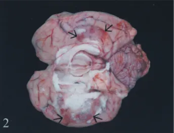
In necropsy, one well circumscribed white-grayish mass, 4x3x2 cm, was found at the caudal edge of right lobe of the pancreas (Figure 1). Regional lymph node of pancreas were bigger than normal, but macroscopically, there were no lesions. Subcortical malacic areas in reddish color was seen especially in occipital and parietal lobes of the brain. (Figure 2). No gross lesion was seen in other organs.
Figure. 1. A white to gray, well-demarcated tumor was in the right lobe of the pancreas (arrow).
Şekil 1. Pankreasın sağ lobunda beyaz–gri renkli, keskin sınırlı tümör dokusu (ok).
Figure. 2. Subcortical malacic areas showing hemorrhage and softening in the brain (arrows).
Şekil 2. Beyinde yumuşama ve hemoraji görülen subkortikal malasik alanlar (oklar).
Microscopically, the tumoral mass consisted of neoplastic cells with round shape and basophilic nucleous, granular eosinophilic cytoplasm arrenged in solid pattern separated by fibrous septa, the cytoplasm of some tumor cells contained large vacuoles. Nuclear atypia was marked, and mitotic figures were infrequent (Figure 3). Ductal or acinar components were not seen within the tumor tissue. Metastasis to the regional lymph node of pancreas was observed, and metastatic tumor
cells was similar to primary tumor cells (Figure 4). Other organ metastases were not seen. Malacia and hemaorrhage were detected in the affected regions of the brain. The liver examination revealed vacuoler to hydropic changes in the hepatocytes. Immmunohistochemically, the tumor cells from both primary and metastatic tissues showed immunoreactivity for insulin antibody. Immunostaining for insulin was diffusely cytoplasmic of the neoplastic cells within in the tumor mass. Insulin immunoreactivity was also seen in some islet’ cells inside the normal pancreatic tissue, but this immunoreactivity was more intense than those in the tumor cells (Figure 5). A functional insulinoma of pancreatic islet with regional lymph node metastasis was diagnosed according to clinical, pathological and immunohistochemical findings.
Figure. 3. The microscopic appearance of the tumor (T) separated by well-defined fibrous capsule from ther normal pancreatic tissue (N), HE, x200. Inset shows a mitotic figure among the tumor cells with vacoulated and granular cytoplasm. HE X800. Şekil 3. Normal pankreatik dokudan (N) belirgin fibröz kapsül ile ayrılan tümörün mikroskobik görüntüsü (T). HE, x200. Ekli küçük resim, vakuollü ve granüler sitoplazmalı tümör hücreleri arasındaki mitotik figürü göstermektedir. HE x800.
Figure. 4. Tumor metastasis (arrows) in regional lymph node, HE, x200.
Şekil 4. Bölgesel lenf düğümündeki tümör metastazı (oklar), HE, x200.
Ankara Üniv Vet Fak Derg, 59, 2012 73
Figure. 5. Immunolabelling for insulin in the normal pancreatic islet cells (arrows) and tumor cells. T: Tumoral tissue, N: Normal pancreatic tissue, SABP-DAB, x400.
Şekil 5. İnsülinpozitif normal pankreatik ada hücreleri (oklar) ve tümör hücreleri, T: Tümör dokusu, N: Normal pankreas dokusu, SABP-DAB, x400.
Insulinomas have been reported in various dog breeds without sex predisposition. Some large breed dogs, such as Collies, Irish Setters, boxers and German shepherds are most often affected (3,4,7). Insulinomas usually metastase to lymph nodes, liver, mesentery and omentum (1,3,5). The dog was boxer breed and the metastasis was observed only to the regional lymph node of the pancreas. Insulinomas are usually 1-1,5 cm in
diameter (1,2,4),but in this case, size of the tumor was
4x3x2 in diamater. This condition may relate to time elapsed between onset of symptoms and animal's death.
Because of low blood glucose levels in animals with insulinoma, dysfunction may occur in many system. Many clinical signs such as mental dullness, collapse, tremors, hind-limb weakness, ataxia, depression, and seizures have been seen. In the present case, low blood glucose levels and other clinical symptoms such as tonic-clonic convulsions and hind-limb paresis as well as brain lesions were harmonious previously cases with insulinoma (4,5). Constant hypoglycemia may result in irreversible brain damage (3,5). Cerebro-cortical layers tend to neuronal death in hypoglycemia (5). In this report, subcortical malacic areas were detected because of hypoglycaemia associated with high blood insulin levels, and this lesions seems to support these report (3,5).
Diagnosis of insulinomas can be difficult, because clinical signs of hypoglycemia are often intermittent and unremarkable (5,6). Usually thoracic and abdominal radiographs are normal (6). In the current case, radiographic findings were normal, but low blood glucose levels together with high insulin levels were detected.
In conclusion, hypoglycemia has many etiologies and insulinomas may be regarded in similar conditions. In the present report, the diagnosis of pancreatic insulinoma by clinical, histopathological and immunohistochemical findings was confirmed in a boxer dog in Turkey.
Acknowledgments
We would like to thank Assistant Professor Ahmet KOÇ, Mustafa Kemal University Faculty of Veterinary Medicine, for kindly supplying monoclonal anti-insulin antibody.
References
1. Katsuo M, Hiroyuki N, Nobuhiko O, Ken-ichiro O, Kazuo Y, Kunuhiko S, Reiji T, Kosaku F (1987): Two
cases of canine insulinoma. Jpn J Vet Sci, 49, 1151-1153.
2. Lamb CR, Simpson KW, Boswood A, Matthewman LA (1995): Ultrasonography of pancreatic neoplasia in the
dog: a retrospective review of 16 cases. Vet Rec, 137,
65-68.
3. Leifer, CE, Peterson ME, Matus RE (1986):
Insulin-secreting tumor: Diagnosis and medical and surgical management in 55 dogs. J Am Vet Med Assoc, 188, 60-64.
4. Petar D, Vesna M, Zoran S, Sven S, Branka A, Zeljko G (2000): Insulinoma in a dog; case report. Veterinarski Arhiv, 70, 13-20.
5. Shimada A, Morita T, Ikeda N, Torii S, Haruna A (2000): Hypoglycaemic brain lesions in a dog with
insulinoma. J Comp Path, 122, 67-71.
6. Steiner JM, Bruyette DS (1996): Canine insulinoma. Compend Contin Educ Pract Vet, 18, 13-22.
7. Vallee KI (2003): Insulin-secreting beta cell neoplasia in
a 10-year-old dog. Can Vet J, 44, 592-59. Geliş tarihi: 01.07.2010 / Kabul tarihi: 07.04.2011
Address for correspondence:
Mehmet Önder Karayiğit
Ondokuz Mayis University, Faculty of Veterinary Medicine Department of Pathology
55139 Kurupelit- SAMSUN, TURKEY. e-mail: [email protected]