Davetli Derleme /Invited Paper
Skrotal kalsinozis (SK) skrotum derisinde çok sayıda küçük kalsifik nodüllerle karakterli, nadir görü-len iyi seyirli bir hastalıktır. Nodüller zaman içinde sayıca ve boyut olarak artış gösterir. Hastalar ge-nellikle uzun seneler bu hastalıkla yaşarlar ve kozmetik kaygıları dışında doktora başvurmazlar. Lez-yonların histopatolojik incelemesinde, dermisde karakteristik amorf homojen kalsiyum depolanma-ları içeren globuler bazofilik nodüller ve bundepolanma-ları çevreleyen histiositik dev hücrelerin eşlik ettiği kro-nik granülomatöz inflamasyon izlenir. SK gelişimine ait birçok hipotez ortaya atılmıştır. Bunların en popüler olanları epitelyal kist zemininde gelişmiş distrofik kalsifikasyon teorisi ile idyopatik etyolo-ji teorisidir. Bu yayında, tesbit edildiği andan itibaren 3 ay gibi kısa bir sürede hızla büyüyen skrotal nodüllere sahip 51 yaşında bir SK hastası sunmaktayız. Skrotal eksizyon materyelinin histopatolojik incelenmesinde, tipik SK lezyonlarına eşlik eden 3 adet küçük epitelyal kist ile bir kenarında epitel komponenti içeren 1 adet kalsifik nodül izlenmiştir. Bulgularımız, kalsifik nodüllerin epidermal kist-lerin distrofik kalsifikasyonu ile geliştiği tezini destekler nitelikte olup, histopatolojik tablonun lez-yonun yaşına göre değiştiği anlaşılmaktadır. Nodüllerin zaman içinde sayısının artması altta yatan nüks eden bir patolojinin varlığını düşündürmektedir. Bulgularımız, bu hazırlayıcı faktörün epitel-yal kistler olduğunu düşündürmüştür. SK’de farklı safhalardaki lezyonların birlikte bulunması, sıklık-la kronik dönemde eksize edilmeleri nedeniyle de lezyonsıklık-larda epitelyal komponent izlenememesi şaşırtıcı bir bulgu olmamalıdır.
Anahtar Sözcükler: Kalsifikasyon, Kalsinozis, Distrofik, Skrotum
Scrotal calcinosis (SC) is a benign disease characterized by multiple calcified nodules localized in scrotal dermis. Scrotal nodules develope slowly over many years and patients usually do not seek for treatment, until they care about their appearance. Histopathologic evaluation reveals multiple calcified nodules surrounded by a chronic granulomatous reaction including histiocytic giant cells. Numerous theories have been reported regarding the possible etiology of SC; the most popular ones including dystrophic calcification of preexisting epithelial cysts and purely idiopathic origin. We report a case of 51 years old man with a rapid onset and spreading scrotal nodules in a period of three months, and discuss whether SC is idopathic or not. We observed three epithelial cysts and a calcified nodule with partially epithelial lining. Our findings support the theory that SC is the result of the calcification of preexisting epithelial cysts. We think that histopathological findings vary with the age of the cysts, and there might be a repetitious cause which leads to new developing active lesions. It is not surprising that because the lesions survive for many years and the treatment is usu-ally performed at late stages, lesions could not demonstrate epithelial component until there is a newly developing cyst.
Key Words: Calcification,Calcinosis,Dystrophic,Scrotum
1 Ankara Üniversitesi Tıp Fakültesi Tıbbi Patoloji Anabilim Dalı 2 Ankara Üniversitesi Tıp Fakültesi Üroloji Anabilim Dalı
Scrotal Calcinosis: A Case Report And Review Of Literature
Skrotal Kalsinozis: Vaka Sunumu ve Literatürün Gözden Geçirilmesi
Saba Kiremitci
1, Seher Yüksel
1, Kadri Anafarta
2, Özden Tulunay
1Received: 22.10.2009 • Accepted: 17.05.2011 Corresponding author
Uz.Dr. Saba Kiremitçi
Ankara Üniversitesi Tıp Fakültesi Tıbbi Patoloji Anabilim Dalı Mor-foloji Binası 01.Kat Sıhhiye /ANKARA
Phone : 0312 595 82 18 GSM : 0505 386 59 35 E-mail Address: [email protected]
Calcinosis cutis, characterized by deposi-tion of hydroxyapatite crystals of cal-cium phosphate in skin, is commonly encountered as a conseqence of con-nective tissue disease or metabolic ab-normalities. However it could be idio-pathic. There have been case reports of calcinosis cutis in various localizations, exhibiting none of preexisting meta-bolic or connective diseases. Calcified nodules in the labia majora of vulva (1), skin of penis (2), areola of nipple
(3) and skin of scrotum (4-38) have been reported previously. SC, first de-scribed by Lewinsky in 1883 (4) is a rare benign condition characterized by multiple calcified nodules within scro-tal dermis. There have been numerous theories about the etiology, including dystrophic calcification of epidermoid cysts and/or eccrine duct milia, degen-eration of dartoic muscle, calcifica-tion secondary to minor trauma and idiopathic calcification within normal
scrotal collagen. But the main debate focus on whether the calcifications were secondary to a preexisting epithe-lial cysts or they are truly idiopathic.
Case Presentation
We report patient a 51 year old male with a three month history of painless nodular lesions within scrotum which increased rapidly in number and size. He was not a smoker and had no his-tory of trauma, surgery, inflammahis-tory or metabolic disease, and drug abuse. None of the members of his family had such a complaint before. The le-sions were palpable by physical exami-nation and there were about 10 to 15 skin-colored nodules with 1,5 cm di-ameter. Ultrasonographic examination revealed multiple variable sized calci-fied nodules located in scrotal skin, without any calcification in testis. Bio-chemical parameters; serum calcium, calcitonin and parathyroid hormon levels, renal and liver function analy-sis, were within normal limits and no evidence of calcification was detected elsewhere. Urine analysis revealed 1000 mg/dl glucose in spot urine. The patient was treated by the excision of nodules en bloc above the Dartos facia with a horizontal section on the scrotal
skin, under spinal anestesia. Primary closure was achieved without compli-cation.
Material and Methods
Gross evaluation of the specimen re-vealed a scrotal skin material; 5,5 cm in length and 2,5 cm in thickness, exhibiting round firm nodules. On the cut surface there were 20 calcified nodules with yellowish white appear-ance measuring between 1 to 20 mm in diameter. Some of them included chulky material. Decalcification was performed and all of the lesions were serially sectioned, and histologically Hematoxylen-Eosin, Prussian Blue and Masson Trichrome stained slides were examined. Microscopic exami-nation of the whole specimen revealed multiple calcified nodules, stained deeply basophilic with HE stain sur-rounded by a foreign body type chron-ic granulomatous reaction. All of the nodules were located within dermis, and an intact epidermis was overly-ing the lesions. Most of the nodules revealed similar appearance with more or less granulamatous reaction around them (Figure 1). However there were multiple punctate pure calcifications (Figure 2a), and large basophilic
mass-es exhibiting comprmass-essed collagen of pseudocapsule (Figure 2b); without any accompanying inflammation, in-dividually. Chronic granulomatous reaction composed of mostly epithe-loid cells and giant cells with a lesser amount of lymphocytes. No organ-isms or parasites, and no hemosiderin was detected in the lesions. With se-rial sections we identified three small epithelial cysts with flattened stratified squamous epithelium and luminal keratin (Figure 3a-c), and a small hair follicle with some keratin in the center (Figure 4), without any calcification or inflammation, individually. Eventually we also managed to find an epithelial cyst, just beneath the epidermis, exhib-iting calcification and granulomatous inflammation (Figure 5). Its epithelial lining of flattened stratified squamous cells was restricted to just one side of the nodule. Except this focus no definite epithelial lining was detected within or around any calcified mass.
Discussion
SC is a rare benign disease characterised by cutaneous nodular calcifications in scrotal skin result from deposits of cal-cium and phosphorus. Scrotal nodules, typically begin to appear during child-hood or early adultchild-hood can be soli-tary or grouped, and they are usually bilateral. Pedinculated (5) or polipoid variants were also reported. Calcified nodules not only increase in number, but also become larger throughout life, and they may encircle almost en-tire scrotum. It is an exception that the lesions undergo spontaneous re-gression ; once reported by Hwang et al (6). While the lesions are initially skin-colored, they become yellowish as they grow. This process takes a long period of time that patients represent a history of slow growing nodules usu-ally in a period of more than 10 years. The duration of the lesions varies from 3 months to 46 years in the literature (7,8). Our case is the second one (7) in the literature displaying a rapid growth within a period of three months from the date of onset. Most cases are as-figure 1: Multiple amorphous basophilic deposits of calcium without epithelial lining within
dermis (Hematoxylin and eosin; original magnification x4). Note foreign body reaction showing numerous histiocytes (HE: x100, x200)
ympthomatic, and patients consult a physician if a secondary infection or discharging of chulky white material from the nodules develop which may be annoying. These complainments are occasional and patients usually do not seek medical help until they care about their external appearance that sometimes nodules become noticable even underneath the patient’s under-wear. Most of the patients consider the disease as a problem not for phyhsical health but particularly for their sexual activity which may lead to embarrass-ment and social problems. Chiumma-riello et al (9) reported a patient who did not seek treatment at any time during 42 years eventhough the lesions were aesthetically so unpleasant, untill he began a new relationship.
Because of the locality and the benign nature of the disease, treatment is rec-ommended for only annoying symp-tomatic situations and aesthetic rea-sons. The treatment is always surgical. Removing these numerous lesions one by one is not recommended because
of time wasting and potential of leav-ing relatively unaesthetic long scars. The most safe and effective surgical procedure for multiple nodules of the scrotum seems to be subtotal excision of the affected part of the scrotal skin above the dartos fascia. By the laxity of scrotal skin, en block excision with primary closure usually leads up to good cosmetic results without postop-erative complications (10). There has also been an alternative method for the patients with limited number of le-sions smaller than 4 mm, called “pinch and punch excision technique”, that is similar with the “minimal excision tec-nique” used for removing epidermoid cysts and steatocystoma multiplex (11). While some authors are con-vinced that surgery is curative, others insist on the high risk of relapse (10). Anyhow patients should be informed about the risk of recurrence, although it is rare. In the present case a follow up period of 3 months is not satisfac-tory to determine the recurrence. SC is definitely diagnosed with histologic
examination by a pathologist. The principal finding is the deposition of calcium in the dermis of the scrotum. Calcified nodules may be surrounded by histiocytes and an inflammatory gi-ant cell reaction or may be surrounded by a fibrous capsule with no inflam-mation. Some of the nodules may ex-hibit epithelial structures in or around which makes observers think of a preexisting true cyst wall. The etiol-ogy of the SC is a very controversial issue. Numerous theories have been challenged to display the cause of the calcified nodules in the dermis. Calci-fication in the cutis is an evident fact which may progress in one of four ma-jor processes; dystrophic, metastatic, iatrogenic and idiopathic, according to the etiology (12). “idiopathic” term is used for the lesions with no defini-tively established etiologic factor such as tissue injury or systemic metabolic defect. Calcific process with the his-tory of hypercalcemia and/or hyper-phosphatemia, is generally attributed to “metastatic calcification”. The most figure 2a: Multiple small “buckshot” deposits scattered throughout
the dermis (HE, x40) figure 2b: A calcified nodule surrounded by a compressed collagen forming pseudocyst (HE, x40)
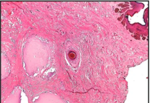
figure 3a, b, c: Three of the epidermal inclusion cysts next to fully developed lesion of scrotal calcinosis. Note the flattened squamous
common form, “dystrophic calcifica-tion”, implies an alteration in dermal collagen, induced by a predisposition like trauma, inflammation or collagen vascular disease, with normal calcium and phosphate levels in the blood. Literature revealed mostly single case reports, but series, consisting of 100 cases in the largest one (8), were also reported . However none of the re-ported cases showed abnormal cal-cium or phosphate levels in the blood chemistry. By positively exclusion of “metastatic calcification” theory the discusssion has made progress around “dystrophic” and “idiopathic” patho-genesis. While some authors believe it is truly idiopathic, others insist on dystrophic nature of the lesions with the evidence of preexisting epithelial structures in the nodules. But there is also a controversy between the authors who agree with the dystrophic process of the disease. The possible causes that may lead dystrophic calcification in the scrotal dermis were classified by Saladi et al (13) as; (i), calcific degen-eration of epidermoid cysts ; (ii), dys-trophic calcification of dartoic muscle and; (iii), calcification of ecrine sweat ducts. Although there have been
rela-tively much support, few authors (14) believe that degeneration and necrosis of dartoic muscle, creating an acido-philic amorphous necrotic mass trig-gers the distrophic calcific process. Role of epidermal cysts leads in the
patho-genesis of SC. Most authors have doc-umented that epithelial cysts them-selves calcify and turn into calcified nodules (15, 16, 17, 18). In a study of Shah et al (15) with 20 SC cases; 14 patients revealed epidermal cysts with a variable amount of keratin and calcification in the vicinity of scrotal nodules, and 6 revealed nodules with abundant calcification and remnant epithelial structures in them. Swinhart and Golitz (19) observed epidermal cysts in three cases of SC; some were calcified with partial or total preexist-ing epithelial walls with an inflama-tory reaction. Song et al (18) studied a patient with 51 scrotal nodules which 3 of epidermal cyts, 1 of calcified pilar cyst, 1 of calcified hibrid cyst, and the remaining indeterminate cysts some with calcification, and there were also purely calcified keratinous material associated with an inflammatory reac-tion and simple granular deposireac-tions of calcium within dermis. The largest study of SC, a combined prospective and retrospective study of 100 cases, conducted over a period of 15 years, was reported by Dubey et al (8). They detected a true cyst wall composed of compressed stratified squamous epi-thelium around calcium deposits in 60 of 100 patients. In the study the epithelium surrounded the deposits circumpherentially in 53, and partial-ly in 11 cases. They stressed both the frequency of epithelial components especially accompanying with smaller nodules, and the naked appearance of larger nodules exhibiting pure calcifi-cation. Immunohistochemical (IHC) examination was also performed in the studies to make sure that the nod-ules have epithelial components in or around their content. Ito et al (16) reported a SC case originating from eccrine cysts with the support of IHC evaluation that revealed a positive re-action for CEA and EMA in the
lu-minal cells and in the contents of a large cyst and ductal structures. Dini and Colafranceschi (17) also used an-tibodies against LMWCK, CAM5.2, cocktail of cytokeratin AE1/AE3 and CEA; and only a slight positivity for CK A1/A3 within one calcified nodule was observed within the whole lesion. Diversity of the lesions directed the ob-servers to suggest a multistage pro-cess for the development of clinically appearent SC from epidermal cysts. Swinhart and Golitz (19) targeted to inflammation of epidermal cysts as the initial event, and carried the process by order of dystrophic calcification of the cyst content, subsequent rup-ture of the cyst wall, a granulomatous proliferation, and with the cyst wall destruction remaining solely calcium deposits in the dermis in the end stage. Shah (15) and Song et al (18) defines a similar stepwise model with some discrapencies and details in each step. They presumed the presence of cysti-cally dilated epidermal cysts or hair follicles with a variable amount of keratin in the center as early or precur-sor lesion. The next step is the devel-opment of head specks of calcification around and within this dilated follicle or the epidermal cyst. Shah et al (15) consider that the laxity or shaving of the scrotal skin causes tethering of hair follicles with obstructive cysts, and when these coupled with the warmer temperature, calcification occurs. As the calcification increases the epithe-lium starts to disappear, producing cystic calcified lesions. Epithelial rem-nants in the form of ghost squames or keratin flakes are seen within the le-sions at this stage. They (15) proved the keratin flakes with CK stain by IHC. Calcific accumulation triggers a mononuclear inflammation and/or foreign body granuloma and the le-sions are surrounded by foamy mac-rophages and foreign body giant cells which resorbe the cyst walls and of ke-ratinous material. Finally with progres-sive calcification, total disappearance of the epithelial structures occur and large residual areas of calcification left behind with the classic picture of SC. figure 4: A dilated hair follicle with keratin
plugging (HE, x40)
figure 5: Calcium deposits partially lined
by flattened stratified squamous epithelium. Resorption of the cyst wall and keratinous material by mononuclear inflammation and foreign body granuloma. (HE, x100, x400)
RefeReNces
1. Bernardo BD, Huettner PC, Merritt DF, et al. Idiaopathic calcinosis cutis presenting as labial lesions in children: report of two cases with literature review. J Pediatr Adolesc Gy-necol. 1999; 12: 157-60
2. Lucke T, Fallowfield M, McHenry P. Idi-aopathic calcinosis cutis of the penis. Br J Dermatol. 1997; 137: 1025-6
3. Oh CK, Kwon KS, Cho SH, et al. Idio-pathic calcinosis of the areola of the nipple. J Dermatol. 2000; 27: 121-2
4. Shapiro L, Platt N, Torres-Rodriquez VM. Idiopathic calcinosis of the scrotum. Arch Dermatol. 1970; 102: 199-204
5. Schoeller T, Schubert HM, Wechselberger G. Idiopathic scrotal calcinosis – a pedin-culated rare variant. Journal of Plastic, Reconstructive & Aesthetic Surgery. 2008 April;61(4):466-7
6. C Hwang, Y J Kim, Y Lee, et al. Clin Exp Dermatol. 2009; 35: 159-60
7. J Hicheri, T Badri, B Fazaa, et al. Scrotal calcinosis: pathogenesis and case report. Acta Dermatoven APA. 2005; 14(2): 53-6 8. Dubey S, Sharma R, Maheshwari V.
Scro-tal calcinosis: Idiopathic or dystrophic? Dermatology Online Journal. 2010 Feb; 16(2):
5-9. S Chiummariello, A Figus, G Menichini, et al. Scrotal calcinosis: a very rare multiple clinical presentation. Clin Exp Dermatol. 2009; 34: 795-7
10. Ruiz Genao DP, Rios-Buceta L, Herrero L, et al. Massive scrotal calcinosis. Dermatol Surg. 2002; 28: 745
11. Chang C-H, Yang C-H, Hong H-S. Sur-gical pearl: Pinch-punch excisions for scrotal calcinosis. J Am Acad Dermatol. 2004; 50: 780-1
12. Angamuthu N, Pais AV, Chandrakala SR. Calcinosis cutis: A report of four cases. Trop Doct. 2003; 33: 50-2
13. Saladi R N, Persaud A N, Phelps R G, et al. Scrotal calcinosis: Is tha cause still un-known? J Am Acad Dermatol. 2004; 51: 97 14. Pabuccuoglu U, Canda MS, Guray M, et
al. The possible role of dartoic muscle de-generation in the pathogenesis of idiopathic scrotal calcinosis. Br J Dermatol. 2003; 148 :827-9
15. Shah V, and Shet T. Scrotal calcinosis results from calcification of cysts derived from hair follicles: A series of 20 cases evaluating the spectrum of changes resulting in scrotal cal-cinosis. Am J Dermatopathol. 2007 April; 29(2): 172-5
16. Ito A, Sakamoto F, Ito M. Dystrophic scrotal calcinosis originating from benign ecrine epithelial cysts. Br J Dermatol. 2001; 144: 146-50
With the composition of the findings Song et al (18) implied that the kera-tin content might be more resistant to inflammation than the attenuated cell wall. In this way after the rupture of cysts, the resorption of the wall is rapid and calcified keratinous material sur-rounded by inflammatory cells may be seen even when the cyst wall could not be observed anymore.
On the other hand, some of the authors insist on the idiopathic origin be-cause of the failure to show epithelial components even with IHC analysis. Wright et al (20); by performing an-tikeratin antibodies in multiple sec-tions of 63 lesions from nine patients, couldn’t show any epithelial structure in or around the calcified nodules. Ka-raca et al (21); also reported two cases with no remnants of epithelial cysts. Some other reports also emphasized the absence of true cyst wall (22,23). These authors concluded that SC is not a secondary phenomenon, and is truly idiopathic. There has been also a minority group of authors reporting different etiologic conditions such as; parasites (24), foreign body (4), vari-ous tumors (lymphangiomas,
xantho-mas, fibroxantho-mas, teratomas or gonado-blastomas)(25), and repetative trauma caused by using tractor continuously as a farmer (26) were reported. In the present case, it was realy confusing
that although the lesions did not ex-ist for a long period of time, we could hardly find just one focus of epithelial cyst wall within 20 nodules. Most of the nodules showed severe granuloma-tous reaction. Solely calcified nodules with fibrotic pseudocapsule were rela-tively rare. These findings support the hypothesis of Song et al (23) that the resolution of cyst wall occurs rapidly. We think that hence the calcification occurs, the epithelium disappears in a very short time, and the main process that takes time is the following steps. Whether the duration of lesions is short or long, if the observer defines an epi-thelial component in any of the nod-ules; then this lesion might be regarded as a newly developing epithelial cyst. It is clear that these lesions survive for many years, and certainly increase in number. There should be a repetitious predisposition, that we can observe both acute and chronic phases of the lesions simultaneously . Scrotum is a
common site where multiple epidermal cyst occur, and we think that epithe-lial cysts may be the certain repetitious cause. The absence of epithelial compo-nent in the lesions should not exclude the possibility of preexisting epithelial cyst. Rather than this; the absence of epithelium may correspond to the en-tirely chronic phase of the nodules with no new developing lesion. It is consid-ered particularly for long standing le-sions. We refer a special histopathologic evaluation of the lesions with multiple serial sections to multiply the chance of finding an epithelial component in the nodules. Neverthless no epithe-lium may be observed. Hence, the term “idiopathic” should be used for cases demonstrating no tip lesion that would facilitate the ethiology even with serial multiple sections. On particular occas-sions with preexisting leoccas-sions; epithelial cysts or alternatives, dystrophic calcifi-cation would be related to the distinct etiology. But in our opinion the main debate should continue in the direction of what is the principle cause that gives rise to calcification of the epithelial cysts in the scrotum.
17. Dini M, Colafrancesci M. Should scrotal calcinosis still be termed idiopathic? Am J Dermatopathol. 1998; 20: 399-402 18. Song DH, Lee KH, Kang WH. Idiopathic
calcinosis of the scrotum: histopathologic observations of fifty-one nodules. J Am Acad Dermatol. 1988; 19: 1095-101 19. Swinehart JM, Golitz LE. Scrotal
calcino-sis: dystrophic calcifications of epidermoid cysts. Arch Dermatol. 1982; 118: 985-8 20. Wright S, Navsaria H, Leigh M. Idiopathic
scrotal calcinosis is idiopathic. J Am Acad Dermatol. 1991; 24: 727-30
21. Karaca M, Taylan G, Akan M, et al. Idio-pathic scrotal calcinosis: Surgical treatment and histopathologic evaluation of etiology. Urology. 2010 April 8
22. Mehregan AH. Calcinosis cutis: A review of the clinical forms and report of 75 cases. Semin Dermatol. 1984; 3: 53-61
23. Yahya H, Rafindadi AH. Idiopathic scrotal calcinosis: a report of four cases and review
of the literature. Int J Dermatol. 2005 Mar; 44(3): 206-9
24. Browne SG. Calcinosis circumscripta of the scrotal wall. The etiological role Onchocer-ca volvulus. Br J Dermat. 1962; 74: 136-40 25. Saad AG, Zaatari GS. Scrotal calcinosis: is
it idiopathic? Urology. 2001 Feb;57(2):365 26. Feinstein A, Kahana M, Schewach-Millet
M, et al. İdiopathic calcinosis and vitiligo of the scrotum. J Am Acad Dermatol. 1984 Sep; 11(3): 519-20