352 © 2015 Indian Journal of Nuclear Medicine | Published by Wolters Kluwer - Medknow
352
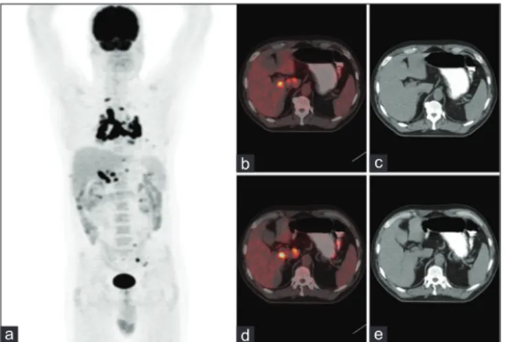
A 56‑year‑old man was admitted to the hospital due to cough, chest and back pain during 4‑month. A computed tomography (CT) of the chest revealed mediastinal lymphadenopathy and lung parenchymal lesion. The patient underwent 18F‑fluorodeoxyglucose positron emission tomography/CT (18F‑FDG PET/CT) imaging due to suspected lung cancer. The image showed abnormal 18F‑FDG uptake in the mediastinal, abdominal, and pelvic lymph nodes, and lungs [Figure 1a]. The images also demonstrated hypermetabolic activity in the inferior vena cava (IVC) and portal vein (PV) consistent with lesions of thrombosis [Figure 1a‑e]. These images were highly suggestive of lung cancer, widespread lymph nodes metastases, and tumor thrombosis in the IVC and PV. Biopsy of the mediastinal lymph node by mediastinoscopy revealed a noncaseating epithelial granulomatous lesion [Figure 2]. The patient was diagnosed as sarcoidosis and venous thrombosis (VT) in the IVC and PV. As the final diagnosis of a benign disease was determined, regular follow‑up by plain radiography was planned, the patient recovered spontaneously without treatment [Figure 3].
Venous thrombosis of sarcoidosis as an unusual
incidental finding on 18F-fluorodeoxyglucose
positron emission tomography/computed
tomography
Seval Erhamamci, Mehmet Reyhan, Ali Fuat Yapar, Tuba Canpolat1
Departments of Nuclear Medicine and 1Pathology, Faculty of Medicine, Baskent University, Turkey
Sarcoidosis is defined as a multisystem granulomatous disorder of unknown cause. Venous thrombosis (VT) in the sarcoidosis is rare. The routine use of 18F‑fluorodeoxyglucose positron emission tomography/computed tomography (18F‑FDG PET/CT) has resulted in clinicians detecting many incidental findings, which have proven to be clinically significant such as thrombosis. Here, we present a case with VT of sarcoidosis in the inferior vena cava and portal vein as an unusual incidental finding on 18F‑FDG PET/CT.
Keywords: 18F‑fluorodeoxyglucose, positron emission tomography/computed tomography, sarcoidosis, venous
thrombosis
Address for correspondence: Dr. Seval Erhamamci,
Department of Nuclear Medicine, Faculty of Medicine, Baskent University, Saray Street No. 1, 42080, Selcuklu, Konya, Turkey. E-mail: [email protected]
This is an open access article distributed under the terms of the Creative Commons Attribution-NonCommercial-ShareAlike 3.0 License, which allows others to remix, tweak, and build upon the work non-commercially, as long as the author is credited and the new creations are licensed under the identical terms.
For reprints contact: [email protected] ABSTRACT
How to cite this article: Erhamamci S, Reyhan M, Yapar AF, Canpolat T.
Venous thrombosis of sarcoidosis as an unusual incidental finding on 18F-fluorodeoxyglucose positron emission tomography/computed tomography. Indian J Nucl Med 2015;30:352-4.
Interesting
Images
Figure 1: Maximum intensity projection (a), transaxial fusion positron emission
tomography/computed tomography (b and d) and computed tomography (c and e) images show 18F‑fluorodeoxyglucose abnormal uptake in the mediastinal, abdominal, and pelvic lymph nodes and lungs due to sarcoidosis. These images show increased linear 18F‑fluorodeoxyglucose uptake in the inferior vena cava and portal vein (SUVmax: 21.7 and 12.7 respectively) consistent with lesions of
venous thrombosis
d
c b
a e
Access this article online Quick Response Code:
Website:
www.ijnm.in
DOI:
10.4103/0972-3919.164027
Erhamamci, et al.: Venous thrombosis of sarcoidosis on 18F-FDG PET/CT
Indian Journal of Nuclear Medicine | Vol. 30: Issue 4 | October-December, 2015 353
Sarcoidosis is defined as a multisystem granulomatous disorder of unknown cause. Lungs and thoracic lymph nodes are most commonly involved. Extrathoracic manifestations, usually associated with thoracic involvement, are seen in 25–50% of cases.[1] VT in the sarcoidosis is rare.[2‑6] In the literature, there are
few reported cases of sarcoidosis with VT, such as pulmonary embolism, thrombosis of brachiocephalic vein, upper and lower extremities veins, dural sinüs, renal vein, retinal veins, PV, and intracardiac thrombi.[2‑6] Also, both arterial and VT, multiple
venous thrombi, widespread and recurrent thrombophlebitis and the antiphospholipid syndrome has also been described in sarcoidosis.
VT results from a combination of hereditary and acquired factors. All these etiological factors are found among Virchow’s triad of hypercoagulability, endothelial injury, and venous stasis.[7] Hypercoagulability related to hematological or
neoplastic processes, venous stasis secondary to compression from a tumor, hematoma, or infectious process, and endothelial injury due to trauma or foreign body have all been implicated in the pathophysiology of VT. The list of known acquired risk factors has also grown in recent years.[7] Other acquired predispositions to VT include acquired
thrombophilias (antiphospholipid antibody syndrome), as well as environmental factors (combined oral contraceptive pill, hormone replacement therapy, pregnancy, obesity, malignancy, and chronic inflammatory conditions). Recent reports have further shown that a majority of autoimmune and immune‑mediated disorders are linked to an increased risk of VT.[7] Vena cava and PV thrombosis is frequently associated
with neoplastic disease. We present a case of sarcoidosis as an uncommon benign cause of IVC and PV thrombosis. Although the exact mechanism of thrombus formation in sarcoidosis is not yet known, venous stasis secondary to lymph node compression, local tissue thrombophilia in involved organs, and granulomatous phlebitis are potential mechanisms.[4] Although prothrombotic conditions workup
was not performed, there was no any predisposing history
for thrombosis in the present case. The development of the PV and IVC thrombosis in the present case may be owing to extrinsic compression.
18F‑FDG PET/CT has been widely used in the diagnosis and follow‑up of suspected or known malignancy.[8] However, infection
or inflammation such as osteomyelitis, tuberculosis, and sarcoidosis can also exhibit increased 18F‑FDG uptake. Thus, 18F‑FDG PET/ CT have also been increasingly used for the assessment of infectious and inflammatory diseases, including sarcoidosis.[1,8‑10] The routine
use of 18F‑FDG PET/CT has resulted in clinicians detecting many incidental findings, which have proven to be clinically significant such as thrombosis.[8‑10] The recognition of this rare complication
by 18F‑FDG PET/CT is essential for the accurate management of patients. We report, to our knowledge, the first case of sarcoidosis with VT as an incidental finding on 18F‑FDG PET/CT. VT is a well‑recognized, relatively common complication in cancer patients and a significant cause of morbidity and mortality. VT is managed with anticoagulant therapy. In contrast to tumor thrombosis is another known cause of thrombosis in patients with cancer requires aggressive multimodality management.[8] Therefore, the correct
diagnosis of VT can change patient management and might facilitate the start of anticoagulant therapy.[8] However, so far, there is no
definitive strategy to manage thrombosis in sarcoidosis. There are no officially approved guidelines or consensus management available regarding the treatment of sarcoidosis as it is a rare disease with rarer clinical manifestations.[11] Therefore, most of the management
decisions have to be individualized depending on the local expertise. In our case, there was no specific treatment initiated for IVC and PV thrombosis as he is currently asymptomatic. It was planned to keep him in regular follow‑up. Further research is needed to elucidate the exact cause of thrombosis and defining management modalities of thrombosis in sarcoidosis. We think that any reported similar case will be of great interest as a guide to better assess the outcomes of patients with sarcoidosis.
Financial support and sponsorship
Nil.
Figure 2: Sarcoidosis granulomas are noncaseating granuloma, containing
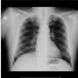
multinucleated giant cell, lymphocytes, and epithelioid cells (H and E, ×200) Figure 3: Follow‑up plain radiography of the chest revealing normal findings
Erhamamci, et al.: Venous thrombosis of sarcoidosis on 18F-FDG PET/CT
354 Indian Journal of Nuclear Medicine | Vol. 30: Issue 4 | October-December, 2015
354
Conflicts of interest
There are no conflicts of interest. REFERENCES
1. Sobic‑Saranovic D, Artiko V, Obradovic V. FDG PET imaging in sarcoidosis. Semin Nucl Med 2013;43:404‑11.
2. Rebeiz TJ, Mahfouz R, Taher A, Charafeddine KH, Kanj N. Unusual presentation of a sarcoid patient: Multiple arterial and venous thrombosis with chest lymphadenopathy. J Thromb Thrombolysis 2009;28:245‑7.
3. Selvi A, Diakou M, Giannopoulos S, Zikou AK, Argyropoulou MI, Kyritsis AP. Cerebral venous thrombosis in a patient with sarcoidosis. Intern Med 2009;48:723‑5.
4. Vahid B, Wildemore B, Marik PE. Multiple venous thromboses in a young man with sarcoidosis: Is there a relation between sarcoidosis and venous thrombosis? South Med J 2006;99:998‑9.
5. McLaughlin AM, McNicholas WT. Sarcoidosis presenting as upper
extremity venous thrombosis. Thorax 2003;58:552.
6. Satti SD, Bartholomew J, Gordon SM, Longworth DL, Adal KA. Antiphospholipid antibody syndrome in a patient with neurosarcoidosis. Vasc Med 1999;4:37‑9.
7. Zöller B, Li X, Sundquist J, Sundquist K. Autoimmune diseases and venous thromboembolism: A review of the literature. Am J Cardiovasc Dis 2012;2:171‑83.
8. Erhamamci S, Reyhan M, Nursal GN, Torun N, Yapar AF. Incidental diagnosis of tumor thrombosis on FDG PET/CT imaging. Rev Esp Med Nucl Imagen Mol 2015;pii: S2253‑654X00054‑2.
9. Ambrosini V, Zompatori M, Fasano L, Nanni C, Nava S, Rubello D,
et al. (18) F‑FDG PET/CT for the assessment of disease extension and
activity in patients with sarcoidosis: Results of a preliminary prospective study. Clin Nucl Med 2013;38:e171‑7.
10. Rubini G, Cappabianca S, Altini C, Notaristefano A, Fanelli M, Stabile Ianora AA, et al. Current clinical use of 18FDG‑PET/CT in patients with
thoracic and systemic sarcoidosis. Radiol Med 2014;119:64‑74.
11. Krishnamoorthy G, Ray G, Agarwal I, Kumar S. Unusual presentation of sarcoidosis in a child. Rheumatol Int 2012;32:1453‑5.